1
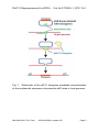
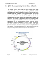
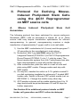
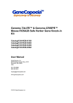
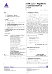
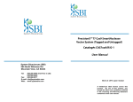
PhiC31 Reprogrammer & miPSCs Catalog#s FC300A-1, SC211A-1 User Manual System Biosciences (SBI) 265 North Whisman Rd. Mountain View, CA 94043 Tel: 888.266.5066 (Toll Free in US) 650.968.2200 Fax: 650.968.2277 E-mail: [email protected] Web: www.systembio.com (ver. 4 - 140425) Please see product certificate for proper storage conditions (ver. 4 - 140425) A limited-use label license covers this product. By use of this product, you accept the terms and conditions outlined in the Licensing and Warranty Statement contained in this user manual. PhiC31 Reprogrammer & miPSCs Cat. #s FC300A-1, SC211A-1 Contents I. Introduction .............................................................................2 A. Background of ϕC31 Integrase Technology .......................2 B. ϕC31 Reprogramming Vector Maps & Details ......................4 C. List of Components ................................................................5 II. Protocol for Deriving Mouse-Induced Pluripotent Stem Cells using the ϕC31 Reprogrammer on MEF source cells .....................7 A. Mouse Induced Pluripotent Stem Cell Generation ................7 III. Non-Viral ϕC31 Integrase-derived Mouse iPS Cells ...........8 A. ϕC31 Generated Mouse Induced Pluripotent Cell Line ..........8 B. Culture Conditions for MEF Feeder Cells ...............................9 C. Growth Conditions for PhiC31 Mouse iPS cells ...................13 D. Validation of PhiC31 Mouse iPS cells ...............................17 IV. References ........................................................................19 V. Technical Support .................................................................19 VI. Licensing and Warranty Statement ...................................20 888-266-5066 (Toll Free) 650-968-2200 (outside US) Page 1 System Biosciences (SBI) I. User Manual Introduction A. Background of ϕC31 Integrase Technology The ϕC31 integrase is a site-specific recombinase encoded within the genome of the bacteriophage ϕC31. The ϕC31 integrase mediates recombination between two ~34 base pair sequences termed attachment sites (att), one found in the phage and the other in the bacterial host. This serine integrase has been show to function efficiently in many different cell types including mammalian cells. In the presence of ϕC31 integrase, an attB containing donor plasmid can be unidirectionally integrated into a target genome through recombination at sites with sequence similarity to the native attP site. These sites are termed pseudo-attP sites. ϕC31 integrase can integrate a donor plasmid of any size, as a single copy, and requires no cofactors. The integrated transgenes are stably expressed and heritable. Page 2 ver. 4-042514 www.systembio.com PhiC31 Reprogrammer & miPSCs Cat. #s FC300A-1, SC211A-1 Fig. 1. Schematic of the ϕC31 Integrase mediated recombination of donor plasmid sequence into pseudo-attP sites in host genome 888-266-5066 (Toll Free) 650-968-2200 (outside US) Page 3 System Biosciences (SBI) User Manual B. ϕC31 Reprogramming Vector Maps & Details The classic Oct3/4, Sox2, Klf4 and cMyc factors have been adapted to the integrase system. The pCOBLW phiC31 reprogramming vector (catalog# FC305A-1) features the iPSC factors driven by a potent CAGs promoter with the factors separated by P2A elements. HS4 insulators flank the expression cassette and a functional attB site directs the integration of the entire plasmid into pseudo attP sites in any genome. The vector is engineered with FRT sites for later excision using standard FLP recombinase methods. After cotransfection of the reprogramming vector with the phiC31 expression plasmid (catalog# FC200PA-1) and integration, you can monitor for successful transfection with GFP fluorescence and select for positive colonies using the built-in Neo marker. The reprogramming iPSC factor donor vector and the phiC31 expression plasmid maps are shown below. Page 4 ver. 4-042514 www.systembio.com PhiC31 Reprogrammer & miPSCs Cat. #s FC300A-1, SC211A-1 C. List of Components List of Components for FC300A-1: Reprogramming Kit Each Non-Viral PhiC31 Integrase Mouse iPSC Reprogramming Kit includes 10ug of plasmid DNA for FC200PA-1 and FC305A-1. FC300A-1 PhiC31 Integrase Plasmid (FC200PA-1) & PhiC31 Reprogrammer (FC305A-1) 888-266-5066 (Toll Free) 650-968-2200 (outside US) 10ug each Page 5 System Biosciences (SBI) User Manual List of Components for SC211A-1: PhiC31 Mouse iPS Cells Each iPS cell line comes as one vial of MEF-derived PhiC315 generated Mouse iPS cells containing ~2 x 10 cells. SC211A-1 Mouse iPS Cell Line [PhiC31 miPSCs] 2x10^5 cells The product is shipped on dry ice and should be immediately stored in the gas phase of liquid nitrogen. In general, iPS cells are challenging to culture and should only be operated by researchers experienced in the intricacies of mouse embryonic stem (mES) cell culture. The methods for culture are nearly identical to mES cell culture, although more careful maintenance will be required. Appropriate feeder cells for mouse iPS cell culture can be obtained from commercial sources, or generated through Mitomycin C treatment of mouse embryonic fibroblasts (MEFs). See protocol on Pgs. 9-12 for more details on generating feeder cells. Page 6 ver. 4-042514 www.systembio.com PhiC31 Reprogrammer & miPSCs II. Cat. #s FC300A-1, SC211A-1 Protocol for Deriving MouseInduced Pluripotent Stem Cells using the ϕC31 Reprogrammer on MEF source cells A. Mouse Generation Induced Pluripotent Stem Cell The following protocol has been optimized for mouse embryonic fibroblasts (MEF) cells as described in Karow M. et al. Other source cells may require transfection optimization for efficient reprogramming. In general, reprogramming requires a single transfection of approximately 3 µg per well in a six-well plate. 1) Use the MEF nucleofector kit I (Lonza) and the program T20 according to the manufacturer’s instructions. 2) Nucleofect with 3 µg total DNA using the PhiC31 Integrase Plasmid (FC200PA-1) & PhiC31 Reprogrammer Donor Plasmid (FC305A-1) at a 1:1 ratio by mass. The Neon transfection system from Life Technologies has also been demonstrated to work with similar efficiency. 3) After nucleofection, plate the cells in a well of a six-well plate using MEF media. Change media each day with MEF media. 4) On day two, transfer the cells to a 0.1% gelatin coated10 cm dish containing irradiated or mitomycin C-treated MEF feeder cells. Switch to Mouse iPSC growth media (cat. no. #SC200M-1). Change the media every other day. 5) GFP positive colonies will be visible starting from 8 to 12 days and should be picked between 20 and 26 days for further expansion. See Section III for additional protocol details on MEF feeder cell generation and iPSC culture techniques. 888-266-5066 (Toll Free) 650-968-2200 (outside US) Page 7 System Biosciences (SBI) III. User Manual Non-Viral ϕC31 Integrase-derived Mouse iPS Cells A. ϕC31 Generated Mouse Induced Pluripotent Cell Line Mouse induced pluripotent stem cells (iPSCs, Cat# SC211A-iPSC) were generated by nucleofecting genetically unmodified C57BL/6 mouse embryonic fibroblasts with the pCOBLW C31 Integrase Mouse Reprogramming Donor plasmid (Cat# FC305A-1) and the C31 Integrase expression plasmid (Cat# FC200PA-1) for stable integration into the mouse genome (Karow 2011). The Reprogramming Donor plasmid encodes the four murine transcription factors (Oct4, Sox2, Klf4, and c-Myc) that have been shown to induce the reprogramming of somatic cells to a pluripotent state. See Section II of this User Manual for more details on the reprogramming protocol used to make this cell line. Due to the specificity of recombination using the C31Integrase system, the iPSCs are expected to have, on average, one copy of the OSKM plasmid per cell, which reduces the risk of insertional mutagenesis effects [integration analysis data available on Pg. 18]. The cells were derived using morphological selection criteria and GFP expression without drug selection. When cultured under standard mouse ES cell culture conditions, the morphology of SBI mouse iPSCs are identical to that of mouse ES cells. The cells also express the pluripotency markers SSEA-1, Nanog, Sox2, and Oct4 and demonstrate strong endogenous AP activity. Mouse iPS cells from SBI are provided at passage 10 and can be passaged 50 times before differentiation. Page 8 ver. 4-042514 www.systembio.com PhiC31 Reprogrammer & miPSCs Cat. #s FC300A-1, SC211A-1 B. Suggested Conditions for MEF Feeder Cells Mouse iPS cells should be grown on a feeder cell layer. Appropriate feeder cells for mouse iPS cell culture can be obtained from commercial sources, or generated through Mitomycin C treatment of mouse embryonic fibroblasts (MEFs). We have included a suggested protocol for MEF recovery, culturing, and freezing, however, we would suggest following the MEF manufacturer’s recommendations if provided with the MEF cells. 1. Required media and reagents Reagent Information MEF Medium DMEM containing 10% FBS, 2 mM glutamine, -4 1x10 M nonessential amino acids and 50 U and 50 µg/ml penicillin and streptomycin. 2x Cold Freezing Media 20% DMSO and 80% FBS Mitomycin C solution 1 mg/ ml 2. Gelatin treatment of plates for MEF feeder cells 1) Add enough sterile/ autoclaved 0.1% gelatin to cover the bottom of the wells. Approximate amounts: 10cm plate – 5 ml 6 well – 1.5 ml/ well 24 well – 0.5 ml/ well 96 well – 100 µl/ well 888-266-5066 (Toll Free) 650-968-2200 (outside US) Page 9 System Biosciences (SBI) User Manual 2) Incubate the gelatin-coated dishes for at least 15 min at 37°C. 3) Aspirate excess gelatin solution before use. 3. Thawing MEF cells To insure the highest level of viability, be sure to warm medium to 37°C before using it on the cells. Cells should be plated at a 4 2 minimum cell density of 10 cells/ cm . 1) Remove the vial from liquid nitrogen and thaw quickly in a 37°C water bath. 2) Remove the vial from the water bath as soon as the cells are half-thawed, and sterilize the tube by spraying with 70% ethanol. 3) Transfer the cells with 10 ml of MEF medium to a 15 mL conical tube and pellet the cells by centrifugation at 200 g for 5 min. 4) Discard the supernatant and resuspend the cells with 10 ml fresh MEF medium and plate the cells on gelatin4 2 coated plates at seed density of 10 cells/ cm . 5) Incubate at 37°C with 5% CO2, until the cells reach 8090% confluency. 6) Change medium twice a week or when pH decreases. 4. Passaging MEF cells Cells should be split when they reach confluency. We recommend 4 2 splitting the cells based on 0.5x10 cells/ cm . 1) Discard the medium and wash the cells once with PBS. 2) Aspirate PBS, and add 2 ml per T75 flask of 0.25% trypsin-EDTA, and incubate for 2 min. Page 10 ver. 4-042514 www.systembio.com PhiC31 Reprogrammer & miPSCs Cat. #s FC300A-1, SC211A-1 3) Add 5 ml of MEF medium, and break up the cell clumps by gently pipetting up and down several times. 4) Transfer cells into a conical tube and centrifuge at 200 g for 5 min. 5) Discard the supernatant and resuspend the cell pellet in 10 ml MEF medium. 4 6) Count the number of cells, plate cells at 0.5x10 cells/ cm 2 and incubate at 37°C with 5% CO2. 5. Freezing MEF cells 1) Follow steps 1-4 from the Passaging MEF cells protocol (above). 2) Discard the supernatant, and resuspend the pellet in MEF medium. 3) Count the number of cells and adjust the cell suspension 6 to 4 x 10 cells/ ml. 4) Add equal volume of cold 2X Freezing Media to the cell suspension. 6 5) Aliquot 1 ml of suspension into each cryovial (2 x 10 cells/ vial). 6) Place the vials in a cell-freezing container and keep it at -80°C overnight. 7) Transfer the vials to a liquid nitrogen tank for long-term storage. 888-266-5066 (Toll Free) 650-968-2200 (outside US) Page 11 System Biosciences (SBI) User Manual 6. Mitomycin C treatment of MEF Mitomycin C acts to halt the division of MEF cells so that they can be used as a feeder layer for miPS cells. MEF cells should be at confluence when treated with mitomycin C. 1) Add 6 ml of fresh MEF medium contain 50 µl of mitomycin C solution (1 mg/ ml) to one T75 flask of confluent MEF cells, and swirl it briefly. The final concentration of mitomycin C is 8 µg/ ml. 2) Incubate at 37°C for at least 3 hrs. 3) Aspirate the mitomycin C-containing medium off the cells and wash the cells twice with 10 ml PBS. 4) Aspirate PBS and add 1 ml of 0.25% trypsin-EDTA, swirl to cover the entire surface, and incubate for 2 min at room temperature. 5) Add 5 ml MEF medium and break up the cells to a singlecell suspension by pipetting up and down. Count the number of cells. 6 6) Seed the cells on gelatin-coated dishes (3 x 10 cells per 5 100-mm dish, or 5 x 10 cells per well of a 6-well plate). 7) Cells should be ready to use by the next day. Page 12 ver. 4-042514 www.systembio.com PhiC31 Reprogrammer & miPSCs Cat. #s FC300A-1, SC211A-1 C. Growth Conditions for PhiC31 Mouse iPS cells 1. Required Media and Reagents Reagent Information Mouse iPSC Growth Medium SC200M-1 2x Cold Freezing Media 20% DMSO and 80% ES-FBS Trypsin-EDTA GIBCO 1. Thawing Mouse iPS cells To insure the highest level of viability, be sure to warm medium to 37°C before using it on the cells. 1) Remove the vial from liquid nitrogen and thaw quickly in a 37°C water bath. 2) Remove the vial from the water bath as soon as the cells are half-thawed, and sterilize by spraying with 70% ethanol. 3) Transfer the cells with 10 ml of mouse iPSC medium to a 15 mL conical tube and pellet the cells by centrifugation at 200 g for 5 min. 4) While centrifuging, remove MEF medium from the feeder cell plates, and wash the wells twice with DMEM. Then add 1 ml of mouse iPSC Medium. 5) Discard the supernatant from the mouse iPS cells, and resuspend cells with 1 ml fresh mouse iPSC medium. 888-266-5066 (Toll Free) 650-968-2200 (outside US) Page 13 System Biosciences (SBI) User Manual Plate the cells on MEF feeder cells in a 6-well plate. 6) Incubate at 37°C with 5% CO2 until the cells reach 80% confluency. The mouse iPSC media must be changed every day. Mouse iPS Cells Express GFP 2. Maintenance of mouse iPS cells It is important not to keep mouse iPS cells in culture for long period of time without passaging, to maintain the pluripotency. 1) Aspirate the medium and wash the cells twice with 1 ml PBS. 2) Remove PBS completely and add 0.7 ml of 0.05% trypsinEDTA solution, and incubate at 37°C for 10 min. 3) While incubating, remove a 6-well plate of feeder cells (mitomycin C-treated MEFs). Aspirate the medium and add 2 ml of mouse iPSC medium to each well. 4) Remove the plate containing mouse iPS cells from the incubator and swirl to dislodge the cells from the bottom of the plate. Page 14 ver. 4-042514 www.systembio.com PhiC31 Reprogrammer & miPSCs Cat. #s FC300A-1, SC211A-1 5) Add 2 ml of mouse iPSC medium, and suspend the cells by pipetting up and down to single cell suspension. 6) Transfer the cell suspension to a 15 ml conical tube and spin the cells at 200 g for 5 min. 7) Add 2 ml of iPSC medium to the plate and suspend the cells by pipetting up and down to single cell suspension. 8) Distribute ~0.2 ml of the mouse iPS cell suspension to each well of the 6-well plate. Right after plating iPS cells, gently swirl the plate back-and-forth and side-to-side and incubate at 37°C with 5% CO2 until the cells reach 80% confluency. 9) The mouse iPSC media must be changed every day and mouse iPS cells subcultured ~1:10 every 2-3 days. Track the passage number of the iPS cells 888-266-5066 (Toll Free) 650-968-2200 (outside US) Page 15 System Biosciences (SBI) 3. User Manual Freezing mouse iPS cells 1) Grow cells to the exponential phase in a 6-well plate. 2) Aspirate the medium, and wash the cells twice with 2 ml of PBS. 3) Aspirate the medium, and wash the cells twice with 2 ml of PBS. 4) Add 0.7 ml 0.05% trypsin-EDTA and incubated for 10 min at 37°C. 5) Add 2 ml of mouse iPSC medium, and suspend the cells by pipetting up and down to single cell suspension. 6) Transfer the cell suspension to a 15 ml conical tube, count the number of cells and spin the cells at 200 g for 5 min. 7) Discard the supernatant, and resuspend the cells with 6 mouse iPSC medium to the concentration of 1x10 cells per ml. 8) Add equal volume of 2x freezing medium and aliquot it at 0.5 ml per vial. 9) Put the vials in a cell-freezing container, and store them at -80°C overnight. 10) Transfer the vials to a liquid nitrogen tank for long-term storage. Page 16 ver. 4-042514 www.systembio.com PhiC31 Reprogrammer & miPSCs Cat. #s FC300A-1, SC211A-1 D. Validation of PhiC31 Mouse iPS cells Phase Sox2 Alkaline Phosphatase Nanog Oct4 SSEA-1 Stem cell markers for Oct4, Sox2, SSEA-1 and Nanog were determined by immunocytochemistry using primary antibodies for SSEA1 (Millipore), Oct4 (Abcam), Sox2 (Abcam), and Nanog (Abcam) followed by Alexa Fluor fluorescent-labeled secondary antibodies (Invitrogen). Detection of Alkaline Phosphatase activity was performed using the AP Detection Kit (Millipore). 888-266-5066 (Toll Free) 650-968-2200 (outside US) Page 17 System Biosciences (SBI) User Manual Integration Analysis A major advantage of using the phiC31 integrase system is that copy number control is in your hands. The majority of clones that undergo integration events have single insertions. To verify the copy numbers in your targeted cells, you can perform Southern blot analysis on their genomic DNA. Shown below is an example of mouse genomic integration analyses using separate restriction digests with Eco01091 and EcoRV with 10ug gDNA. The reason for two separate enzyme digests in to assess the copy number status. The probe used in these Southern blot analyses was for GFP present in the pCOBLW iPSC integrase vector. The data below show the copy number status for 16 different iPSC lines and demonstrate that 14 out of 16 lines show single insertions (88%). Page 18 ver. 4-042514 www.systembio.com PhiC31 Reprogrammer & miPSCs IV. Cat. #s FC300A-1, SC211A-1 References Karow, M. et al.2011. Site-specific recombinase strategy to create induced pluripotent stem cells efficiently with plasmid DNA. Stem Cells 29(11):1696-704. Rossant, J. 2007. Stem cells: The magic brew. Nature 448, 260– 262. Takahashi, K. and Yamanaka, S. 2006. Induction of pluripotent stem cells from mouse embryonic and adult fibroblast cultures by defined factors. Cell 126: 663–676. Takahashi K. et al. 2007. Induction of pluripotent stem cells from adult human fibroblasts by defined factors. Cell. 131: 861–72. Park, IH et al. 2008. Reprogramming of human somatic cells to pluripotency with defined factors. Nature. 451:141–6. Baker, Monya 2007. Adult cells reprogrammed to pluripotency, without tumors. Nature Reports Stem Cells. 2007–12–11. Nakagawa, M et al. 2008. Generation of induced pluripotent stem cells without Myc from mouse and human fibroblasts. Nature Biotechnology. 26: 101–106. Okita, K, et al. 2007. Generation of germline-competent induced pluripotent stem cells. Nature. 448:313–7 Yu, J. et al. 2007. Induced Pluripotent Stem Cell Lines Derived from Human Somatic Cells. Science 318: 1917–1920. V. Technical Support For more information about SBI products or to download manuals in PDF format, please visit our website: 888-266-5066 (Toll Free) 650-968-2200 (outside US) Page 19 System Biosciences (SBI) User Manual http://www.systembio.com For additional information or technical assistance, please call or email us at: [email protected] 650-968-2200 VI. Licensing Statement and Warranty Limited Use License Use of the human and mouse iPS cells (i.e., the “Product”) is subject to the following terms and conditions. If the terms and conditions are not acceptable, return all components of the Product to System Biosciences (SBI) within 7 calendar days. Purchase and use of any part of the Product constitutes acceptance of the above terms. The purchaser of the Product is granted a limited license to use the Product under the following terms and conditions: The Product shall be used by the purchaser for internal research purposes only. The Product is expressly not designed, intended, or warranted for use in humans or for therapeutic or diagnostic use. The Product may not be resold, modified for resale, or used to manufacture commercial products without prior written consent of SBI. This Product should be used in accordance with the NIH guidelines developed for stem cell research. SBI has pending patent applications related to the Product. For information concerning licenses for commercial use, contact SBI. Page 20 ver. 4-042514 www.systembio.com PhiC31 Reprogrammer & miPSCs Cat. #s FC300A-1, SC211A-1 Purchase of the product does not grant any rights or license for use other than those explicitly listed in this Licensing and Warranty Statement. Use of the Product for any use other than described expressly herein may be covered by patents or subject to rights other than those mentioned. SBI disclaims any and all responsibility for injury or damage which may be caused by the failure of the buyer or any other person to use the Product in accordance with the terms and conditions outlined herein. Limited Warranty SBI warrants that the Product meets the specifications described in this manual. If it is proven to the satisfaction of SBI that the Product fails to meet these specifications, SBI will replace the Product or provide the purchaser with a refund. This limited warranty shall not extend to anyone other than the original purchaser of the Product. Notice of nonconforming products must be made to SBI within 30 days of receipt of the Product. SBI’s liability is expressly limited to replacement of Product or a refund limited to the actual purchase price. SBI’s liability does not extend to any damages arising from use or improper use of the Product, or losses associated with the use of additional materials or reagents. This limited warranty is the sole and exclusive warranty. SBI does not provide any other warranties of any kind, expressed or implied, including the merchantability or fitness of the Product for a particular purpose. SBI is committed to providing our customers with high-quality products. If you should have any questions or concerns about any SBI products, please contact us at (888) 266-5066. © 2012 System Biosciences (SBI). 888-266-5066 (Toll Free) 650-968-2200 (outside US) Page 21