1
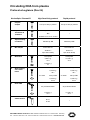
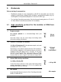
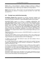
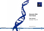
Circulating DNA
from plasma
User manual
NucleoSpin® Plasma XS
June 2014 / Rev. 04
Circulating DNA from plasma
Protocol-at-a-glance (Rev. 04)
NucleoSpin® Plasma XS
1
1a
2
3
4
Prepare
sample
Optional:
Proteinase K
treatment
Adjust binding
conditions
Mix sample
Bind DNA
High Sensitivity protocol
Rapid protocol
Use up to 240 μL plasma
Use up to 200 μL plasma
Add 20 μL Proteinase K
Mix
/
Incubate at 37 °C for 10 min
Add 360 μL BB
Add 300 μL BB
Invert tube 3 x
Invert tube 3 x
Vortex 3 s
Vortex 3 s
Spin down briefly
Spin down briefly
Load lysate
Load lysate
2,000 x g,
30 s
11,000 x g,
30 s
11,000 x g,
5s
5
Wash and dry
silica membrane
1st wash
500 μL WB
1st wash
500 μL WB
11,000 x g,
30 s
2nd wash
250 μL WB
11,000 x g,
30 s
2nd wash
250 μL WB
11,000 x g,
3 min
6
7
11,000 x g,
3 min
Elute DNA
Removal of residual ethanol
20 μL Elution Buffer
20 μL Elution Buffer
11,000 x g,
30 s
11,000 x g,
30 s
90 °C,
8 min
/
MACHEREY-NAGEL GmbH & Co. KG · Neumann-Neander-Str. 6–8 · 52355 Düren · Germany
Tel.: +49 24 21 969-270 · Fax: +49 24 21 969-199 · [email protected] · www.mn-net.com
Circulating DNA from plasma
Table of contents
1 Components 4
1.1 Kit contents
4
1.2 Consumables and equipment to be supplied by user
5
1.3 About this user manual
5
2 Product description
6
2.1 The basic principle
6
2.2 Kit specifications
6
2.3 Handling of sample material
8
2.4 Elution procedures
8
2.5 Removal of residual traces of ethanol for highest PCR sensitivity
9
2.6 Stability of isolated DNA
10
3 Storage conditions and preparation of working solutions
11
4 Safety instructions
12
5Protocols
14
5.1 High sensitivity protocol for the isolation of DNA from plasma
14
5.2Rapid protocol for the isolation of DNA from plasma
17
6Appendix
19
6.1Troubleshooting
19
6.2 Ordering information
20
6.3References
20
6.4 Product use restriction / warranty
23
MACHEREY-NAGEL – 06 / 2014, Rev. 04
3
Circulating DNA from plasma
1
Components
1.1 Kit contents
NucleoSpin® Plasma XS
10 preps
50 preps
250 preps
740900.10
740900.50
740900.250
Binding Buffer BB
5 mL
22 mL
110 mL
Wash Buffer WB
10 mL
50 mL
250 mL
Elution Buffer*
13 mL
13 mL
13 mL
Proteinase K
(lyophilized)**
6 mg
30 mg
2 x 75 mg
1.8 mL
1.8 mL
8 mL
NucleoSpin® Plasma XS
Columns (red rings –
plus Collection Tubes)
10
50
250
Collection Tubes (2 mL)
20
100
500
User manual
1
1
1
REF
Proteinase Buffer PB
* Composition of Elution Buffer: 5 mM Tris/HCl, pH 8.5
**For preparation of working solutions and storage condistions see section 3.
4
MACHEREY-NAGEL – 06 / 2014, Rev. 04
Circulating DNA from plasma
1.2 Consumables and equipment to be supplied by user
Consumables
•
•
1.5 mL microcentrifuge tubes
Disposable pipette tips
Equipment
•
Manual pipettors
•
Vortex mixer
•
•
•
Centrifuge for microcentrifuge tubes
Heating-block for incubation at 90 °C
Personal protection equipment (lab coat, gloves, goggles)
1.3 About this user manual
The manual provides two procedures differing in the number of handling steps, speed
and performance. The High sensitivity procedure is recommended if highest DNA
yield and concentration is required. The Rapid procedure is recommended if shortest
preparation time is required.
It is strongly recommended reading the detailed protocol sections of this user manual if
the NucleoSpin® Plasma XS kit is used for the first time. Experienced users, however,
may refer to the Protocol-at-a-glance instead. The Protocol-at-a-glance is designed
to be used only as a supplemental tool for quick referencing while performing the
purification procedure.
All technical literature is available on the internet at www.mn-net.com.
Please contact Technical Service regarding information about changes of the current
user manual compared to previous revisions.
MACHEREY-NAGEL – 06 / 2014, Rev. 04
5
Circulating DNA from plasma
2
Product description
2.1 The basic principle
The NucleoSpin® Plasma XS kit is designed for the efficient isolation of circulating DNA
from human blood plasma. Fragmented DNA as small as 50–1000 bp can be purified with
high efficiency. Due to a special funnel design the NucleoSpin® Plasma XS Columns
allow very small elution volumes (5–30 μL) which results in highly concentrated DNA.
The protocol follows state-of-the-art bind-wash-elute procedures: After mixing of
a plasma sample with the binding buffer, the mixture is applied to the NucleoSpin®
Plasma XS Column. Upon loading of the mixture DNA binds to a silica membrane.
Two subsequent washing steps efficiently remove contaminations and highly pure DNA
is finally eluted with 5–30 μL of a slightly alkaline elution buffer of low ionic strength
(5 mM Tris-HCl, pH 8.5).
2.2 Kit specifications
6
•
The NucleoSpin® Plasma XS kit is recommended for the isolation of fragmented
cell-free DNA from human EDTA plasma, serum, and bronchial lavage.
•
The NucleoSpin® Plasma XS kit is designed for high recovery, especially of
fragmented DNA in a range of 50–1000 bp.
•
Up to 240 μL plasma can be used as sample material with a single column
loading step. DNA yield strongly depends on the individual sample, but is
typically in the range of 0.1 to 100 ng DNA per mL plasma. Up to 720 μL plasma
can be used with three column loadings.
•
Elution can be performed with as little as 5–30 μL elution buffer. DNA is ready
to use for downstream applications like real-time PCR or others.
•
The preparation time is approximately 15–30 min for 6–12 plasma samples.
MACHEREY-NAGEL – 06 / 2014, Rev. 04
Circulating DNA from plasma
Table 1: Kit specifications at a glance
Parameter
NucleoSpin® Plasma XS
Sample material
Up to 240 μL EDTA plasma (single column loading)
Average yield
Typically in a range of 0.1–100 ng per mL plasma,
depending on sample
(depending on kind of patient samples, yield can be much higher).
Elution volume
5–30 μL
Preparation time
High sensitivity procedure: 22–27 min/6 preps
Rapid procedure: 15–20 min/6 preps
Format
XS spin column
DNA yield from human plasma
DNA amounts from less than 0.1 ng DNA per mL of plasma up to several 100 ng DNA
per mL of plasma have been reported (Chiu et al. 2006; Chun et al. 2006; Fatouros et
al. 2006; Lazar et al. 2006; Rainer et al. 2006; Rhodes et al. 2006; Schmidt et al. 2005).
The content of DNA in plasma depends on: condition of the donor, sampling and
handling of the blood, plasma preparation and DNA isolation method, DNA quantification
method, and others.
Size of circulating DNA
A good portion of the cell-free DNA in plasma results from apoptotic cells. Therefore,
a considerable percentage of this circulating nucleosomal DNA is known to be highly
fragmented. However, the degree of fragmentation and the ratio of fragmented DNA to
high molecular weight DNA depends on several parameters like origin of the DNA (e.g.,
fetal, tumor, microbial DNA), health of the blood donor, blood sampling procedure, and
handling of the sample.
The performance of many downstream applications depends on the efficient isolation
even of smallest DNA fragments (Chan et al. 2006, 2005, 2004, 2003; Deligezer et al.
2006; Giacona et al. 1998; Hanley et al. 2006; Hromadnikova et al. 2006; Jiang et al.
2006; Koide et al. 2005; Li et al. 2006, 2005, 2004; Wang et al. 2004). According to this
the NucleoSpin® Plasma XS purification system is designed for the efficient isolation
of highly fragmented DNA in a range of 50–1000 bp. Within this range fragments are
recovered with similar high efficiency.
MACHEREY-NAGEL – 06 / 2014, Rev. 04
7
Circulating DNA from plasma
2.3 Handling of sample material
Several publications indicate strong influence of blood sampling, handling, storage,
and plasma preparation on DNA yield and DNA quality (Page et al. 2006; Sozzi et
al. 2005; Chan et al. 2005; Lam et al. 2004; Jung et al. 2003). Therefore it is highly
recommended keeping blood sampling procedure, handling, storage, and plasma
preparation method constant in order to achieve highest reproducibility.
Plasma can be isolated according to protocols described in literature (e.g., Chiu and Lo
2006; Birch et al. 2005) or according to the following recommendation:
Preparation of plasma from human EDTA blood
1
Centrifuge fresh blood sample for 10 min at 2,000 x g.
2
Remove the plasma without disturbing sedimented cells.
3
Freeze plasma at -20 °C for storage upon DNA isolation.
4
Thaw frozen plasma samples prior to DNA isolation and centrifuge for 3 min at
≥ 11,000 x g in order to remove residual cells, cell debris, and particulate matter.
Use the supernatant for DNA isolation.
2.4 Elution procedures
The recommended standard elution volume is 20 μL. A reduction of the elution volume
to 5–15 μL will increase DNA concentration, the total DNA yield is decreased by this
reduction however. An increase of the elution volume to 30 μL or more will only slightly
increase total DNA yield, but reduce DNA concentration. Figure 1 gives a graphic
description of the correlation between elution volume and DNA concentration to help
finding the optimized elution volume for your individual application.
2.0
0.4
1.5
0.3
1.0
0.2
0.1
Yield [µg]
Concentration [µg/µL]
0.5
0.5
20 µL
10 µL
5 µL
Decreasing elution volume
Figure 1: Correlation between elution volume and DNA concentration (NucleoSpin® Plasma XS
Columns)
8
MACHEREY-NAGEL – 06 / 2014, Rev. 04
Circulating DNA from plasma
2.5 Removal of residual traces of ethanol for highest PCR
sensitivity
A reduction of the 20 μL standard elution volume will increase the concentration of
residual ethanol in the eluate. For 20 μL elution volume a heat incubation of the elution
fraction (incubate eluate with open lid for 8 min at 90 °C) is recommended if the eluate
comprises more than 20 % of the final PCR volume, in order to avoid an inhibition of
sensitive downstream reactions. In this context, please mind the remarks below:
•
An incubation of the elution fraction at higher temperatures will increase signal
output in PCR. This is of importance especially if the template represents
more than 20 % of the total PCR reaction volume (e.g., more than 4 μL
eluate used as template in a PCR reaction with a total volume of 20 μL).
The template may represent up to 40 %* of the total PCR reaction volume, if the
eluate is incubated at increased temperature as described.
•
A volume of 20 μL used for elution will evaporate to 12–14 μL during a heat
incubation for 8 min at 90 °C. If a higher final volume is required, please increase
the initial volume of elution buffer, for example from 20 μL to 30 μL.
•
An incubation of the elution fraction for 8 min at 90 °C will denature DNA. If
non denatured DNA is required (e.g., for downstream applications other than
PCR; like ligation or cloning), we recommend an incubation for longer time at a
temperature below 80 °C as most of the DNA has a melting point above 80 °C.
Suggestion: Incubate for 17 min at 75 °C.
•
The incubation of the eluate at higher temperatures may be adjusted according
to Figure 2. The incubation times and conditions shown will reduce an initial
elution volume of 20 μL to about 12–14 μL and will effectively remove traces of
ethanol as described above.
•
If the initial volume of elution buffer applied to the column is less than 20 μL,
time of heat incubation should be reduced to avoid complete dryness.
* The maximum percentage of template volume in a PCR reaction may vary depending on the robustness of the
PCR system; 40 % template volume were tested using LightCyclerTM PCR (Roche) with DyNAmoTM Capillary
SYBR® Green qPCR Kit (Finnzymes).
MACHEREY-NAGEL – 06 / 2014, Rev. 04
9
Circulating DNA from plasma
25
without shaking
700 rpm
1400 rpm
Incubation time [min]
20
15
10
Incubation time [min]
5
0
65
70
75
80
85
90
95
Incubation temperature [°C]
Figure 2: Removal of residual ethanol from the elution fraction by heat treatment.
In order to obtain highest PCR sensitivity, heat incubation of the eluate is recommended.
Heat incubation may be performed at temperatures of 70–90 °C in a heat block with or
without shaking. Effective conditions (temperature, time, and shaking rate) for ethanol
removal can be read from the diagram; an initial volume of 20 μL will evaporate to
12–14 μL during the described incubation.
2.6 Stability of isolated DNA
Due to the typically low DNA content in plasma and the resulting low total amount of
isolated DNA, its fragmentation, and the absence of DNase inhibitors (the elution buffer
does NOT contain EDTA) the eluates should be placed on ice for short term and frozen
at -20 °C for long term storage.
10
MACHEREY-NAGEL – 06 / 2014, Rev. 04
Circulating DNA from plasma
3
Storage conditions and preparation of working
solutions
Attention: The Buffer BB contains chaotropic salt and ethanol! Wear gloves and
goggles!
CAUTION: Buffer BB contains guanidinium thiocyanate which can form highly reactive
compounds when combined with bleach (sodium hypochlorite). DO NOT add bleach or
acidic solutions directly to the sample-preparation waste.
Storage conditions:
•
All kit components can be stored at room temperature (18–25 °C) and are
stable for at least one year.
•
If there is any precipitate present in the buffers, warm the buffer up to 25–37 °C
to dissolve the precipitate before use.
Before starting any NucleoSpin® Plasma XS protocol prepare the following:
•
Before first use of the kit, add the indicated volume of Proteinase Buffer PB
to dissolve lyophilized Proteinase K (see bottle or table below). Proteinase K
solution is stable at -20 °C for at least 6 months.
NucleoSpin® Plasma XS
REF
Proteinase K
(lyophilized)
10 preps
50 preps
250 preps
740900.10
740900.50
740900.250
6 mg
30 mg
2 x 75 mg
Add 260 μL
Proteinase Buffer
Add 1.35 mL
Proteinase Buffer
Add 3.35 mL
Proteinase Buffer
to each vial
MACHEREY-NAGEL – 06 / 2014, Rev. 04
11
Circulating DNA from plasma
4
Safety instructions
The following components of the NucleoSpin® Plasma XS kits contain hazardous
contents.
Wear gloves and goggles and follow the safety instructions given in this section.
GHS classification
Only harmful features do not need to be labeled with H and P phrases up to 125 mL
or 125 g.
Mindergefährliche Eigenschaften müssen bis 125 mL oder 125 g nicht mit H- und P-Sätzen gekennzeichnet
werden.
Component Hazard contents
GHS symbol
Hazard Precaution
phrases phrases
Inhalt
Gefahrstoff
GHS Symbol
H-Sätze
P-Sätze
BB
Guanidinium thiocyanate 30–60 % + ethanol
35–55 %
Warning
Guanidiniumthiocyanat
30–60 % + Ethanol 35–55 %
Achtung
226, 302,
412,
EUH031
210, 233, 260D,
273, 301+312, 330,
403+235
Ethanol 55–75 %
Warning
225
210, 233, 403+235
Ethanol 55–75 %
Achtung
Proteinase K, lyophilized
Danger
315, 317,
319, 334,
335
261, 280, 302+352,
304+340,
305+351+338, 312,
332+313, 337+313,
342+311, 363,
403+233
WB
Proteinase K
Proteinase K, lyophilisiert
Gefahr
Hazard phrases
H 225
Highly flammable liquid and vapour.
H 226
Flammable liquid and vapour.
H 302
Harmful if swallowed.
H 315
Causes skin irritation.
H 317
May cause an allergic skin reaction.
H 319
Causes serious eye irritation.
H 334
May cause allergy or asthma symptoms or breathing difficulties if inhaled.
H 335
May cause respiratory irritation.
12
Flüssigkeit und Dampf leicht entzündbar.
Flüssigkeit und Dampf entzündbar.
Gesundheitsschädlich bei Verschlucken.
Verursacht Hautreizungen.
Kann allergische Hautreaktionen verursachen.
Verursacht schwere Augenreizung.
Kann bei Einatmen Allergie, asthmaartige Symptome oder Atembeschwerden verursachen.
MACHEREY-NAGEL – 06 / 2014, Rev. 04
Circulating DNA from plasma
Hazard phrases
Kann die Atemwege reizen.
H 412
Harmful to aquatic life with long lasting effects.
EUH031
Contact with acids liberates toxic gas.
Schädlich für Wasserorganismen, mit langfristiger Wirkung.
Entwickelt bei Berührung mit Säure giftige Gase.
Precaution phrases
P 210
Keep away from heat / sparks /open flames / hot surfaces – No smoking.
P 233
Keep container tightly closed.
P 260D
Do not breathe vapours.
P 261
Avoid breathing dust.
P 273
Avoid release to the environment.
P 280
Wear protective gloves / eye protection.
P 301+312
IF SWALLOWED: Call a POISON CENTER/ doctor/…/if you feel unwell.
P 302+352
IF ON SKIN: Wash with plenty of water/…
P 304+340
IF INHALED: If breathing is difficult, remove to fresh air and keep at rest in a
position comfortable for breathing.
Von Hitze / Funken / offener Flamme / heißen Oberflächen fernhalten. Nicht rauchen.
Behälter dicht verschlossen halten.
Dampf nichht einatmen.
Einatmen von Staub vermeiden.
Freisetzung in die Umwelt vermeiden.
Schutzhandschuhe / Augenschutz tragen.
BEI VERSCHLUCKEN: Bei Unwohlsein GIFTINFORMATIONSZENTRUM / Arzt /…
anrufen.
BEI KONTAKT MIT DER HAUT: Mit viel Wasser/… waschen.
BEI EIANTMEN: Bei Atembeschwerden an die frische Luft bringen und in einer Position
ruhigstellen, die das Atmen erleichtert.
P 305+351+338 IF IN EYES: Rinse continuously with water for several minutes. Remove
contact lenses if present and easy to do – continue rinsing.
BEI KONTAKT MIT DEN AUGEN: Einige Minuten lang behutsam mit Wasser spülen.
Vorhandene Kontaktlinsen nach Möglichkeit entfernen. Weiter spülen.
P 312
Call a POISON CENTER/ doctor/…/if you feel unwell.
P 330
Rinse mouth.
P 332+313
If skin irritation occurs: Get medical advice / attention.
P 337+313
Get medical advice / attention.
P 342+311
If experiencing respiratory symptoms: Call a POISON CENTER/ doctor/…
P 363
Wash contaminated clothing before reuse.
P 403+233
Store in a well ventilated place. Keep container tightly closed.
P 403+235
Store in a well ventilated place. Keep cool.
Bei Unwohlsein GIFTINFORMATIONSZENTRUM / Arzt /… anrufen.
Mund ausspülen.
Bei Hautreizung: Ärztlichen Rat einholen / ärztliche Hilfe hinzuziehen.
Bei anhaltende Augenreizung: Ärztlichen Rat einholen / ärztliche Hilfe hinzuziehen.
Bei Symptomen der Atemwege: GIFTINFORMATIONSZENTRUM /Arzt/… anrufen.
Kontaminierte Kleidung vor erneutem Tragen waschen.
Behälter dicht verschlossen an einem gut belüfteten Ort aufbewahren.
Kühl an einem gut belüfteten Ort aufbewahren.
MACHEREY-NAGEL – 06 / 2014, Rev. 04
13
NucleoSpin® Plasma XS
5
Protocols
Before starting the preparation:
•
Equilibrate sample to room temperature (18–25 °C) and make sure that the
sample is cleared from residual cells, cell debris, and particular matter (e.g., by
centrifugation of the plasma sample for 3 min at ≥ 11,000 x g).
•
For the High Sensitivity procedure: Set the thermal heating block to 75–90 °C
for final ethanol removal (see section 2.6 for details).
5.1 High sensitivity protocol for the isolation of DNA from
plasma
1
Prepare sample
Add 240 μL plasma to a microcentrifuge tube (not
provided).
240 μL
plasma
Less than 240 μL may be used. Adopt the binding buffer
volume accordingly (see below).
1 a
Optional: Proteinase K treatment
Add 20 μL Proteinase K to the plasma sample, mix, and
incubate at 37 °C for 10 min.
Depending on the plasma sample and the PCR conditions,
the proteinase treatment of the plasma sample provokes a
increase of the PCR signal of 0.5–1.5 cycles, i.e. the cycle
threshold (Ct-value) / crossing point (Cp-value) is reached
0.5–1.5 cycles earlier. The proteinase treatment may
however alter the ratio of high to low molecular weight DNA.
2
Adjust DNA binding conditions
Add 360 μL Buffer BB.
If less than 240 μL plasma is used, adjust the binding buffer
volume accordingly. A ratio of 1:1.5 (v / v) for plasma and
binding buffer has to be ensured.
3
+ 360 μL BB
Mix sample
Invert the tube 3 x and vortex for 3 s. Centrifuge the tube
briefly to clean the lid.
14
Optional:
+ 20 μL
Proteinase K
MACHEREY-NAGEL – 06 / 2014, Rev. 04
Mix sample
NucleoSpin® Plasma XS
4
Bind DNA
For each sample, load the mixture (720 μL) to a
NucleoSpin® Plasma XS Column placed in a Collection
Tube (2 mL).
Centrifuge at 2,000 x g for 30 s, increase centrifuge force
to 11,000 x g for further 5 s. Discard Collection Tube with
flow-through and place column into new Collection Tube
(provided).
The maximal column volume is approximately 600 μL.
Do not apply a higher volume in order to avoid spillage. If
larger plasma sample volumes have to be processed, the
loading step may be repeated. Be aware of an increased risk
of membrane clogging in case of multiple column loading
steps. If the solution has not completely passed the column,
centrifuge for an additional 60 s at 11,000 x g.
5
2,000 x g,
30 s
11,000 x g,
5s
Wash and dry silica membrane
1st wash
+ 500 μL WB
Pipette 500 μL Buffer WB onto the NucleoSpin
Plasma XS Column. Centrifuge for 30 s at 11,000 x g.
Discard Collection Tube with flow-through and place the
column into new Collection Tube (provided).
®
2nd wash
11,000 x g,
30 s
+ 250 μL WB
Add 250 μL Buffer WB to the NucleoSpin® Plasma XS
Column. Centrifuge for 3 min at 11,000 x g. Discard
Collection Tube with flow-through and place the column
into a 1.5 mL microcentrifuge tube for elution (not
provided).
6
Load lysate
Elute DNA
Add 20 μL Elution Buffer to the NucleoSpin® Plasma XS
Column. Centrifuge for 30 s at 11,000 x g.
Elution volume may be varied in range of 5–30 μL. For a
correlation of elution volume, DNA concentration, and DNA
amount eluted from the column see section 2.4.
MACHEREY-NAGEL – 06 / 2014, Rev. 04
11,000 x g,
3 min
+ 20 μL
Elution Buffer
11,000 x g,
30 s
15
NucleoSpin® Plasma XS
7
Removal of residual ethanol
Incubate elution fraction with open lid for 8 min at 90 °C.
See section 2.5 for further comments and alternative
incubation times and temperatures for a removal of residual
ethanol.
16
MACHEREY-NAGEL – 06 / 2014, Rev. 04
90 °C,
8 min
NucleoSpin® Plasma XS
5.2 Rapid protocol for the isolation of DNA from plasma
The rapid procedure represents a good compromise between DNA yield and
concentration as well as ease and speed of nucleic acid extraction.
1
Prepare sample
Add 200 μL plasma to a microcentrifuge tube (not
provided).
200 μL
plasma
Less than 240 μL may be used. Adopt the binding buffer
volume accordingly (see below).
2
Adjust DNA binding conditions
Add 300 μL Buffer BB.
If less than 200 μL plasma is used, adjust the binding buffer
volume accordingly. A ratio of 1:1.5 (v / v) for plasma and
binding buffer has to be ensured.
3
Mix sample
Invert the tube 3 x and vortex for 3 s. Centrifuge the tube
briefly to clean the lid.
4
+ 300 μL BB
Mix sample
Bind DNA
For each sample, load the mixture (500 μL) to a
NucleoSpin® Plasma XS Column placed in a Collection
Tube (2 mL).
Centrifuge at 11,000 x g for 30 s. Discard Collection Tube
with flow-through and place column into new Collection
Tube (provided).
Load lysate
The maximal column volume is approximately 600 μL.
Do not apply a higher volume in order to avoid spillage. If
larger plasma sample volumes have to be processed, the
loading step may be repeated. Be aware of an increased risk
of membrane clogging in case of multiple column loading
steps. If the solution has not completely passed the column,
centrifuge for an additional 60 s at 11,000 x g.
11,000 x g,
30 s
MACHEREY-NAGEL – 06 / 2014, Rev. 04
17
NucleoSpin® Plasma XS
5
Wash and dry silica membrane
1st wash
+ 500 μL WB
Pipette 500 μL Buffer WB onto the NucleoSpin
Plasma XS Column. Centrifuge for 30 s at 11,000 x g.
Discard Collection Tube with flow-through and place the
column into new Collection Tube (provided).
®
2nd wash
+ 250 μL WB
Add 250 μL Buffer WB to the NucleoSpin Plasma XS
Column. Centrifuge for 3 min at 11,000 x g. Discard
Collection Tube with flow-through and place the column
into a 1.5 mL microcentrifuge tube for elution (not
provided).
®
6
11,000 x g,
3 min
Elute DNA
Add 20 μL Elution Buffer to the NucleoSpin® Plasma XS
Column. Centrifuge for 30 s at 11,000 x g.
Elution volume may be varied in range of 5–30 μL. For a
correlation of elution volume, DNA concentration, and DNA
amount eluted from the column see section 2.4.
18
11,000 x g,
30 s
MACHEREY-NAGEL – 06 / 2014, Rev. 04
+ 20 μL
Elution Buffer
11,000 x g,
30 s
Circulating DNA from plasma
6
Appendix
6.1 Troubleshooting
Problem
Possible cause and suggestions
Low DNA content of the sample
Low DNA yield
•
The content of cell-free DNA in human plasma may vary
over several orders of magnitude. DNA contents from
approximately 0.1–1000 ng DNA per mL of plasma have
been reported (see remarks in section 2.2).
•
If the DNA concentration is measured with double strand
specific dyes, e.g., PicoGreen, and step 7 “Removal of
residual ethanol” with incubation at 90 °C is performed, the
measured yield is below the actual value. This is due to
the denaturation of DNA during the heat incubation step
and the double strand specificity of certain DNA dyes, e.g.,
PicoGreen.
Sample contains residual cell debris or cells
Column clogging
•
No increase
of PCR signal
despite of an
increased volume
of eluate used as
template in PCR
Residual ethanol in eluate
•
The plasma sample may have contained residual cells or
cell debris. Make sure to use only clear plasma samples
(see remarks in section 2.3).
Please see the detailed description of removal of residual
traces of ethanol in section 2.5.
MACHEREY-NAGEL – 06 / 2014, Rev. 04
19
Circulating DNA from plasma
Silica abrasion from the membrane
•
Discrepancy
between A260
quantification
values and PCR
quantification
values
Due to the typically low DNA content in plasma and
the resulting low total amount of isolated DNA, a DNA
quantification via A260 absorption measurement is often
hampered due to the low sensitivity of the absorption
measurement. When performing absorption measurements
close to the detection limit of the photometer, the
measurement may be influenced by minor amounts of silica
abrasion. In order to prevent incorrect A260-quantification
of small DNA amounts, centrifuge the eluate for 30 s at
> 11.000  x  g and take an aliquot for measurement without
disturbing any sediment. Alternatively, use a silica abrasion
insensitive DNA quantification method (e.g., PicoGreen®
fluorecent dye).
Measurement not in the range of photometer detection limit
Unexpected
A260 / A280 ratio
•
In order to obtain a significant A260 / A280 ratio, it is necessary
that the initially measured A260 and A280 values are
significantly above the detection limit of the photometer
used. An A280 value close to the background noise of the
photometer will cause unexpeced A260 / A280 ratios.
6.2 Ordering information
Product
REF
Pack of
740900.10 / .50 / .250
10 / 50 / 250
Buffer BB
740394.22
22 mL
Buffer WB
740331
250 mL
Collection Tubes (2 mL)
740600
1000
NucleoSpin® Plasma XS
6.3 References
Birch L, English CA, O‘Donoghue K, Barigye O, Fisk NM, Keer JT: Accurate and robust
quantification of circulating fetal and total DNA in maternal plasma from 5 to 41 weeks
of gestation. Clin Chem. 2005 Feb;51(2):312-20. Epub 2004 Dec 17.
20
MACHEREY-NAGEL – 06 / 2014, Rev. 04
Circulating DNA from plasma
Chan KC, Lo YM: Clinical applications of plasma Epstein-Barr virus DNA analysis and
protocols for the quantitative analysis of the size of circulating Epstein-Barr virus DNA.
Methods Mol Biol. 2006;336:111-21.
Chan KC, Yeung SW, Lui WB, Rainer TH, Lo YM: Effects of preanalytical factors on
the molecular size of cell-free DNA in blood. Clin Chem. 2005 Apr;51(4):781-4. Epub
2005 Feb 11.
Chan KC, Zhang J, Chan AT, Lei KI, Leung SF, Chan LY, Chow KC, Lo YM: Molecular
characterization of circulating EBV DNA in the plasma of nasopharyngeal carcinoma
and lymphoma patients. Cancer Res. 2003 May 1;63(9):2028-32.
Chan KC, Zhang J, Hui AB, Wong N, Lau TK, Leung TN, Lo KW, Huang DW, Lo YM:
Size distributions of maternal and fetal DNA in maternal plasma. Clin Chem. 2004
Jan;50(1):88-92.
Chiu RW, Lo YM: Noninvasive prenatal diagnosis by analysis of fetal DNA in maternal
plasma. Methods Mol Biol. 2006;336:101-9.
Chiu TW, Young R, Chan LY, Burd A, Lo DY: Plasma cell-free DNA as an indicator of
severity of injury in burn patients. Clin Chem Lab Med. 2006;44(1):13-7.
Chun FK, Muller I, Lange I, Friedrich MG, Erbersdobler A, Karakiewicz PI, Graefen M,
Pantel K, Huland H, Schwarzenbach H: Circulating tumour-associated plasma DNA
represents an independent and informative predictor of prostate cancer. BJU Int. 2006
Sep;98(3):544-8.
Deligezer U, Erten N, Akisik EE, Dalay N: Circulating fragmented nucleosomal DNA
and caspase-3 mRNA in patients with lymphoma and myeloma. Exp Mol Pathol. 2006
Feb;80(1):72-6. Epub 2005 Jun 15.
Fatouros IG, Destouni A, Margonis K, Jamurtas AZ, Vrettou C, Kouretas D, Mastorakos
G, Mitrakou A, Taxildaris K, Kanavakis E, Papassotiriou I: Cell-free plasma DNA as
a novel marker of aseptic inflammation severity related to exercise overtraining. Clin
Chem. 2006 Sep;52(9):1820-4. Epub 2006 Jul 13.
Giacona MB, Ruben GC, Iczkowski KA, Roos TB, Porter DM, Sorenson GD: Cell-free
DNA in human blood plasma: length measurements in patients with pancreatic cancer
and healthy controls. Pancreas. 1998 Jul;17(1):89-97.
Hanley R, Rieger-Christ KM, Canes D, Emara NR, Shuber AP, Boynton KA, Libertino
JA, Summerhayes IC: DNA integrity assay: a plasma-based screening tool for the
detection of prostate cancer. Clin Cancer Res. 2006 Aug 1;12(15):4569-74.
Hromadnikova I, Zejskova L, Doucha J, Codl D: Quantification of fetal and total
circulatory DNA in maternal plasma samples before and after size fractionation by
agarose gel electrophoresis. DNA Cell Biol. 2006 Nov;25(11):635-40.
Jiang WW, Zahurak M, Goldenberg D, Milman Y, Park HL, Westra WH, Koch W,
Sidransky D, Califano J: Increased plasma DNA integrity index in head and neck
cancer patients. Int J Cancer. 2006 Dec 1;119(11):2673-6.
MACHEREY-NAGEL – 06 / 2014, Rev. 04
21
Circulating DNA from plasma
Jung M, Klotzek S, Lewandowski M, Fleischhacker M, Jung K: Changes in concentration
of DNA in serum and plasma during storage of blood samples. Clin Chem. 2003
Jun;49(6 Pt 1):1028-9.
Koide K, Sekizawa A, Iwasaki M, Matsuoka R, Honma S, Farina A, Saito H, Okai T:
Fragmentation of cell-free fetal DNA in plasma and urine of pregnant women. Prenat
Diagn. 2005 Jul;25(7):604-7.
Lam NY, Rainer TH, Chiu RW, Lo YM: EDTA is a better anticoagulant than heparin
or citrate for delayed blood processing for plasma DNA analysis. Clin Chem. 2004
Jan;50(1):256-7.
Lazar L, Nagy B, Ban Z, Nagy GR, Papp Z: Presence of cell-free fetal DNA in plasma
of women with ectopic pregnancies. Clin Chem. 2006 Aug;52(8):1599-601. Epub 2006
Jun 1.
Li Y, Di Naro E, Vitucci A, Zimmermann B, Holzgreve W, Hahn S: Detection of paternally
inherited fetal point mutations for beta-thalassemia using size-fractionated cell-free
DNA in maternal plasma. JAMA. 2005 Feb 16;293(7):843-9. Erratum in: JAMA. 2005
Apr 13;293(14):1728.
Li Y, Holzgreve W, DI Naro E, Vitucci A, Hahn S: Cell-free DNA in maternal plasma: is
it all a question of size? Ann N Y Acad Sci. 2006 Sep;1075:81-7.
Li Y, Holzgreve W, Page-Christiaens GC, Gille JJ, Hahn S: Improved prenatal detection
of a fetal point mutation for achondroplasia by the use of size-fractionated circulatory
DNA in maternal plasma--case report. Prenat Diagn. 2004 Nov;24(11):896-8.
Li Y, Wenzel F, Holzgreve W, Hahn S: Genotyping fetal paternally inherited SNPs
by MALDI-TOF MS using cell-free fetal DNA in maternal plasma: influence of size
fractionation. Electrophoresis. 2006 Oct;27(19):3889-96.
Li Y, Zimmermann B, Rusterholz C, Kang A, Holzgreve W, Hahn S: Size separation of
circulatory DNA in maternal plasma permits ready detection of fetal DNA polymorphisms.
Clin Chem. 2004 Jun;50(6):1002-11. Epub 2004 Apr 8.
Page K, Powles T, Slade MJ, DE Bella MT, Walker RA, Coombes RC, Shaw JA: The
Importance of Careful Blood Processing in Isolation of Cell-Free DNA. Ann N Y Acad
Sci. 2006 Sep; 1075:313-317.
Rainer TH, Lam NY, Man CY, Chiu RW, Woo KS, Lo YM: Plasma beta-globin DNA as
a prognostic marker in chest pain patients. Clin Chim Acta. 2006 Jun;368(1-2):110-3.
Epub 2006 Feb 14.
Rhodes A, Wort SJ, Thomas H, Collinson P, Bennett ED: Plasma DNA concentration
as a predictor of mortality and sepsis in critically ill patients. Crit Care. 2006;10(2):R60.
Schmidt B, Weickmann S, Witt C, Fleischhacker M: Improved method for isolating cellfree DNA. Clin Chem. 2005 Aug;51(8):1561-3.
22
MACHEREY-NAGEL – 06 / 2014, Rev. 04
Circulating DNA from plasma
Sozzi G, Roz L, Conte D, Mariani L, Andriani F, Verderio P, Pastorino U: Effects of
prolonged storage of whole plasma or isolated plasma DNA on the results of circulating
DNA quantification assays. J Natl Cancer Inst. 2005 Dec 21;97(24):1848-50.
Wang M, Block TM, Steel L, Brenner DE, Su YH: Preferential isolation of fragmented
DNA enhances the detection of circulating mutated k-ras DNA. Clin Chem. 2004
Jan;50(1):211-3.
6.4 Product use restriction / warranty
NucleoSpin® Plasma XS kit components are intended, developed, designed, and
sold FOR RESEARCH PURPOSES ONLY, except, however, any other function of the
product being expressly described in original MACHEREY-NAGEL product leaflets.
MACHEREY-NAGEL products are intended for GENERAL LABORATORY USE
ONLY! MACHEREY-NAGEL products are suited for QUALIFIED PERSONNEL ONLY!
MACHEREY-NAGEL products shall in any event only be used wearing adequate
PROTECTIVE CLOTHING. For detailed information please refer to the respective
Material Safety Data Sheet of the product! MACHEREY-NAGEL products shall
exclusively be used in an ADEQUATE TEST ENVIRONMENT. MACHEREY-NAGEL
does not assume any responsibility for damages due to improper application of our
products in other fields of application. Application on the human body is STRICTLY
FORBIDDEN. The respective user is liable for any and all damages resulting from such
application.
DNA/RNA/PROTEIN purification products of MACHEREY-NAGEL are suitable for INVITRO-USES ONLY!
ONLY MACHEREY-NAGEL products specially labeled as IVD are also suitable for INVITRO-diagnostic use. Please pay attention to the package of the product. IN-VITROdiagnostic products are expressly marked as IVD on the packaging.
IF THERE IS NO IVD SIGN, THE PRODUCT SHALL NOT BE SUITABLE FOR INVITRO-DIAGNOSTIC USE!
ALL OTHER PRODUCTS NOT LABELED AS IVD ARE NOT SUITED FOR ANY
CLINICAL USE (INCLUDING, BUT NOT LIMITED TO DIAGNOSTIC, THERAPEUTIC
AND/OR PROGNOSTIC USE).
No claim or representations is intended for its use to identify any specific organism
or for clinical use (included, but not limited to diagnostic, prognostic, therapeutic, or
blood banking). It is rather in the responsibility of the user or - in any case of resale of
the products - in the responsibility of the reseller to inspect and assure the use of the
DNA/RNA/protein purification products of MACHEREY-NAGEL for a well-defined and
specific application.
MACHEREY-NAGEL shall only be responsible for the product specifications and the
performance range of MN products according to the specifications of in-house quality
control, product documentation and marketing material.
MACHEREY-NAGEL – 06 / 2014, Rev. 04
23
Circulating DNA from plasma
This MACHEREY-NAGEL product is shipped with documentation stating specifications
and other technical information. MACHEREY-NAGEL warrants to meet the stated
specifications. MACHEREY-NAGEL´s sole obligation and the customer´s sole remedy
is limited to replacement of products free of charge in the event products fail to perform
as warranted. Supplementary reference is made to the general business terms and
conditions of MACHEREY-NAGEL, which are printed on the price list. Please contact
us if you wish to get an extra copy.
There is no warranty for and MACHEREY-NAGEL is not liable for damages or defects
arising in shipping and handling (transport insurance for customers excluded), or
out of accident or improper or abnormal use of this product; defects in products or
components not manufactured by MACHEREY-NAGEL, or damages resulting from
such non-MACHEREY-NAGEL components or products.
MACHEREY-NAGEL makes no other warranty of any kind whatsoever, and
SPECIFICALLY DISCLAIMS AND EXCLUDES ALL OTHER WARRANTIES OF
ANY KIND OR NATURE WHATSOEVER, DIRECTLY OR INDIRECTLY, EXPRESS
OR IMPLIED, INCLUDING, WITHOUT LIMITATION, AS TO THE SUITABILITY,
REPRODUCTIVITY, DURABILITY, FITNESS FOR A PARTICULAR PURPOSE OR
USE, MERCHANTABILITY, CONDITION, OR ANY OTHER MATTER WITH RESPECT
TO MACHEREY-NAGEL PRODUCTS.
In no event shall MACHEREY-NAGEL be liable for claims for any other damages,
whether direct, indirect, incidental, compensatory, foreseeable, consequential, or
special (including but not limited to loss of use, revenue or profit), whether based upon
warranty, contract, tort (including negligence) or strict liability arising in connection with
the sale or the failure of MACHEREY-NAGEL products to perform in accordance with
the stated specifications. This warranty is exclusive and MACHEREY-NAGEL makes
no other warranty expressed or implied.
The warranty provided herein and the data, specifications and descriptions of this
MACHEREY-NAGEL product appearing in MACHEREY-NAGEL published catalogues
and product literature are MACHEREY-NAGEL´s sole representations concerning
the product and warranty. No other statements or representations, written or oral, by
MACHEREY-NAGEL´s employees, agent or representatives, except written statements
signed by a duly authorized officer of MACHEREY-NAGEL are authorized; they should
not be relied upon by the customer and are not a part of the contract of sale or of this
warranty.
Product claims are subject to change. Therefore please contact our Technical Service
Team for the most up-to-date information on MACHEREY-NAGEL products. You
Trademarks / Disclaimer:
may also contact your local distributor for general scientific information. Applications
DyNAmo is ain
trademark
of Finnzymes Oyliterature are provided for informational purposes
mentioned
MACHEREY-NAGEL
LightCycler
is
a
trademark
of a does
member
the Roche
Group
only. MACHEREY-NAGEL
notofwarrant
that
all applications have been tested in
NucleoSpin is a trademark
of MACHEREY-NAGEL
GmbH & Co KG
MACHEREY-NAGEL
laboratories
using MACHEREY-NAGEL
products. MACHEREYPicoGreen
is a registered
trademark
of Molecular
Inc. applications.
NAGEL
does
not warrant
the correctness
of Probes,
any of those
SYBR is a registered trademark of Molecular Probes, Inc.
Last
updated:
07 / 2010,
Rev.can
03 be brands, trademarks, or registered labels of their respective
All used
names and
denotations
owner – also if they are not special denotation. To mention products and brands is only a kind of
Please
contact:
information (i.e., it does not offend against trademarks and brands and can not be seen as a kind
MACHEREY-NAGEL
GmbH & Co.
KG
of recommendation or assessment).
Regarding
these products or services we can not grant any
guarantees regarding selection, efficiency, or operation.
24
MACHEREY-NAGEL – 06 / 2014, Rev. 04
Circulating DNA from plasma
Tel.: +49 24 21 969-270
e-mail: [email protected]
MACHEREY-NAGEL – 06 / 2014, Rev. 04
25