1
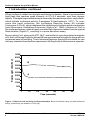
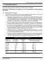
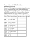
Luciferase Reporter
Assay Kit
User Manual
Cat. No. K2039-1
PT3392-1 (PR2Y278)
Published 11/22/2002
Luciferase Reporter Assay Kit User Manual
Table of Contents
I. Introduction
3
II. List of Components
5
III. Additional Materials Required
5
IV. General Considerations
6
V. Assay Procedure
7
A. Eukaryotic Cell Lysis
7
B. Bacterial Cell Lysis
8
C. Luciferase Assay
8
VI. Troubleshooting Guide
9
VII. References
10
VIII. Related Products
11
Notice to Purchaser
This product is intended to be used for research purposes only. It is not to be used for drug or diagnostic
purposes nor is it intended for human use. BD Biosciences Clontech products may not be resold,
modified for resale, or used to manufacture commercial products without written approval of BD
Biosciences Clontech.
BD Adeno-X™, BD Creator™, BD Living Colors™, BD Mercury™, BD RevTet-Off™,
BD RevTet-On™,BD™ Tet-Off, and BD™Tet-On are trademarks of Becton, Dickinson and Company.
© 2002, BD
BD Biosciences Clontech
2
www.bdbiosciences.com
Protocol # PT3392-1
Version # PR2Y278
Luciferase Reporter Assay Kit User Manual
I. Introduction
Firefly beetle (Photinus pyralis) luciferase is one of the most popular reporter
molecules used in molecular biology and biochemistry (Gould & Subramani,
1988; Vieites et al., 1994; Gailey et al., 1997). Luciferase can be used to monitor
promoter response activity in bacteria, cultured cells, and transgenic plants or
animals. By providing faster results, lower costs, and over a 1,000-fold increase
in sensitivity, the luciferase assay has largely replaced the standard 14C chloramphenicol acetyltransferase (CAT) assay.
Our Luciferase Reporter Assay Kit provides a simple means for detecting
luciferase activity in transformed bacteria or transfected eukaryotic cells. The kit
includes a firefly luciferase substrate formulation and an optimized cell lysis
buffer. This buffer enhances luciferase recovery and activation when used with
in vitro assays. The unique formulation of this kit provides high sensitivity,
constant light output, as well as convenience and consistency when working with
multiple samples.
Background
Firefly luciferase has been reliably expressed as a reporter gene from many
expression vectors and in a variety of organisms. Its major use has been to
characterize gene regulation—primarily transcriptional control—by correlating
variations in luciferase activity with the regulation of promoter and enhancer
elements. Performed under optimal conditions, the peak height and integrated
total light output from a reaction is proportional to the amount of functional
luciferase enzyme. This results in a direct relationship between the amount of
light emitted from the sample and the transcriptional activity of the regulatory
elements.
Firefly luciferase catalyzes the oxidative carboxylation of luciferin, a reaction with
the highest efficiency of any known bioluminescence reaction (Seliger & McElroy,
1960). At the optimal reaction pH of 7.8, light emission peaks at 562 nm. This form
of light emission yields a very sensitive non-radioactive assay.
The Assay
Firefly luciferase is a 62,000 dalton protein which is active as a monomer and
does not require subsequent processing for its activity. However, several factors
may affect the sensitivity and success of the assay including pH, temperature,
and substrate concentration. To ensure maximum sensitivity, the assay is
performed in the presence of excess ATP, luciferin and Mg2+ in a buffer that will
maintain a pH of 7.8.
For measurement of expressed luciferase activity in vitro, luciferase is extracted
from transfected cells through cell lysis. A typical firefly luciferase assay is then
carried out in an assay cuvette. ATP, Mg2+ and buffer are added to the lysate
either separately or as a preformulated solution. The luminescent reaction is then
triggered by an injection of luciferin, and the emitted light is recorded.
Protocol # PT3392-1
Version # PR2Y278
www.bdbiosciences.com
BD Biosciences Clontech
3
Luciferase Reporter Assay Kit User Manual
I. Introduction continued
When luciferin is added to a sample containing luciferase , there is an immediate
light flash that reaches peak intensity at 0.3–0.5 seconds, and then decays
rapidly. This rapid exponential decay is caused by the reaction product, oxyluciferin,
which inhibits luciferase activity (Lemasters & Hackenbrock, 1977). To overcome this rapid extinction, the Luciferase Reporter Assay Kit includes
Coenzyme-A (CoA), which displaces the inhibiting oxyluciferin product substrate
from the enzyme, facilitating its turnover (Airth et al., 1958). Inclusion of CoA in
the luciferase assay yields a nearly constant light emission rather than the typical
flash kinetics (Figure 1), resulting in a more sensitive assay.
By providing CoA, along with ATP, Mg2+ and buffer in a preformulated substrate
mix, the Luciferase Reporter Assay Kit ensures maximal sensitivity along with an
increased ease of handling. The Luciferase Reporter Assay Kit is suitable for use
with any standard transfection experiment utilizing firefly luciferase as a reporter.
5000
Relative Light Units (RLU)
4500
4000
B
3500
3000
2500
A
2000
1500
1000
500
0
1
10
20
30
40
Time (seconds)
50
60
Figure 1. Effect of CoA on firefly luciferase kinetics. Basic luciferase assay (A) and luciferase
assay modified by the addition of CoA (B).
BD Biosciences Clontech
4
www.bdbiosciences.com
Protocol # PT3392-1
Version # PR2Y278
Luciferase Reporter Assay Kit User Manual
II. List of Components
Prior to reconstitution, store all reagents at –20°C.
Substrates A and B must each be reconstituted in 10 ml deionized or distilled
water. After reconstitution, Substrates A and B may be stored as aliqouts either
at –20°C for 6 months or at 4°C for 5 days. Before starting an assay, dilute an
aliquot of 3X Cell Lysis Buffer to 1X with deionized or distilled water (see Section
V.A).
Reagents provided are sufficient for 100 assays.
•
10 ml
Substrate A
•
10 ml
Substrate B
•
50 ml
3X Cell Lysis Buffer
III. Additional Materials Required
The following materials are required but not supplied:
• Phosphate buffered saline (PBS; pH 7.4)
Final conc.
Na2HPO4
58 mM
17 mM
NaH2PO4
NaCl
68 mM
To prepare 2 L of solution
16.5 g
4.1 g
8.0 g
Dissolve the above components in 1.8 L of deionized H2O. Adjust to pH 7.4
with 0.1 N NaOH. Add deionized H2O to final volume of 2 L. Store at room
temperature.
• Centrifuge for collecting cells
• 1.5-ml microcentrifuge tubes
• 0.5-ml microcentrifuge tubes or 96-well flat-bottom microtiter plate
Chemiluminescence assays are generally performed in 0.5-ml microcentrifuge
tubes. Alternatively, reactions can be performed in white opaque 96-well
flat-bottom microtiter plates such as those from Xenopore or Costar.
• Luminometer (tube or plate), scintillation counter or x-ray film
• Lysozyme for bacterial cell lysis
Protocol # PT3392-1
Version # PR2Y278
www.bdbiosciences.com
BD Biosciences Clontech
5
Luciferase Reporter Assay Kit User Manual
IV. General Considerations
• Ensure that all reagents have reached room temperature before performing
assays.
• Do not repeatedly freeze/thaw sample extracts. Loss of luciferase activity will
result.
• We strongly recommend using the Cell Lysis Buffer and protocols supplied
with this kit. Sonication or other methods of cell lysis may reduce the sensitivity
of the assay.
• Optimization of the Luciferase Assay Kit may be necessary for use with your
equipment or samples. Signal detection may become saturated when measuring very high light-emitting samples in a luminometer or scintillation
counter. If this occurs, dilute your sample with 1X Lysis Buffer and repeat the
assay.
• Chemiluminescent detection of luciferase activity can be performed with a
luminometer (tube or plate) or a liquid scintillation counter (LSC). Use of an
LSC may result in lowered sensitivity and increased variability between
samples due to the need for manual addition of Substrate B (Nguyen et al.,
1988). In addition, it is necessary to make specific adjustments to the LSC for
the correct detection of the luciferase signal (Fulton & Van Ness, 1993).
• It is also possible to measure luciferase expression via exposure of x-ray film
to reactions performed in a white opaque 96-well flat-bottom microtiter plate
(Xenopore or Costar).
• Measured levels of luciferase activity are normally stated in relative light units
(RLUs), which do not represent an absolute value. If you wish to correlate your
relative experimental luciferase activities with an absolute value, you must
generate a standard curve for your measuring equipment using purified
luciferase. However, it is important to be aware that a purified luciferase
standard does not necessarily represent the exact amount of luciferase
produced by transfected cells, since the specific activity of the expressed
luciferase may differ from the purified luciferase.
BD Biosciences Clontech
6
www.bdbiosciences.com
Protocol # PT3392-1
Version # PR2Y278
Luciferase Reporter Assay Kit User Manual
V. Assay Procedure
PLEASE READ ENTIRE PROTOCOL BEFORE STARTING.
Important: Equilibrate all reagents to room temperature before starting
the assay.
A. Eukaryotic Cell Lysis
This protocol is optimized for use with eukaryotic cell cultures. For a lysis
protocol for bacterial cells, please see Part B below.
1. Prepare an adequate amount of 1X Cell Lysis Buffer by diluting 1 part
3X Cell Lysis Buffer into 2 parts distilled or deionized water (see Table I).
The following protocol is designed for use with adherent cultures
growing in 35-mm tissue culture plates. If you are using plates, wells,
or flasks of a different size, adjust the volume proportionally.
2. Remove media from cell culture plates and rinse twice with phosphate
buffered saline (PBS without Ca2+ and Mg2+; see Section III for recipe).
3. Add 200 µl 1X Cell Lysis Buffer to cells and shake at room temperature
for 15–20 min. Alternatively, cells may be lysed at 4°C to minimize
protease activity. If performing lysis at 4°C, allow cell lysate to reach
room temperature before continuing with protocol.
4. Dislodge cells by scraping or pipetting and transfer to a 1.5-ml
microcentrifuge tube. Spin cells at 14,000 rpm at room temperature for
1 min to remove cellular debris.
5. Samples should be assayed within 20 min. For measurements that
require longer time points or for assays that are to be completed at a
later date, extracts may be stored for up to one month at –70°C.
TABLE I. CULTURE PLATE CONVERSION
Plate or
Flask Size
96 well
24 well
12 well
6 well
35 mm
60 mm
10 cm
T25
T75
Growth
Area (cm2)
0.32
1.88
3.83
9.4
8.0
21
55
25
75
Relative
Area*
0.04 X
0.25 X
0.50 X
1.20 X
1.00 X
2.60 X
7.00 X
3.00 X
9.00 X
Recommended Volume
1X Cell Lysis Buffer
20 µl
50 µl
100 µl
200 µl
200 µl
500 µl
1.0 ml
500 µl
1.2 ml
* Relative area is expressed as a factor of the growth area of a 35-mm culture plate.
Protocol # PT3392-1
Version # PR2Y278
www.bdbiosciences.com
BD Biosciences Clontech
7
Luciferase Reporter Assay Kit User Manual
V. Assay Procedure continued
B. Bacterial Cell Lysis
1. Prepare 1 ml of 1X Cell Lysis Buffer for each 1–10 ml aliquot of bacterial
culture by making a 1:3 dilution of 1 part 3X Cell Lysis Buffer to 2 parts
distilled or deionized water. Add lysozyme to a final concentration of
1 mg/ml.
2. Centrifuge a 1–10 ml aliquot of bacterial culture. If required, an optimal
volume may be determined after initial measurement of activity.
3. Remove and discard supernatant without disturbing cell pellet.
4. Add 1 ml 1X Cell Lysis Buffer and vortex to resuspend cell pellet.
5. Let stand at room temperature for 5–10 min, then centrifuge lysate at
14,000 rpm at room temperature for 1 min to remove insoluble debris.
6. As with eukaryotic samples, assays should be performed within 20 min.
For measurements that require longer time points or for assays that are
to be completed at a later date, extracts may be stored for up to one
month at –70°C.
C. Luciferase Assay
1. Place 20–100 µl cell extract into an assay cuvette. Be sure to use the
same volume for each sample.
2. If measurement will be performed on a luminometer or scintillation
counter, the recommended measurement time is 10–30 sec (photographic or CCD-type instruments typically require exposures as long as
5 min). Follow the step below that is appropriate for your instrument:
a. If your luminometer contains a single automatic injector, manually
add 100 µl of Substrate A to the assay cuvette and automatically
inject 100 µl of Substrate B within 10 min. Set the delay after the
injection and before measurement to 1 or 2 sec.
b. If your luminometer contains two automatic injectors, inject 100 µl of
Substrate A first, followed by 100 µl of Substrate B. Set the delay
between injections to 1 or 2 sec. Set the delay after the second
injection and before measurement to 1 or 2 sec.
c. If no automatic injectors are used, manually add 100 µl of Substrate
A to the assay cuvette. Immediately before measurement, manually
add 100 ml of Substrate B to the assay cuvette. The time between
adding Substrate B and start of measurement should be as short as
possible and consistent from sample to sample.
BD Biosciences Clontech
8
www.bdbiosciences.com
Protocol # PT3392-1
Version # PR2Y278
Luciferase Reporter Assay Kit User Manual
VI. Troubleshooting Guide
A. Intra-assay Variability
Pipetting error
Use larger sample volumes to minimize variability
caused by pipetting error.
Temperature
changes
Be sure all reagents have reached room temperature
before performing assay.
Allowing sample
and buffer to sit
for extended
periods of time.
Work quickly to minimize the time between adding
and initiating the reaction.
Reagent
degradation
Store all reagents at –20°C.
B. Abnormally Low Light from Assay
Improper pH
Test pH of each reagent and adjust to 7.8 if necessary.
Improper substrate
concentrations
Check that the correct volume of each reagent is
being added to the assay reaction and adjust if
necessary.
Reagent
degradation
Store all reagents at –20°C.
Presence of
interfering
substances
Be sure to wash cells thoroughly with PBS 2–3
times before performing lysis.
C. High Background
Contaminated
reagents
Contaminated
injector lines
Protocol # PT3392-1
Version # PR2Y278
Reagents may become contaminated by carry-over
from pipette tips. Be sure to change tips between
reaction components and/or samples. Replace component if necessary.
Flush injector lines thoroughly with distilled water.
www.bdbiosciences.com
BD Biosciences Clontech
9
Luciferase Reporter Assay Kit User Manual
VII. References
Airth, R. L., Rhodes, W. C. & McElroy, W. D. (1958) The function of coenzyme A in luminescence.
Biochem. et Biophys. Acta 27:519–532.
Gailey, P. C., Miller, E. J. & Griffin, G. D. (1997) Low-cost system for real-time monitoring of
luciferase gene expression. BioTechniques 22:528–534.
Gould, S. J. & Subramani, S. (1988) Firefly luciferase as a tool in molecular and cell biology. Analyt.
Biochem. 175:5–13
Fulton, R. & Van Ness, B. (1993) Luminescent reporter gene assays for luciferase and
β-galactosidase using a liquid scintillation counter. BioTechniques 14:762-763.
Lemasters, J. J. & Hackenbrock, C. R. (1977) Kinetics of product inhibition during firefly luciferase
luminescence. Biochemistry 16(3):445–447.
Nguyen, V. T., Morange, M. & Bensaude, O. (1988) Firefly luciferase luminescence assays using
scintillation counters for quantitation in transfected mammalian cells. Analyt. Biochem. 171:404–
408.
Seliger, H. H. & McElroy, W. D. (1960) Spectral emission and quantum yield of firefly bioluminescence. Arch. Biochem. Biophys. 88:136–141.
Vieites, J. M., Navarro-García, F., Pérez-Diaz, R., Pla, J. & Nombela, C. (1994) Expression and in
vivo determination of firefly luciferase as gene reporter in Saccharomyces cerevisiae. Yeast
10:1321–1327.
BD Biosciences Clontech
10
www.bdbiosciences.com
Protocol # PT3392-1
Version # PR2Y278
Luciferase Reporter Assay Kit User Manual
VIII. Related Products
For the latest and most complete listing of all
BD Biosciences Clontech products,
please visit www.bdbiosciences.com
BD Mercury™ Reporter Systems
• BD Mercury™ Pathway Profiling Luciferase System 1
K2049-1
• BD Mercury™ Pathway Profiling Luciferase System 2
K2052-1
• BD Mercury™ Pathway Profiling Luciferase System 3
K2053-1
• BD Mercury™ Pathway Profiling Luciferase System 4
K2056-1
• BD Mercury™ Pathway Profiling Luciferase System 5
K2057-1
Tet Expression Systems
• BD™ Tet-Off Gene Expression System
K1620-1
• BD™ Tet-On Gene Expression System
K1621-1
• pBI-L Bidirectional Tet Vector
6151-1
Retroviral Expression Systems
• BD pRevTet-Off™ System
K1626-1
• BD pRevTet-On™ System
K1627-1
• BD Adeno-X™ Tet-Off Expression System
K1651-1
• BD Adeno-X™ Tet-On Expression System
K1652-1
BD Creator™ DNA Cloning Kits
• BD Creator™ pDNR-1 Cloning Kit
K1670-1
BD Living Colors™ Vectors
• pEGFPLuc Vector
Protocol # PT3392-1
Version # PR2Y278
6169-1
www.bdbiosciences.com
BD Biosciences Clontech
11