1
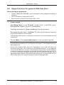
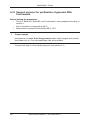
Genomic DNA
from Tissue
User Manual
NucleoSpin® Tissue
January 2010 / Rev. 11
MACHEREY-NAGEL
Genomic DNA Purification from Tissue
Protocol-at-a-glance (Rev. 11)
NucleoSpin® Tissue
1
Prepare sample
Cut 25 mg into small pieces
2
Pre-lyse sample
180 µl T1
25 µl Proteinase K
56 °C
1–3h
3
Lyse sample
200 µl B3
70 °C
10 min
4
5
Adjust DNA
binding conditions
210 µl 96 – 100 % ethanol
Bind DNA
Load all
11,000 x g
1 min
6
Wash silica membrane
1st and 2nd
1st wash
500 µl BW
2nd wash
600 µl B5
11,000 x g
1 min
7
Dry silica membrane
11,000 x g
1 min
8
Elute highly pure DNA
100 µl BE
(70 °C)
RT
1 min
11,000 x g
1 min
MACHEREY-NAGEL GmbH & Co. KG • Neumann-Neander-Str. 6-8 • D-52355 Düren • Germany
Tel.: +49 (0) 24 21 969 270 • www.mn-net.com • e-mail: [email protected]
MN
Genomic DNA from Tissue
Table of contents
1
Components
5
1.1 Kit contents
5
1.2 Reagents, consumables, and equipment to be supplied by user
6
1.3 About this User Manual
6
Product description
7
2.1 The basic principle
7
2.2 Kit specifications
7
2.3 Elution procedures
8
3
Storage conditions and preparation of working solutions
9
4
Safety instructions – risk and safety phrases
10
5
Standard protocol for human or animal tissue and cultured cells
11
6
Support protocols
14
6.1 Support protocol for mouse or rat tails
14
6.2 Support protocol for bacteria
15
2
6.3 Support protocol for yeast
16
6.4 Support protocol for dried blood spots (e.g., NucleoCards, FTA cards,
Guthrie cards)
17
6.5 Support protocol for genomic DNA and viral DNA from blood samples
18
6.6 Support protocol for hair roots
19
6.7 Support protocol for paraffin-embedded tissue
20
6.8 Support protocol for genomic DNA from stool
21
6.9 Support protocol for viral DNA (e.g., CMV) from stool
22
6.10 Support protocol for detection of Mycobacterium tuberculosis or
Legionella pneumophila in sputum or bronchoalveolar lavage
24
6.11 Support protocol for detection of EHEC bacteria in food
(e.g., fresh cows‘ milk)
25
6.12 Support protocol for purification of bacterial DNA (e.g., Chlamydia
trachomatis) from cultures, biological fluids, or clinical specimens
26
6.13 Support protocol for purification of bacterial DNA
(e.g., Borrelia burgdorferi) from urine
27
6.14 Support protocol for purification of viral DNA (e.g., CMV) from urine
28
®
MACHEREY-NAGEL – 01/ 2010, Rev. 11
3
Genomic DNA from Tissue
7
4
6.15 Support protocol for purification of genomic DNA from insects
30
6.16 Support protocol for purification of genomic DNA from dental swabs
31
6.17 Support protocol for purification of genomic DNA from buccal swabs
32
Appendix
34
7.1 Troubleshooting
34
7.2 Ordering information
36
7.3 Product use restriction / warranty
37
MACHEREY-NAGEL – 01 / 2010, Rev.11
Genomic DNA from Tissue
1
Components
1.1
Kit contents
NucleoSpin® Tissue
10 preps
50 preps
250 preps
740952.10
740952.50
740952.250
Lysis Buffer T1
5 ml
20 ml
100 ml
Buffer B1*
6 ml
12 ml
60 ml
Buffer B2*
1.5 ml
3 ml
15 ml
Wash Buffer BW
6 ml
30 ml
2 x 75 ml
Wash Buffer B5
(Concentrate)*
4 ml
2 x 7 ml
2 x 40 ml
Elution Buffer BE**
3 ml
15 ml
75 ml
Proteinase K
(lyophilized)*
6 mg
30 mg
2 x 75 mg
Proteinase Buffer PB
0.8 ml
1.8 ml
8 ml
NucleoSpin® Tissue
Columns (light green rings)
10
50
250
Collection Tubes (2 ml)
20
100
500
Labels for Lysis Buffer B3
1
1
1
User Manual
1
1
1
Cat. No.
* For preparation of working solutions and storage conditions see section 3.
** Composition of Elution Buffer BE: 5 mM Tris/HCl, pH 8.5
MACHEREY-NAGEL – 01/ 2010, Rev. 11
5
Genomic DNA from Tissue
1.2
Reagents, consumables, and equipment to be supplied
by user
Reagents
•
96 – 100% ethanol
Consumables
•
1.5 ml microcentrifuge tubes for sample lysis and DNA elution
•
Disposable tips
Equipment
•
Manual pipettors
•
Centrifuge for microcentrifuge tubes
•
Vortex mixer
•
Heating-block for incubation at 70 °C
•
Equipment for sample disruption and homogenization (see section 2.5)
•
Personal protection equipment (lab coat, gloves, goggles)
1.3
About this User Manual
It is strongly recommended reading the detailed protocol sections of this User Manual
if the NucleoSpin® Tissue kit is used for the first time. Experienced users, however,
may refer to the Protocol-at-a-glance instead. The Protocol-at-a-glance is designed to
be used only as a supplemental tool for quick referencing while performing the purification procedure.
All technical literature is available on the internet at www.mn-net.com.
6
MACHEREY-NAGEL – 01 / 2010, Rev.11
Genomic DNA from Tissue
2
Product description
2.1
The basic principle
With the NucleoSpin® Tissue method genomic DNA can be prepared from tissue, cells
(e.g., bacteria), and many other sources. Lysis is achieved by incubation of the sample
material in a proteinase K / SDS solution. Appropriate conditions for binding of DNA to
the silica membrane in the NucleoSpin® Tissue Columns are created by the addition
of chaotropic salts and ethanol to the lysate. The binding process is reversible and
specific to nucleic acids. Contaminations are removed by subsequent washing with two
different buffers. Pure genomic DNA is finally eluted under low ionic strength conditions
in a slightly alkaline elution buffer.
2.2
Kit specifications
•
NucleoSpin® Tissue is designed for the rapid, small-scale preparation of highly
pure genomic DNA from any tissue, cells, bacteria, yeast, forensic samples,
serum, plasma, or other body fluids. It is also suitable for preparation of DNA
from human or animal blood. The purified DNA can be used directly for PCR,
Southern blotting, or any kind of enzymatic reactions.
•
The kit allows purification of up to 35 µg of pure genomic DNA with an A260/A280
ratio between 1.7 and 1.9. The NucleoSpin® Tissue Column is capable of
binding up to 60 µg of genomic DNA.
•
For lysis of certain bacterial and yeast strains, additional enzymes may be
necessary which are not part of this kit. See the relevant support protocol for
details.
Table 1: Kit specifications at a glance
NucleoSpin® Tissue
Parameter
Sample material
1 – 25 mg / 102 – 107 cells
Typical yield
20 – 35 µg
Elution volume
60 – 100 µl
Binding capacity
60 µg
Preparation time
20 min / 4 – 6 preps (after lysis)
Format
Mini spin column
MACHEREY-NAGEL – 01/ 2010, Rev. 11
7
Genomic DNA from Tissue
2.3
Elution procedures
In addition to the standard method (recovery rate about 70 – 90 %) several modifications are possible to increase yield, concentration, and convenience. Use elution buffer
preheated to 70 °C for one of the following procedures:
•
High yield: Perform two elution steps with the volume indicated in the individual
protocol. About 90 – 100 % of bound nucleic acid can be eluted.
•
High concentration: Perform one elution step with 60 % of the volume indicated in the individual protocol. Concentration of DNA will be ca. 30 % higher than
with standard elution. The yield of eluted nucleic acid will be about 80 %.
•
High yield and high concentration: Apply half the volume of elution buffer
as indicated in the individual protocol, incubate for 3 min and centrifuge. Apply
a second aliquot of elution buffer, incubate and centrifuge again. Thus, about
85 – 100 % of bound nucleic acid is eluted in the standard elution volume at a
high concentration.
•
Convenient elution: For convenience, elution buffer of ambient temperature
may be used. This will result in a somewhat lower yield (approximately 20 %)
compared to elution with heated elution buffer.
Elution may also be performed with Tris-EDTA-buffer (TE) of pH equal or higher than 8.
This will increase DNA stability especially during long term and / or multi use storage
at 4 °C or ambient temperature by inhibition of omnipresent DNases. However, EDTA
interferes, depending on the final concentration, with certain downstream applications.
Note: Elution Buffer BE (5 mM Tris/HCl, pH 8.5) provided with the kit does not contain
EDTA.
For optimal performance of isolated DNA in downstream applications we recommend
elution with the supplied elution buffer and storage, especially long term, at - 20 °C.
Freeze-thaw cycles will have no effect on most downstream applications. (Possible
exceptions are detection of trace amounts of DNA or long-range PCR (e.g., > 10 kbp).
Multiple freeze-thaw cycles or storing DNA at + 4 °C or room temperature may influence
detection sensitivities or reaction efficiencies due to DNA shearing or adsorption to
surfaces.)
8
MACHEREY-NAGEL – 01 / 2010, Rev.11
Genomic DNA from Tissue
3
Storage conditions and preparation of
working solutions
Attention:
Buffers B1, B3, and BW contain guanidine hydrochloride! Wear gloves and goggles!
•
All kit components can be stored at room temperature (18 – 25 °C) and are
stable up to one year.
•
During storage, especially at low temperatures, a white precipitate may form in
Buffer T1, B1, or B3. Such precipitates can be easily dissolved by incubating
the bottle at 50 – 70 °C before use.
Before starting any NucleoSpin® Tissue protocol prepare the following:
•
Lysis Buffer B3: Transfer the total contents of Buffer B1 to Buffer B2 and mix
well. Place the labels for Lysis Buffer B3 on the bottle. The resulting Lysis Buffer
B3 is stable for up to one year at room temperature.
•
Wash Buffer B5: Add the indicated volume of ethanol (96 – 100 %) to Wash
Buffer B5 Concentrate. Mark the label of the bottle to indicate that ethanol
was added. Store Wash Buffer B5 at room temperature (18 – 25 °C) for up to
one year.
•
Proteinase K: Add the indicated volume of Proteinase Buffer PB to dissolve
lyophilized Proteinase K. Proteinase K solution is stable at - 20 °C for up to 6
months.
NucleoSpin® Tissue
10 preps
50 preps
250 preps
740952.10
740952.50
740952.250
Wash
Buffer B5
(Concentrate)
4 ml
Add 16 ml ethanol
2 x 7 ml
Add 28 ml ethanol
to each bottle
2 x 40 ml
Add 160 ml ethanol
to each bottle
Proteinase K
6 mg
Add 260 µl
Proteinase Buffer
30 mg
Add 1.35 ml
Proteinase Buffer
2 x 75 mg
Add 3.35 ml
Proteinase Buffer
to each vial
Cat. No.
MACHEREY-NAGEL – 01/ 2010, Rev. 11
9
NucleoSpin® Tissue
4
Safety instructions – risk and safety phrases
The following components of the NucleoSpin® Tissue kits contain hazardous contents.
Wear gloves and goggles and follow the safety instructions given in this section.
Component
Hazard
contents
Hazard
symbol
Risk
phrases
Safety
phrases
B1
Guanidine
hydrochloride
Xn*
Harmful if swallowed - Irritating to
eyes and skin
R 2236/38
BW
Guanidine
hydrochloride
+ isopropanol
< 25%
Xn*
Flammable - Harmful
if swallowed Irritating to eyes and
skin.
R 10-2236/38
S 7-16-25
Proteinase K
Proteinase K,
lyophilized
Xn
Xi*
Irritating to eyes,
respiratory system
and skin, may cause
sensitization by
inhalation
R
36/37/3842
S 22-2426-36/37
Risk phrases
R 10
Flammable
R 22
Harmful if swallowed
R 36/37/38
Irritating to eyes, respiratory system and skin
R 36/38
Irritating to eyes and skin
R 42
May cause sensitization by inhalation
Safety phrases
S7
Keep container tightly closed
S 16
Keep away from sources of ignition - No smoking!
S 22
Do not breathe dust
S 24
Avoid contact with the skin
S 25
Avoid contact with the eyes
S 26
In case of contact with eyes, rinse immediately with plenty of water
and seek medical advice
S 36/37
Wear suitable protective clothing and gloves
* Hazard labeling not necessary if quantity per bottle below 125 g or ml (certificate of exemption
according to 67/548/EEC Art. 25, 1999/45/EC Art. 12 and German GefStoffV § 20 (3) and TRGS 200 7.1).
For further information see Material Safety Data Sheet.
10
MACHEREY-NAGEL – 01 / 2010, Rev.11
NucleoSpin® Tissue
5
Standard protocol for human or animal tissue
and cultured cells
Before starting the preparation:
•
•
•
1
Check if Buffer B3, Buffer B5 and Proteinase K were prepared according to
section 3.
Set an incubator or water bath to 56 °C.
Preheat Elution Buffer BE to 70 °C.
Prepare sample
Tissue
Cut 25 mg human or animal tissue into small pieces.
Place the sample in a microcentrifuge tube (not provided).
Proceed with step 2.
Samples that are difficult to lyse can be ground under liquid
nitrogen or may be treated in a mechanical homogenizer
(Polytron, Ultra Turrax): Add 25 mg of tissue to a 1.5 ml
microcentrifuge tube (not provided), add 50 – 75 µl phosphate buffered saline (PBS) and homogenize.
Cultured cells
Resuspend up to 107 cells in a final volume of 200 µl
Buffer T1. Add 25 µl Proteinase K solution and 200 µl
Buffer B3. Incubate the sample at 70 °C for 10 – 15 min.
Proceed with step 4.
2
Pre-lyse sample
Add 180 µl Buffer T1 and 25 µl Proteinase K solution.
Vortex to mix. Be sure that the samples are completely
covered with lysis solution.
If processing several samples, Proteinase K and Buffer T1
may be premixed directly before use. Do never mix Buffer T1
and Proteinase K more than 10 – 15 min before addition to
the sample: Proteinase K tends to self-digestion in Buffer T1
without substrate.
MACHEREY-NAGEL – 01/ 2010, Rev. 11
+ 180 µl T1
+ 25 µl
Proteinase K
11
NucleoSpin® Tissue
Incubate at 56 °C until complete lysis is obtained (at least
1 – 3 h). Vortex occasionally during incubation or use a
shaking incubator.
Samples can be incubated overnight as well. If RNA-free
DNA is crucial for downstream applications, an RNase digest
may be performed: Add 20 µl RNase A (20 mg/ml) solution
(not included; see ordering information) and incubate for an
additional 5 min at room temperature.
3
or
56°C
overnight
Lyse sample
Vortex the samples. Add 200 µl Buffer B3, vortex vigorously and incubate at 70 °C for 10 min. Vortex briefly.
If insoluble particles are visible, centrifuge for 5 min at high
speed (e.g., 11,000 x g) and transfer the supernatant to a new
microcentrifuge tube (not provided).
4
56 °C
1–3h
+ 200 µl B3
70°C
10 min
Adjust DNA binding conditions
Add 210 µl ethanol (96 – 100 %) to the sample and
vortex vigorously.
+ 210 µl
ethanol
After addition of ethanol a stringy precipitate may become
visible. This will not affect the DNA isolation. Be sure to load
all of the precipitate on the column in the following step.
5
Bind DNA
For each sample, place one NucleoSpin® Tissue
Column into a Collection Tube. Apply the sample to
the column. Centrifuge for 1 min at 11,000 x g. Discard
the flow-through and place the column back into the
Collection Tube.
If the sample is not drawn completely through the matrix,
repeat the centrifugation step at 11,000 x g. Discard flowthrough.
12
MACHEREY-NAGEL – 01 / 2010, Rev.11
Load samples
11,000 x g
1 min
NucleoSpin® Tissue
6
Wash silica membrane
+ 500 µl BW
1 wash
st
Add 500 µl Buffer BW. Centrifuge for 1 min at 11,000 x g.
Discard flow-through and place the column back into the
Collection Tube.
+ 600 µl B5
2nd wash
Add 600 µl Buffer B5 to the column and centrifuge for
1 min at 11,000 x g. Discard flow-through and place the
column back into the Collection Tube.
6
11,000 x g
1 min
11,000 x g
1 min
Dry silica membrane
Centrifuge the column for 1 min at 11,000 x g.
11,000 x g
1 min
Residual ethanol is removed during this step.
7
Elute highly pure DNA
Place the NucleoSpin® Tissue Column into a 1.5 ml
microcentrifuge tube (not provided) and add 100 µl prewarmed Buffer BE (70°C). Incubate at room temperature for 1 min. Centrifuge 1 min at 11,000 x g.
For alternative elution procedures see section 2.3.
MACHEREY-NAGEL – 01/ 2010, Rev. 11
+ 100 µl BE
(70 °C)
RT
1 min
11,000 x g
1 min
13
NucleoSpin® Tissue
6
Support protocols
6.1
Support protocol for mouse or rat tails
Before starting the preparation:
1
•
Check if Buffer B3, Buffer B5, and Proteinase K were prepared according to
section 3.
•
Set an incubator or water bath to 56 °C.
•
Before elution, preheat Elution Buffer BE to 70 °C.
Prepare sample
Cut two 0.6 cm-pieces of mouse tail and place them in a 1.5 ml centrifuge tube
(not provided).
If processing rat tails, one 0.6 cm-piece is sufficient.
2
Pre-lyse sample
Add 180 µl Buffer T1 and 25 µl Proteinase K and vortex. Incubate at 56 °C
overnight or until complete lysis is obtained. Vortex occasionally during incubation or use a shaking water bath. To remove residual bones or hair, centrifuge
for 5 min at high speed (e.g., 11,000 x g). Transfer 200 µl supernatant to a
new tube.
If processing several samples, Proteinase K and Buffer T1 may be premixed directly
before use. Do never mix Buffer T1 and Proteinase K more than 10 – 15 min before
addition to the sample: Proteinase K tends to self-digestion in Buffer T1 without substrate.
3
Lyse sample
Add 200 µl Buffer B3 to the lysate and vortex vigorously.
Buffer B3 and ethanol (see step 4) can be premixed before addition to the lysate.
4
Adjust DNA binding conditions
Add 210 µl ethanol to the lysate and vortex vigorously.
Proceed with step 5 of the standard protocol (see section 5.1).
14
MACHEREY-NAGEL – 01 / 2010, Rev.11
NucleoSpin® Tissue
6.2
Support protocol for bacteria
Before starting the preparation:
1
•
Check if Buffer B3, Buffer B5, and Proteinase K were prepared according to
section 3.
•
Set an incubator or water bath to 56 °C.
•
Before elution, preheat Elution Buffer BE to 70 °C.
Prepare sample
Up to 1 ml of bacterial culture can be used for the preparation depending on, for
example, density of culture, culture medium, and bacterial strain.
Centrifuge up to 1 ml culture for 5 min at 8,000 x g. Remove supernatant.
2
Pre-lyse sample
Resuspend the pellet in 180 µl Buffer T1 by pipetting up and down. Add 25 µl
Proteinase K. Vortex vigorously and incubate at 56 °C until complete lysis is
obtained (at least 1 – 3 h). Vortex occasionally during incubation or use a shaking incubator.
Samples can be incubated overnight as well.
If RNA-free DNA is crucial for downstream applications, an RNase digest may be
performed: Add 20 µl RNase A (20 mg / ml) solution (not included; see ordering information) and incubate for an additional 5 min at room temperature.
Hard-to-lyse bacteria: Some strains, especially Gram-positive bacteria, are more
difficult to lyse. In such cases, a preincubation with a lytic enzyme is necessary:
Resuspend the pelleted cells in 20 mM Tris/HCl; 2 mM EDTA; 1% Triton X-100; pH
8 (instead of Buffer T1) supplemented with 20 mg/ml lysozyme or 0.2 mg/ml lysostaphin and incubate for 30 – 60 min at 37 °C. Add 25 µl Proteinase K, incubate at
56 °C until complete lysis is obtained.
Proceed with step 3 of the standard protocol (see section 5.1).
MACHEREY-NAGEL – 01/ 2010, Rev. 11
15
NucleoSpin® Tissue
6.3
Support protocol for yeast
Before starting the preparation:
•
•
1
Check if Buffer B3, Buffer B5, and Proteinase K were prepared according to
section 3.
Check that sorbitol buffer and lyticase or zymolase (not provided with the kit) is
available for sample pre-lysis.
•
Set an incubator or water bath to 30 °C and 56 °C.
•
Before elution, preheat Elution Buffer BE to 70 °C.
Prepare sample
Harvest 3 ml YPD yeast culture (OD600 ≤ 10) by centrifugation for 10 min at
5,000 x g. Wash the cells once with 1 ml 10 mM EDTA, pH 8. Remove the supernatant and pellet the cells by centrifugation (5,000 x g, 10 min).
2
Pre-lyse sample
Resuspend the pellet in 600 µl sorbitol buffer (1.2 M sorbitol; 10 mM CaCl2;
0.1 M Tris/HCl pH 7.5; 35 mM ß-mercaptoethanol). Add 50 U lyticase or
zymolase*. Incubate at 30 °C for 30 min. This step degrades the yeast cell wall
creating spheroplasts. Spheroplast formation may be checked microscopically.
Centrifuge the mixture for 10 min at 2,000 x g remove supernatant and resuspend the pelleted spheroplasts in 180 µl Buffer T1. Add 25 µl Proteinase K
solution and vortex vigorously. Incubate at 56 °C until complete lysis is obtained
(at least 1 – 3 h). Vortex occasionally during incubation or use a shaking water
bath.
Samples can be incubated overnight as well.
If RNA-free DNA is crucial for downstream applications, an RNase digest may be
performed: Add 20 µl RNase A (20 mg / ml) solution (not included; see ordering information) and incubate for an additional 5 min at room temperature.
Proceed with step 3 of the standard protocol (see section 5.1).
* Other protocols use 5 – 200 U lyticase or zymolase depending on enzyme quality or brand. Increasing the
enzyme concentration may be required if spheroplasts are not formed.
16
MACHEREY-NAGEL – 01 / 2010, Rev.11
NucleoSpin® Tissue
6.4
Support protocol for dried blood spots
(e.g., NucleoCards, FTA® cards, Guthrie cards)
Before starting the preparation:
1
•
Check if Buffer B3, Buffer B5, and Proteinase K were prepared according to
section 3.
•
Set an incubator or water bath to 56 °C.
•
Before elution, preheat Elution Buffer BE to 70 °C.
Prepare sample
Cut out one or two dried blood spots as accurately as possible. Cut spots into
small pieces and place them in a 1.5 ml microcentrifuge tube (not provided).
The area of the dried blood spots should be between 15 and 30 mm2.
2
Pre-lyse sample
Add 180 µl Buffer T1 and mix by vortexing. Place the samples in a water bath or
heating block and heat for 10 min at 94 °C. Let the sample cool down. Add 25 µl
Proteinase K solution. Spin the samples briefly, vortex and incubate at 56 °C for
1 h. Vortex occasionally during incubation or use a shaking water bath.
Make sure that the samples are completely covered with lysis buffer during incubation.
3
Lyse sample
Add 200 µl Buffer B3, vortex vigorously to mix and incubate at 56 °C for
10 min.
Proceed with step 4 of the standard protocol (see section 5.1).
MACHEREY-NAGEL – 01/ 2010, Rev. 11
17
NucleoSpin® Tissue
6.5
Support protocol for genomic DNA and viral DNA from
blood samples
Before starting the preparation:
•
Check if Buffer B3, Buffer B5, and Proteinase K were prepared according to
section 3.
•
Before elution, preheat Elution Buffer BE to 70 °C.
1-2
Prepare sample / Pre-lyse sample
Not necessary
3
Lyse blood sample
Pipette 25 µl Proteinase K and up to 200 µl blood, buffy coat or body fluid
sample (equilibrated to room temperature) into 1.5 ml microcentrifuge tubes
(not provided).
For sample volumes less than 200 µl, add PBS to adjust the volume to 200 µl.
If purifying DNA viruses, we recommend starting with 200 µl serum or plasma. If
cultured cells are used, resuspend up to 5 x 10⁶ cells in a final volume of 200 µl
PBS.
Add 200 µl Buffer B3 to the samples and vortex the mixture vigorously
(10 – 20 s).
Incubate samples at room temperature for 5 min. Mix.
Incubate samples at 70 °C for 10 – 15 min.
The lysate should become brownish during incubation with Buffer B3. Increase
incubation time with Proteinase K (up to 30 min) and vortex once or twice vigorously during incubation if processing older or clotted blood samples.
4
Adjust DNA binding conditions
Add 210 µl ethanol (96 – 100 %) to each sample and vortex again.
5
Bind DNA
For each preparation, take one NucleoSpin® Tissue Column placed in a
Collection Tube and load the sample. Centrifuge 1 min at 11,000 x g. If the
samples are not drawn through the matrix completely, repeat the centrifugation
at higher g-force (< 15,000 x g). Discard Collection Tube with flow-through.
Proceed with step 6 (Wash silica membrane) of the standard protocol (see
section 5.1).
18
MACHEREY-NAGEL – 01 / 2010, Rev.11
NucleoSpin® Tissue
6.6
Support protocol for hair roots
Before starting the preparation:
1
•
Check if Buffer B3, Buffer B5, and Proteinase K were prepared according to
section 3.
•
Set an incubator or water bath to 56 °C.
•
Before elution, preheat Elution Buffer BE to 70 °C.
Prepare sample
Cut off the hair roots from the hair sample (up to 100) and collect them in a 1.5 ml
microcentrifuge tube (not provided).
2
Pre-lyse sample
Add 180 µl Buffer T1 to the hair roots and freeze the samples in liquid nitrogen.
Thaw samples in a 56 °C water bath. Repeat this procedure 4 times. Add 25 µl
Proteinase K solution, mix by vortexing, and incubate 6 – 8 h or overnight at
56 °C. Use a shaking water bath or vortex occasionally.
Proceed with step 3 of the standard protocol (see section 5.1).
MACHEREY-NAGEL – 01/ 2010, Rev. 11
19
NucleoSpin® Tissue
6.7
Support protocol for paraffin-embedded tissue
Before starting the preparation:
1
•
Check if Buffer B3, Buffer B5, and Proteinase K were prepared according to
section 3.
•
Set an incubator or water bath to 37 °C and 56 °C.
•
Before elution, preheat Elution Buffer BE to 70 °C.
Prepare sample
Prepare small sections (up to 25 mg) from blocks of fixed, embedded tissue. If
possible, trim excess paraffin from the block before slicing. Handle the sections
with tweezers or toothpicks and place the samples into microcentrifuge tubes.
Add 1 ml n-octane or xylene to each tube. Vortex vigorously and incubate at
room temperature for about 30 min. Vortex occasionally.
Centrifuge at 11,000 x g for 3 min. Pipette off supernatant.
Add 1 ml ethanol (96 – 100 %) to each tube. Close and mix by inverting several
times. Centrifuge at 11,000 x g for 3 min. Pipette off supernatant.
Repeat the ethanol washing step. Pipette off as much of the ethanol as
possible.
Incubate the open tube at 37 °C until the ethanol has evaporated (~ 15 min).
Proceed with step 2 of the standard protocol (see section 5.1).
20
MACHEREY-NAGEL – 01 / 2010, Rev.11
NucleoSpin® Tissue
6.8
Support protocol for genomic DNA from stool
Before starting the preparation:
1
•
Check if Buffer B3, Buffer B5, and Proteinase K were prepared according to
section 3.
•
Set an incubator or water bath to 56 °C.
•
Before elution, preheat Elution Buffer BE to 70 °C.
Prepare sample
Add 250 mg feces to 1 ml TE buffer (10 mM Tris / Cl; 1 mM EDTA, pH 8).
Resuspend the sample by vigorous vortexing (30 s).
Centrifuge the sample for 15 min at 4,000 x g. Discard supernatant.
Resuspend the pellet in 0.2 – 1 ml Buffer T1. Use as much buffer as necessary
for good resuspension of the sample.
The prepared pellet contains, among other constituents, cells from the digestive tract
and bacteria.
Transfer 200 µl of the resuspended sample to a new microcentrifuge tube.
Proceed with the addition of 25 µl Proteinase K in step 2 of the standard protocol
(see section 5.1).
Human cells, bacterial cells, and cells of pathogens in the stool lyse during the
incubation step at 56 °C with Proteinase K with different efficiency. For the detection
of cells that are difficult to lyse (e.g., some bacteria and parasites) it can be beneficial
to perform an additional incubation at increased incubation temperature (up to 95 °C;
5 – 10 min). DNA yield will often be higher with such an additional incubation step at
high temperature. However, note that the ratio of human to non-human DNA will typically change due to the increased release of bacterial / pathogen DNA.
MACHEREY-NAGEL – 01/ 2010, Rev. 11
21
NucleoSpin® Tissue
6.9
Support protocol for viral DNA (e.g., CMV) from stool
Before starting the preparation:
1
•
Check if Buffer B3, Buffer B5, and Proteinase K were prepared according to
section 3.
•
Set an incubator or water bath to 56 °C.
•
Before elution, preheat Elution Buffer BE to 70 °C.
Prepare sample
Suspend the stool sample in 0.9 % NaCl (ca. 0.5 g in max. 4 ml)
Centrifuge aliquots of the stool sample for 5 min at 800 x g at RT (e.g., 4 ml:
4 x 1 ml in a 1.5 ml microcentrifuge tube). Carefully reunite supernatant (do not
touch the pellet).
Filtrate supernatant through 0.22 – 0.45 µm sterile filter. Fractionate the filtrate
and centrifuge for 1 min at 11,000 x g.
2
Pre-lyse sample
Carefully remove the supernatant by decanting. Add 400 µl Buffer T1 and 35 µl
Proteinase K and mix by vortexing.
3
Lyse sample
Add 400 µl Buffer B3 and mix by vortexing. Incubate for at least 30 min at
70 °C.
4
Adjust DNA binding conditions
Add 420 µl ethanol (96 – 100 %) and mix by vortexing.
5
Bind DNA
For each sample, place one NucleoSpin® Tissue Column into a Collection
Tube. Load the NucleoSpin® Tissue Column successively. Centrifuge for 1 min
at 4,500 x g. Discard the flow-through and place the column back into the
Collection Tube.
If the sample is not drawn completely through the matrix, repeat the centrifugation
step at 11,000 x g. Discard flow-through.
22
MACHEREY-NAGEL – 01 / 2010, Rev.11
NucleoSpin® Tissue
6
Wash silica membrane
1st wash
Add 600 µl Buffer BW. Centrifuge for 1 min at 4,500 x g. Discard flow-through
and place the column back into the Collection Tube.
2nd wash
Add 600 µl Buffer B5 to the column and centrifuge for 1 min at 4,500 x g.
Discard flow-through and place the column back into the Collection Tube.
3rd wash
Add 600 µl Buffer B5 to the column and centrifuge for 2 min at 11,000 x g.
Discard flow-through.
7
Dry silica membrane
Place The NucleoSpin® Tissue Column into a new Collection Tube and incubate
with open lid for 1 – 2 min at 70 °C.
Residual ethanol is removed during this step.
8
Elute highly pure DNA
Place the NucleoSpin® Tissue Column into a 1.5 ml microcentrifuge tube (not
provided) and add 100 µl prewarmed Buffer BE (70 °C). Incubate with closed
lid for 3 – 5 min at 70 °C. Centrifuge for 1 min at 4,500 x g.
For alternative elution procedures see section 2.3.
Use 10 µl DNA extract for a 20 µl PCR reaction mix.
Add inhibition control mix (10 µl DNA extract with human DNA) and amplify with
for example actin- / ß-globin- / or other human specific primer.
MACHEREY-NAGEL – 01/ 2010, Rev. 11
23
NucleoSpin® Tissue
6.10 Support protocol for detection of Mycobacterium tuberculosis or Legionella pneumophila in sputum or bronchoalveolar lavage
Before starting the preparation:
1
•
Check if Buffer B3, Buffer B5, and Proteinase K were prepared according to
section 3.
•
Set an incubator or water bath to 56 °C.
•
•
Before elution, preheat Elution Buffer BE to 70 °C.
Prepare N-acetyl cystein / NaOH (2 g NaOH; 1.45 g sodium citrate; 0.5 g Nacetyl cystein. Add water to 100 ml).
Prepare sample
Add 200 – 500 µl sputum or bronchoalveolar lavage to an equal volume
N-acetyl cystein / NaOH. Vortex gently to mix.
Incubate the mixture for 25 min at room temperature with shaking.
Adjust the volume to 25 ml with sterile water.
Centrifuge for 30 min at 4,000 x g. Discard the supernatant.
Resuspend the pellet in 0.5 – 1 ml Buffer T1 (depending on sample viscosity).
Transfer 200 µl of the resuspended sample to a new microcentrifuge tube (not
provided).
Proceed with step 2 of the standard protocol (see section 5.1).
24
MACHEREY-NAGEL – 01 / 2010, Rev.11
NucleoSpin® Tissue
6.11 Support protocol for detection of EHEC bacteria in food
(e.g., fresh cows‘ milk)
Before starting the preparation:
1
•
Check if Buffer B3, Buffer B5, and Proteinase K were prepared according to
section 3.
•
Set an incubator or water bath to 56 °C.
•
Before elution, preheat Elution Buffer BE to 70 °C.
Prepare sample
To a sterile 1 liter flask, add 25 ml milk and 225 ml prewarmed (37 °C) mTSB
medium (supplied with Novobiocin). Incubate the mixture in a shaking water
bath for 5 – 6 h or overnight at 37 °C.
Preparation of mTSB medium: 30 g Tryptic Soy Broth (Gibco), 1.5 g bile salts No. 3
(Oxoid), 1.5 g KH2PO4. Add 900 ml H2O. Filter the medium and adjust the pH with 2 M
NaOH to 7.4. Add water to 1 liter and autoclave for 15 min at 121 °C.
Centrifuge 100 ml culture for 40 min at 6,000 x g.
Gently pour off the supernatant and resuspend the pellet in 2 ml sterile water.
Centrifuge for 10 min at 10,000 x g.
2
Pre-lyse sample
Resuspend the pellet in 180 µl Buffer T1 and add 25 µl Proteinase K solution.
Carry out the standard protocol, beginning with step 3 (see section 5.1).
After elution of the DNA, proceed with the following step.
Precipitate the DNA by adding 20 µl 3.2 M sodium acetate and 400 µl ethanol
to 200 µl eluate. Centrifuge for 30 min at 11,000 x g. Discard supernatant and
wash the pellet with 1 ml 70 % ethanol and resuspend in 10 µl sterile water.
MACHEREY-NAGEL – 01/ 2010, Rev. 11
25
NucleoSpin® Tissue
6.12 Support protocol for purification of bacterial DNA (e.g.,
Chlamydia trachomatis) from cultures, biological fluids,
or clinical specimens
Before starting the preparation:
1
•
Check if Buffer B3, Buffer B5, and Proteinase K were prepared according to
section 3.
•
Set an incubator or water bath to 56 °C.
•
Before elution, preheat Elution Buffer BE to 70 °C.
Prepare sample
•
Isolation of bacterial DNA from bacterial cultures or biological fluids:
Pellet bacteria by centrifugation for 5 min at 13,000 x g and proceed
with step 2 of the standard protocol, see section 5.
•
Isolation of bacterial DNA from eye, nasal or pharyngeal swabs: Collect
samples, add 2 ml PBS containing a common fungicide, and incubate for
several hours at room temperature. Pellet bacteria by centrifugation
for 5 min at 13,000 x g.
Proceed with step 2 of the standard protocol (see section 5.1).
26
MACHEREY-NAGEL – 01 / 2010, Rev.11
NucleoSpin® Tissue
6.13 Support protocol for purification of bacterial DNA
(e.g., Borrelia burgdorferi) from urine
Before starting the preparation:
1
•
Check if Buffer B3, Buffer B5, and Proteinase K were prepared according to
section 3.
•
Set an incubator or water bath to 56 °C.
•
Before elution, preheat Elution Buffer BE to 70 °C.
Prepare sample
Centrifuge 1 ml urine sample at 13,000 x g for 30 min. Discard supernatant,
add again 1 ml urine sample to the pellet and centrifuge at 13,000 x g for
30 min. Repeat this steps a third time.
The sample material should be fresh and storage at - 20 °C to - 80 °C is only recommended for a couple of days. After thawing incubate the sample at 40 °C as long as
all precipitates are dissolved. Urine tends to form precipitates when stored at low
temperatures.
Proceed with step 2 of the standard protocol (see section 5.1).
MACHEREY-NAGEL – 01/ 2010, Rev. 11
27
NucleoSpin® Tissue
6.14 Support protocol for purification of viral DNA
(e.g., CMV) from urine
Before starting the preparation:
1
•
Check if Buffer B3, Buffer B5, and Proteinase K were prepared according to
section 3.
•
Set an incubator or water bath to 56 °C.
•
Before elution, preheat Elution Buffer BE to 70 °C.
Prepare sample
Centrifuge aliquots of the urine sample for 10 min at full speed (e.g., 4 ml:
4 x 1 ml in a 1.5 ml microcentrifuge tube). Carefully decant supernatant.
If frozen urine samples are used precipitates may appear after defrosting, which
must be dissolved before the centrifugation step. This can be done through a 30 min
incubation step at 37 - 40 °C. If a complete solution does not happen let the precipitate
sediment and proceed with step 1 of the support protocol using only the supernatant.
2
Pre-lyse sample
Resuspend the pellet in 180 µl Buffer T1 and 25 µl Proteinase K.
Two possible working procedures:
A: Resuspend the first pellet in 180 µl Buffer T1 and 25 µl Proteinase K. Transfer
the resuspended solution of the first tube to the second tube and the resuspended solution of the second tube to the third tube and so on. Finally continue
with step 3.
B: Resuspend each pellet as mentioned above and proceed with step 3. In this
case the solutions are pooled and the spin column has to be loaded successively.
3
Lyse sample
Add 200 µl Buffer B3, vortex and incubate at least for 20 min at 70 °C.
4
Adjust DNA binding conditions
Add 210 µl ethanol (96 – 100 %) to the sample and vortex vigorously.
28
MACHEREY-NAGEL – 01 / 2010, Rev.11
NucleoSpin® Tissue
5
Bind DNA
For each sample, place one NucleoSpin® Tissue Column into a Collection
Tube. Apply the sample to the column. Centrifuge for 1 min at 4,500 x g. Discard
the flow-through and place the column back into the Collection Tube.
6
Wash silica membrane
1st wash
Add 500 µl Buffer BW. Centrifuge for 1 min at 4,500 x g. Discard flow-through
and place the column back into the Collection Tube.
2nd wash
Add 600 µl Buffer B5 to the column and centrifuge for 2 min at 11,000 x g.
Discard flow-through and place the column back into the Collection Tube.
7
Dry silica membrane
Incubate with open lid for 1 – 2 min at 70 °C.
Residual ethanol is removed during this step.
8
Elute highly pure DNA
Add 70 µl prewarmed Buffer BE (70 °C), close the lid and incubate for further
3 – 5 min at 70 °C. Centrifuge for 1 min at 4,500 x g.
MACHEREY-NAGEL – 01/ 2010, Rev. 11
29
NucleoSpin® Tissue
6.15 Support protocol for purification of genomic DNA
from insects
Before starting the preparation:
1
•
Check if Buffer B3, Buffer B5, and Proteinase K were prepared according to
section 3.
•
Set an incubator or water bath to 56 °C.
•
Before elution, preheat Elution Buffer BE to 70 °C.
Prepare sample
Homogenize not more than 50 mg insects under liquid nitrogen and transfer
the powder into a 1.5 ml microcentrifuge tube (not provided).
Proceed with step 2 of the standard protocol (see section 5.1).
30
MACHEREY-NAGEL – 01 / 2010, Rev.11
NucleoSpin® Tissue
6.16 Support protocol for purification of genomic DNA
from dental swabs
Before starting the preparation:
1
•
Check if Buffer B3, Buffer B5, and Proteinase K were prepared according to
section 3.
•
Set an incubator or water bath to 56 °C.
•
Before elution, preheat Elution Buffer BE to 70 °C.
Prepare sample
Place swab material (paper, cotton, brushes, plastic) in a 1.5 ml microcentrifuge
tube (not provided).
2
Pre-lyse sample
Add 180 µl Buffer T1 and 25 µl Proteinase K to each sample. Close the
microcentrifuge tube and spin briefly for 15 s at 1,500 x g in order to get the
swab material completely submerged. Incubate at room temperature for 5 min.
Vortex the tube vigorously for 15 s and spin briefly for 15 s at 1,500 x g.
Incubate the tubes at 70 °C in an incubator for 10 min. Place a weight on top of
the tube in order to prevent the caps from popping off. Shift the temperature to
95 °C for 5 min. Spin briefly for 15 s at 1,500 x g to collect any sample from the
lids. Open the microcentrifuge tubes.
Depending on the bacterial strains that are to be detected, incubation at 95 °C can
be skipped.
2 a Separate lysis solution from buccal swabs
Alternative A:
Place a NucleoSpin® Filter (not provided; see ordering information) into a
Collection Tube (2 ml). Transfer the swab tip (cut off swab shaft) and the remaining
solution onto the NucleoSpin® Filter. Centrifuge for 1 min at 11,000 x g. Discard
the NucleoSpin® Filter. Continue with flow-through.
Alternative B:
Transfer as much as possible of the lysate solution to a 1.5 ml microcentrifuge
tube (not provided). Discard swab and continue with recovered solution.
Proceed with step 3 of the standard protocol (see section 5.1).
MACHEREY-NAGEL – 01/ 2010, Rev. 11
31
NucleoSpin® Tissue
6.17 Support protocol for purification of genomic DNA
from buccal swabs
Before starting the preparation:
1
•
Check if Buffer B3, Buffer B5, and Proteinase K were prepared according to
section 3.
•
Set an incubator or water bath to 56 °C.
•
Before elution, preheat Elution Buffer BE to 70 °C.
Prepare sample
Collect the samples with cotton, dacron® (Daigger), or C.E.P. swabs (Gibco
BRL). Scrape firmly against the inside of each cheek several times and let the
swabs air dry.
The respective individual should not have consumed food or drink within 30 min
before collection of the sample.
2
Pre-lyse sample
Place the dry swab material in 2 ml microcentrifuge tubes (not provided). Add
400 – 600 µl PBS and 25 µl Proteinase K solution to the swabs.
The volume of PBS is depending on the type of swab used: for cotton and dacron‚
swabs, 400 µl are sufficient; for C.E.P. swabs, 600 µl are necessary.
Mix by vortexing 2 x 5 s and incubate 10 min at 56 °C.
2 a Separate lysis solution from buccal swabs
Alternative A:
Place a NucleoSpin® Filter (not provided; see ordering information) into a
Collection Tube (2 ml). Transfer the swab tip (cut off swab shaft) and the remaining
solution onto the NucleoSpin® Filter. Centrifuge for 1 min at 11,000 x g. Discard
the NucleoSpin® Filter. Continue with flow-through.
Alternative B:
Transfer as much as possible of the lysate solution to a 1.5 ml microcentrifuge
tube (not provided). Discard swab and continue with recovered solution.
32
MACHEREY-NAGEL – 01 / 2010, Rev.11
NucleoSpin® Tissue
3
Lyse sample
Add one volume Buffer B3 (400 or 600 µl; depending on the swab type / volume
of PBS buffer used) and vortex vigorously. Incubate the samples at 70 °C for
10 min.
Note: Depending on the number of preparations, additional Buffer B3 might be
needed (see ordering information).
4
Adjust DNA binding conditions
Add one volume 96 – 100 % ethanol (400 or 600 µl, depending on the swab
type) to each sample and mix by vortexing.
5
Bind DNA
Transfer 600 µl of the samples from the 2 ml microcentrifuge tubes into
NucleoSpin® Tissue Columns. Centrifuge at 11,000 x g for 1 min. If the
samples are not drawn through completely, repeat the centrifugation. Discard
flow-through.
Place the columns back into the Collection Tubes and repeat step 5 once or
twice, depending on the lysis volume.
When all of the lysate has been applied to the columns, proceed with step 6 of
the standard protocol (section 5.1).
MACHEREY-NAGEL – 01/ 2010, Rev. 11
33
Genomic DNA from Tissue
7
Appendix
7.1
Troubleshooting
Problem
Possible cause and suggestions
Incomplete lysis
•
Sample not thoroughly homogenized and mixed with Buffer T1 /
Proteinase K. The mixture has to be vortexed vigorously
immediately after the addition of Buffer T1.
•
Decreased Proteinase K activity: Store dissolved Proteinase K
at - 20 °C for 6 months.
Reagents not applied properly
•
No or poor
DNA yield
Prepare Buffer B3, Buffer B5, and Proteinase K solution
according to instructions (see section 3). Add ethanol to the
lysates before loading them onto the columns.
Suboptimal elution of DNA from the column
•
Preheat Buffer BE to 70 °C before elution. Apply Buffer BE
directly onto the center of the silica membrane.
•
Elution efficiencies decrease dramatically, if elution is done with
buffers with a pH < 7.0. Use slightly alkaline elution buffers like
Buffer BE (pH 8.5).
•
Especially when expecting high yields from large amounts of
material, we recommend elution with 200 µl Buffer BE and
incubation of the closed columns in an incubator at 70 °C for
5 min before centrifugation.
Incomplete lysis
•
Sample not thoroughly homogenized and mixed with Buffer T1 /
Proteinase K. The mixture has to be vortexed vigorously
immediately after the addition of Buffer T1.
•
Decreased Proteinase K activity: Store dissolved Proteinase K
at - 20 °C for 6 months.
Poor DNA
quality
34
MACHEREY-NAGEL – 01 / 2010, Rev.11
Genomic DNA from Tissue
Reagents not applied properly
•
Poor DNA
quality
(continued)
Prepare Buffer B3, Buffer B5, and Proteinase K solution according to instructions (see section 3). Add ethanol to the lysates
before loading them on the columns.
RNA in sample
•
If RNA-free DNA is desired, add 10 µl of RNase A solution
(5 mg / ml; not supplied with the kit) before addition of Buffer B3
and incubate at 37 °C for 5 min.
Too much sample material used
•
Do not use more sample material than recommended (25 mg
for most tissue types). If insoluble material like bones or hair
remains in the lysate, spin down the debris and transfer the
clear supernatant to a new microcentrifuge tube before proceeding with addition of Buffer B3 and ethanol.
Incomplete lysis
Clogged
columns
•
Sample not thoroughly homogenized and mixed with Buffer T1 /
Proteinase K. The mixture has to be vortexed vigorously immediately after the addition of Buffer T1.
•
Decreased Proteinase K activity: Store dissolved Proteinase K
at - 20 °C for 6 months.
Reagents not applied properly
•
Prepare Buffer B3, Buffer B5, and Proteinase K solution according to instructions (see section 3). Add ethanol to the lysates
before loading them on the columns.
Carry-over of ethanol or salt
Suboptimal
performance
of genomic
DNA in
enzymatic
reactions
•
Make sure to centrifuge ≥ 1 min at 11,000 x g in order to remove
all of ethanolic Buffer B5 before eluting the DNA. If, for any
reason, the level of Buffer B5 has reached the column outlet
after drying, repeat the centrifugation.
•
Do not chill Buffer B5 before use. Cold buffer will not remove
salt effectively. Equilibrate Buffer B5 to room temperature before
use.
MACHEREY-NAGEL – 01/ 2010, Rev. 11
35
Genomic DNA from Tissue
Suboptimal
performance
of genomic
DNA in
enzymatic
reactions
(continued)
7.2
Contamination of DNA with inhibitory substances
•
Do not elute DNA with TE buffer. EDTA may inhibit enzymatic
reactions. Repurify DNA and elute in Buffer BE.
•
If the A260 /A280 ratio of the eluate is below 1.6, repeat the purification procedure: Add 1 volume Buffer B3 plus 1 volume ethanol
(96 – 100 %) to the eluate. Load the mixture onto a NucleoSpin®
Tissue Column and proceed with step 5 of the standard protocol
(see section 5.1).
Ordering information
Product
Cat. No.
Pack of
NucleoSpin® Tissue
740952.10 / .50 / .250
10 / 50 / 250 preps
NucleoSpin® Tissue XS
740901.10 / .50 / .250
10 / 50 / 250 preps
NucleoSpin® Blood
740951.10 / .50 / .250
10 / 50 / 250 preps
Buffer T1
740940.25
25 ml
Buffer B3
740920
100 ml
740921
20 ml
Buffer BW
740922
100 ml
Proteinase K
740506
100 mg
740505.50
740505
50 mg
NucleoSpin® DNA Trace
740942.4 / .25
4 / 25 preps
Collection Tubes (2 ml)
740600
1000
740403.10 / .100
10 / 100 cards
Buffer B5 Concentrate
(for 100 ml Buffer B5)
RNase A
NucleoCard
Visit www.mn-net.com for more detailed product information.
36
MACHEREY-NAGEL – 01 / 2010, Rev.11
Genomic DNA from Tissue
7.3
Product use restriction / warranty
NucleoSpin® Tissue kit components were developed, designed, distributed, and sold
FOR RESEARCH PURPOSES ONLY. They are suitable FOR IN - VITRO USES ONLY.
No claim or representation is intended for its use to identify any specific organism or for
clinical use (diagnostic, prognostic, therapeutic, or blood banking).
It is rather the responsibility of the user to verify the use of the NucleoSpin® Tissue
kit for a specific application range as the performance characteristic of this kit has not
been verified to a specific organism.
This MACHEREY-NAGEL product is shipped with documentation stating specifications and other technical information. MACHEREY-NAGEL warrants to meet the stated
specifications. MACHEREY-NAGEL´s sole obligation and the customer´s sole remedy
is limited to replacement of products free of charge in the event products fail to perform
as warranted. Supplementary reference is made to the general business terms and
conditions of MACHEREY-NAGEL, which are printed on the price list. Please contact
us if you wish an extra copy.
MACHEREY-NAGEL does not warrant against damages or defects arising in shipping
and handling (transport insurance for customers excluded), or out of accident or improper or abnormal use of this product; against defects in products or components not
manufactured by MACHEREY-NAGEL, or against damages resulting from such nonMACHEREY-NAGEL components or products.
MACHEREY-NAGEL makes no other warranty of any kind whatsoever, and
SPECIFICALLY DISCLAIMS AND EXCLUDES ALL OTHER WARRANTIES OF
ANY KIND OR NATURE WHATSOEVER, DIRECTLY OR INDIRECTLY, EXPRESS
OR IMPLIED, INCLUDING, WITHOUT LIMITATION, AS TO THE SUITABILITY,
REPRODUCTIVITY, DURABILITY, FITNESS FOR A PARTICULAR PURPOSE OR
USE, MERCHANTABILITY, CONDITION, OR ANY OTHER MATTER WITH RESPECT
TO MACHEREY-NAGEL PRODUCTS.
In no event shall MACHEREY-NAGEL be liable for claims for any other damages,
whether direct, indirect, incidental, compensatory, foreseeable, consequential, or special (including but not limited to loss of use, revenue or profit), whether based upon
warranty, contract, tort (including negligence) or strict liability arising in connection with
the sale or the failure of MACHEREY-NAGEL products to perform in accordance with
the stated specifications. This warranty is exclusive and MACHEREY-NAGEL makes
no other warranty expressed or implied.
The warranty provided herein and the data, specifications and descriptions of this
MACHEREY-NAGEL product appearing in MACHEREY-NAGEL published catalogues
and product literature are MACHEREY-NAGEL´s sole representations concerning
the product and warranty. No other statements or representations, written or oral, by
MACHEREY-NAGEL´s employees, agent or representatives, except written statements signed by a duly authorized officer of MACHEREY-NAGEL are authorized; they
should not be relied upon by the customer and are not a part of the contract of sale or
of this warranty.
MACHEREY-NAGEL – 01/ 2010, Rev. 11
37
Genomic DNA from Tissue
Product claims are subject to change. Therefore please contact our Technical Service
Team for the most up-to-date information on MACHEREY-NAGEL products. You
may also contact your local distributor for general scientific information. Applications
mentioned in MACHEREY-NAGEL literature are provided for informational purposes
only. MACHEREY-NAGEL does not warrant that all applications have been tested in
MACHEREY-NAGEL laboratories using MACHEREY-NAGEL products. MACHEREYNAGEL does not warrant the correctness of any of those applications.
Please contact:
MACHEREY-NAGEL Germany
Tel.: +49 (0) 24 21 969 270
e-mail: [email protected]
Last updated: 12 / 2006, Rev. 02
Trademarks:
Dacron is a trademark of E.l. du Pont de Nemours and Company
FTA is a trademark of Whatman Ltd.
NucleoSpin is a trademark of MACHEREY-NAGEL GmbH & Co KG
All used names and denotations can be brands, trademarks, or registered labels of their respective
owner – also if they are not special denotation. To mention products and brands is only a kind of
information (i.e., it does not offend against trademarks and brands and can not be seen as a kind
of recommendation or assessment). Regarding these products or services we can not grant any
guarantees regarding selection, efficiency, or operation.
38
MACHEREY-NAGEL – 01 / 2010, Rev.11