1
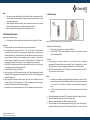
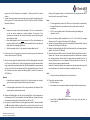
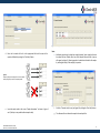
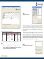
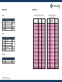
User manual Check-MDR CT101 Rapid Molecular Detection and Identification of Carbapenemases (KPC and NDM), ESBL (CTX-M, TEM and SHV) and AmpC (CMY, DHA, FOX, MOX, ACC, MIR and ACT) 10-0020 Key to symbols used For In Vitro Diagnostic Use Catalog number Batch code Instructions for use number Use before YYYY-MM Consult instructions for use Temperature limitation Contains sufficient for < n > tests Manufacturer 72 INTENDED USE .................................................................................................... 2 INTRODUCTION .................................................................................................. 2 PRINCIPLE OF THE METHOD ................................................................................ 3 KIT CONTENTS (FOR 72 SAMPLES) ....................................................................... 3 SHELF LIFE, STORAGE AND HANDLING ................................................................. 4 MATERIALS REQUIRED BUT NOT SUPPLIED WITH THE KIT .................................... 4 GOOD LABORATORY PRACTICES .......................................................................... 4 PROTOCOL.......................................................................................................... 5 1. DNA RECOGNITION STEP A ....................................................................................... 5 2. DNA AMPLIFICATION STEP B ..................................................................................... 6 3. DETECTION STEP ...................................................................................................... 6 FREQUENTLY ASKED QUESTIONS (FAQ) & TROUBLESHOOTING .......................... 11 APPENDIX 1 ...................................................................................................... 13 APPENDIX 2 ...................................................................................................... 13 LIMITATIONS .................................................................................................... 14 020-01 Check-MDR CT101 User manual Version 2.1, Issued 1 October 2012 1 Intended use Check-MDR CT101 is designed for the rapid molecular detection of a wide range of clinically important β-lactamase genes in gram-negative bacteria. Check-MDR CT101 detects the TEM, SHV and CTX-M extended-spectrum β-lactamase (ESBL) genes, AmpC β-lactamase genes CMY, DHA, FOX, MOX, ACC, MIR and ACT, the Klebsiella Pneumoniae Carbapenemase gene (KPC) and the New Delhi Metallo-β-lactamase gene (NDM). In most cases the presence of the genes indicates the expression of extended or full spectrum β-lactamase activity. For TEM and SHV, ESBL and non-ESBL variants exist and the test employs highly specific DNA markers to allow distinction between these variants. Check-MDR CT101 generates definitive results within the same working day, compared to the 24 hours necessary for analysis with conventional phenotypic methods. Information on gene type yields extra information for outbreak management. Introduction The TEM, SHV and CTX-M genes encode the clinically most prevalent extended-spectrum βlactamases (ESBLs). These three groups of ESBLs are generally capable of hydrolyzing first, second, third and fourth generation cephalosporins, penicillins and monobactams, thereby limiting treatment options. TEM and SHV ESBL subtypes are derived from their parental sequences by point mutations leading to amino acid substitutions (www.lahey.org/studies). These amino acid substitutions may extend the substrate spectrum to hydrolyze a wide range of β-lactam antibiotics, i.e. ESBL. The CTX-M genes originate from Kluyvera species and are presently the most prevalent ESBLs. They can be divided into 5 different groups according to their amino acid sequence: CTX-M-1, CTX-M-2, CTX-M-9, CTX-M-8 and CTX-M25. The AmpC cephalosporinases are able to hydrolyze almost all β-lactam antibiotics including: penicillins, cephalosporins and monobactams. Unlike ESBLs they are not inhibited by clavulanic acid. Many gram-negative rod bacteria carry a chromosomal copy of an AmpC gene and plasmid-encoded AmpC genes have emerged from the chromosomes of these Enterobacteriaceae. Species carrying a chromosomal AmpC gene include Citrobacter freundii, Morganella morganii, Aeromonas caviae, Hafnia alvei, Enterobacter spp. and Aeromonas hydrophilia (see table 1). Generally, plasmid-encoded AmpC genes give rise to significantly increased β-lactamase production compared to the chromosomally-located AmpC genes. Hence, it is useful for the clinician to be able to discriminate between plasmid and chromosomally-encoded AmpC genes. The presence of a plasmidic AmpC gene is indicated when detected in a species that does not have a chromosomal AmpC, e.g. Check-MDR CT101 User manual Version 2.1, Issued 1 October 2012 Klebsiella pneumoniae, or when the genotype of the AmpC gene differs from the inherent chromosomal gene of the species. For example, Morganella morganii contains a chromosomal copy of the DHA family. Detection of this AmpC in a species not being M. morganii will indicate the presence of plasmid-mediated AmpC. Table 1: AmpC genes and the species from which they originate. AmpC gene CMY II DHA FOX ACC ACT/MIR CMY I/MOX Origin of chromosomal gene Citrobacter freundii Morganella morganii Aeromonas caviae Hafnia alvei Enterobacter cloacae & asburiae Aeromonas hydrophilia Bacteria carrying ESBL or AmpC genes generally stay susceptible to carbapenem antibiotics. During the last decade various carbapenemase genes have been reported associated with elevated or complete resistance against carbapenem antibiotics. Strains carrying such genes are generally resistant to all β-lactam antibiotics. They often have additional β-lactamase genes and genes for resistance against quinolones and aminoglycosides, leaving very few therapeutic options. The carbapenemase genes tested for with Check-MDR CT101 are briefly described in the following text. Klebsiella Pneumoniae Carbapenemases (KPC) are plasmid-encoded Ambler class A carbapenemases that were first identified in Klebsiella pneumoniae isolates in North America. Currently, these enzymes have spread to many other parts of the world and are found in several other Enterobacteriaceae species including Escherichia coli. New Delhi Metallo-β-lactamase (NDM) is a new type of carbapenemase that was first reported in 2009 when found in a Klebsiella pneumoniae and Escherichia coli strain from a Swedish patient repatriated from a New Delhi hospital. Recent reports indicate that it has already spread to various European countries, notably the U.K., and that NDM may be prevalent in the Indian subcontinent. Conventional methods for detection of β-lactamases rely on phenotypic identification which is time-consuming, frequently inconclusive and not applicable to all species. Therapeutic failures associated with infections caused by bacteria containing various β-lactamase genes 2 are often due to serious problems with interpretation of phenotypic tests. Therefore, the capability of accurate and objective molecular detection is becoming more and more important. Check-MDR CT101 is a microarray-based diagnostic test that identifies the presence of TEM, SHV, CTX-M, KPC, NDM and AmpC genes as well as mutations leading to extended spectrum types of TEM and SHV enzymes. Check-MDR CT101 detects TEM mutations at positions E104K, R164S/H and G238S and SHV mutations at position G238S/A and E240K covering the clinically most relevant TEM and SHV ESBLs. Principle of the method The principle of the Check-Points diagnostic system is based on specific molecular recognition of DNA target sequences and subsequent amplification with universal primers. Each individual DNA target is recognized by a specific probe that contains a unique ZIP code corresponding to a unique position (address) on the microarray. These ZIP codes are used for detection on the microarray after amplification. Probes consist of two segments (probe arms), which are joined by a DNA ligase when they match perfectly with the target DNA. Only connected probe arms will result in amplification products. Probes that differ from the target DNA will not give amplification products, even in the case of a single nucleotide difference. Amplification products are hybridized to the microarray and visualized by colorimetric detection. The microarrays are contained in so called CP Array Tubes, which are inserted in the Check-Points Tube Reader upon completion of the detection reaction. This generates an array image that is analyzed by dedicated software to yield a definitive and objective assay result. Kit contents (for 72 samples) Components (Mat. No.) Description Storage conditions Detection Buffer (9-0007) 1 bottle 80 ml Room temperature Blocking Buffer (9-0008) 1 bottle 20 ml Room temperature Staining Solution (9-0014) 1 bottle 5 ml Room temperature store in the dark CP Array Tubes (10-0003) 6 bags of 4 each (total 24) Room temperature Manual (9-0036) Leaflet – download from website Not critical Blue tray (9-0031) 24x3-tube strip, 2.5 μl reagent/tube - 20°C Solution A (9-0021) 1 tube (brown cap ) 600 μl - 20°C Solution B (9-0023) 2 tubes (white cap O) 1600 µl - 20°C Solution C (9-0024) 1 tube (red cap ) 120 μl - 20°C Conjugate Solution (9-0027) 1 black tube & cap () 120 μl - 20°C Box Room Temperature Box -20°C Positive and negative controls are built into the system. It is, however, strongly recommended to use a positive and negative control for each series of reactions. Check-MDR CT101 User manual Version 2.1, Issued 1 October 2012 3 Shelf life, Storage and Handling Good laboratory practices Please check the individual components for optimal storage conditions immediately after delivery of the kit and store components accordingly. Reagents stored at the appropriate storage conditions can be used until the expiration date indicated on the boxes. Please visually inspect the boxes upon initial opening to ensure that their content is intact. Do not use when damaged. Please contact the Check-Points office at [email protected] if you have any questions or in case shipping has taken more than 2 days. Recommendations for best results Materials required but not supplied with the kit Pre-PCR Equipment Thermocycler* Vortex Mixer Mini-centrifuge Supplies Disposable laboratory (powder-free) gloves Pipettes & disposable (preferable filter-) tips for volumes of 1 to 1000 µl 1.5 ml tubes (“Eppendorf tubes”) 10 ml tubes *contact your local representative for specifications. Check-MDR CT101 User manual Version 2.1, Issued 1 October 2012 Post-PCR The quality of the results depends on strict compliance with the following good laboratory practices, especially concerning PCR: Thermocycler* Vortex Mixer Mini-centrifuge Thermomixer with active cooling* Check-Points Tube Reader with E-Ads software Computer with USB drive and internet connection Barcode Reader (optional) Disposable laboratory (powder-free) gloves Pipettes & disposable (preferable filter-) tips for volumes of 1 to 1000 µl 1.5 ml tubes (“Eppendorf tubes”) 10 ml tubes The test must be performed by adequately trained personnel. Spinning down for a few seconds is done in the various steps to ensure that all material is collected at the bottom of the tubes. Do not use reagents after their expiration date. Before use, thaw frozen reagents completely at room temperature and vortex briefly to obtain a homogeneous solution. After vortexing briefly, spin down the solution to avoid contamination when opening the lid. Avoid unnecessary freezethawing of the kit content. Periodically, verify the accuracy and precision of pipettes, as well as correct functioning of the instruments. Prevention of contaminations PCR produces a very high quantity of DNA amplification products (amplicons) even from minute quantities of starting material. Check-MDR CT101 may therefore yield unreliable results if samples become contaminated with amplicons from previous amplification reactions prior to the PCR (step B of the protocol). Preventive measures to minimize the risk of amplicon contamination must be taken. Please read carefully and follow the instructions outlined below. Use separate rooms: a pre-PCR room and a post-PCR room. Sample preparation, DNA recognition (step A) and preparation of the amplification step (step B) is carried out in the pre-PCR room. Incubation in the PCR thermocycler of step B is carried out in the post-PCR room. The detection step is also carried out in the post-PCR room. Alternatively, the detection step is carried out in a third room separate from the other two rooms. Never bring the reaction products of step B to the pre-PCR room. Never prepare the ligation and/or amplification steps in the post-PCR room. 4 To keep laboratory free of PCR product contamination: Use pipettes with hydrophobic filter tips. Make sure to always use a new pipette tip when adding solutions or samples to a reaction tube to avoid contamination. Follow proper pipette-dispensing techniques to prevent aerosols. Use separate equipment, pipettes, thermocyclers, sample holders, lab coats, gloves, disposables and reagents, that are assigned to these rooms. Never transfer items from the post-PCR room to the pre-PCR room. Wear a clean lab coat and clean gloves during all steps of the test. Wear clean gloves and a clean lab coat not previously worn while handling amplified PCR products or during sample preparation. Change gloves whenever you suspect that they are contaminated. Keep the tubes of all kit components and samples closed as much as possible. Clean the lab benches and all equipment regularly with a 0.5% sodium hypochlorite solution. Protocol It is strongly recommended that the full protocol is read before using the test. The protocol consists of the following steps: 1. 2. 3. DNA recognition step A DNA amplification step B Detection step 1. DNA recognition step A Important points before starting: Step A is completely carried out in the pre-PCR room. To ensure the best results we recommend using purified nucleic acids isolated from colonies or cells grown in nutrient broth. Methods should be used that purify total DNA, i.e. genomic as well as plasmid DNA. Generally such methods copurify DNA and RNA from bacterial cells. A nucleic acids concentration of 5 - 50 ng/μl should be used compensating for the excess of RNA generally present in DNA preparations using commercially available extraction methods. Column-based isolation methods are recommended. Store DNA extracts according to the manufacturer’s protocol for the DNA extraction method used. Procedure: 1. Thaw all reagents (i.e. Solution A, DNA samples if kept at -20˚C), mix well and keep on ice. 2. First add 5 μl Solution A (brown cap ) to every reaction tube of the strip (supplied with the kit). Next add 10 μl DNA solution of each sample (NB: use 1/100 part of the elution volume of the isolation and add up to 10 μl with MQ-water or 0.1 TE-buffer). Please write down the sample reference for each tube of the strip. 3. Close the tubes and spin down briefly using the mini-centrifuge to collect both sample and Solution A at the bottom of the tubes. Mix well by tapping against each strip; the solution should have a uniform blue color. Spin down briefly again using the minicentrifuge. 4. Place the strip(s) in the thermocycler and run the CP step A progam (total sample volume 18 μl). The program will run for approximately 2.5 hours. (The step A program is outlined in Appendix 1). Check-MDR CT101 User manual Version 2.1, Issued 1 October 2012 5 Note: 3. Detection step The reaction tubes supplied with the kit are prefilled with a small amount of bluecolored reagent. Proper mixing of this reagent, Solution A and the sample is crucial for optimal performance. When closing the tubes of the strip(s), don’t use excessive pressure as the cap may distort and the sample may then evaporate during steps A and B. Refer to Appendix 1 if the settings of the thermocycler are lost. 2. DNA amplification step B Important point before starting: The preparation of the reaction mix for step B is carried out in the pre-PCR room. Procedure: 1. Briefly spin down the reaction tubes when step A has been completed. 2. For amplification step B, prepare BC-mix in a 1.5 ml or 10 ml tube. Take Solution B (white cap O) from the freezer. Thaw properly at room temperature, mix well, and spin down briefly before use. Then take Solution C (red cap ) from the freezer. Prepare the required amount of BC-mix using the pipetting scheme in Appendix 2. First add the required amount of Solution B to the tube. Then dispense Solution C in Solution B by pipetting up and down 3 times. Mix very well by vortexing and spin down briefly. 3. Add 30 μl of the freshly prepared BC-mix to each sample in the strip(s). Close the tubes, mix by tapping each strip and spin down briefly. 4. Transfer the strips to the post-PCR room. 5. Place the strip(s) in the PCR thermocycler and run the CP step B program (total sample volume 48 μl). The program will run for approximately 1.5 hours. (The step B program is outlined in Appendix 1). 6. Briefly spin down the reaction mixture, after step B has been completed. Leave the reaction tubes in the PCR thermocycler at 4˚C (for a maximum period of two hours) until step 5 of the detection step has been completed. 7. Alternatively, store the reaction mixtures at -20°C for a maximum period of two weeks. A Figure 1: A) the CP Array Tube (AT) and B) the Check-Points Tube Reader. Important points before starting: The detection step is carried out in the post-PCR room. TM The ArrayTube DNA microarray platform (see figure 1) is sold under license from Alere Technologies GmbH. Procedure: 1. Start preparing the required number of CP Array Tubes (ATs) for detection approximately 10 minutes before the end of step B. One AT is required for every 3-tube strip. Remove the ATs from their package(s) and place them in the thermomixer. 2. Add 300 μl Detection Buffer to each AT. Switch on the thermomixer and heat to 50˚C. It is not necessary to close the tubes. Note: 3. 4. 5. 6. Check-MDR CT101 User manual Version 2.1, Issued 1 October 2012 B Be careful when removing or adding liquids with the pipette to or from the AT. Do not touch the microarray at the bottom of the tube at any time. Pipette all material in or out of the AT at the side of the bottom of the tube without touching the array. At 50˚C, remove and add solutions to one AT at a time to prevent the ATs from drying out. When the thermomixer has reached 50˚C, incubate the tubes for 2 minutes (400 rpm). Remove the Detection Buffer from the ATs and repeat step 3. Replace the Detection Buffer with 300 μl fresh Detection Buffer. Take the samples from step B. Samples stored for longer than 2 hours after step B was completed should be heated in the PCR thermocycler at 98°C for 2 minutes (CP Melt 6 7. program of the PCR thermocycler see Appendix 1). Briefly spin down the reaction mixture. Transfer 10 μl reaction mixture from each tube of one strip to the corresponding AT (in total 30 μl per AT). The total volume of the AT will be 330 µl. The lid of the AT should be labelled for reference. Note: Samples may contain a white-colored precipitate. This is due to denaturation of one of the reaction components, a protein stabilizer. The presence of this precipitate has no effect on the result of the detection step and may be ignored when adding the sample. When adding samples to the AT do not remove the AT from the thermomixer, to prevent the buffer from cooling down. Add the sample directly into the Detection Buffer of the AT by pipetting up and down. With the completion of step 7, three samples have been added to one AT. 8. Close the lids of the ATs properly to prevent them from drying out and incubate the tubes for 30 minutes at 50°C (400 rpm). 9. After 30 minutes, replace the Detection Buffer with 300 µl Blocking Buffer: do this with one AT at a time! Remove the AT from the thermomixer, discard the Detection Buffer using a pipette and immediately replace with 300 µl Blocking Buffer using a new pipette tip. Place the AT back in the thermomixer at 50°C and proceed with the next AT until all the AT solutions have been replaced with Blocking Buffer. Incubate for 5 minutes at 50°C (400 rpm). Optional: Evaporated water condensed in the lid of the AT may be removed with a pipette, e.g. before removing the Detection Buffer containing the sample. Note: Collect the liquids removed from the ATs in a disposable tube, and dispose of it the same day with the other laboratory waste. 10. Replace the Blocking Buffer with 300 μl fresh Blocking Buffer. Set the temperature of the thermomixer to 30°C and incubate for 10 minutes (400 rpm). During this time the thermomixer containing the ATs will cool down from 50°C to 30°C. 11. During step 10, prepare a dilution of the Conjugate Solution (black 2 ml tube with black cap ) with Detection Buffer using Appendix 2 at the back of this protocol. For this purpose a 1.5 ml tube or a 10 ml tube may be used depending on the amount required. Check-MDR CT101 User manual Version 2.1, Issued 1 October 2012 Dispense the Conjugate Solution in the Detection Buffer by pipetting up and down 3 times. Mix well by vortexing for 30 seconds. Note: The Conjugate Solution is stored at -20°C but is not frozen and can be used directly. Conjugate dilutions have to be made fresh, and should be used on the day of preparation. At 30˚C, remove solutions from all ATs before proceeding with adding new solutions to the ATs. 12. Remove the Blocking Buffer completely from all ATs. Then add 150 μl conjugate dilution to each AT. Incubate for 15 minutes at 30°C (400 rpm). 13. Remove the conjugate dilution from the ATs and add 600 μl Detection Buffer. Incubate the tubes for 2 minutes at 30°C (400 rpm). 14. Replace the Detection Buffer with 600 μl fresh Detection Buffer, and incubate the tubes again for 2 minutes at 30°C (400 rpm). 15. Remove the Detection Buffer from the ATs and add 150 μl Staining Solution to each AT. Incubate for 15 minutes at room temperature outside the thermomixer to complete the staining procedure. Continue with the image analysis immediately after the 15 minutes incubation time. Do not incubate with Staining Solution for more than 15 minutes, because images may become too dark. During the 15 minutes incubation time, complete the required sample information and relevant test data in the Check-Points software as outlined in point 16 a-g. Note: Store the bottle with Staining Solution in the dark after use. 16. Filling out the experimental data: a. Start the computer b. Start software on the computer by double-clicking the Check-Points desktop icon: Check-Points c. Double Click on “Check-MDR CT101.arr” in the first screen, “Array selection”, as shown in figure 2. 7 Figure 4: ”Sample information”. Figure 2: “Array Selection”. d. Enter the lot number of the kit in the appropriate field and the name of the operator, followed by pressing the “Next Step” button. Note: Additional remarks may be added per sample (optional), when sample references have been filled out. Double click on one of the sample reference fields. A pop-up will appear (see figure 5) allowing remarks to be added to individual or all samples by marking the box(es) of the sample(s) in question. 1 Figure 3: Example of filling out sample references in the software and assigning samples to the corresponding tubes. 1 Figure 5: Pop-up for additional sample information. e. Insert the sample codes in the screen “Sample information” as shown in figures 3 and 4. (Analysis is not possible without sample codes) Check-MDR CT101 User manual Version 2.1, Issued 1 October 2012 f. Go to the “Detection step” screen (see figure 6) by clicking the “Next Step” button. g. The software will now indicate the samples to be analyzed first. 8 Figure 6: “Detection step”. Figure 7: Presentation of the final result. 17. Enter the AT lot number in the appropriate field when the 15 minutes incubation time has been completed (optional: use the Barcode Reader to scan the AT lot numbers). Next, insert the AT with open cap into the reader, close the lid of the reader, and click on the “Confirm” button in the software. Then click on “Scan image”: the results will be displayed immediately (see figure 7). Finally click on “Save Results” to save the results in the database. The software will now indicate which sample should be analyzed next. Repeat this step until all ATs are analyzed. Note: Check-MDR CT101 User manual Version 2.1, Issued 1 October 2012 It is important to adhere to the 15 minutes incubation time with Staining Solution as much as possible (step 15). A shorter incubation time may lead to faint images; exceeding the 15 minutes incubation time may lead to overstaining. In both cases incorrect results may be obtained. 18. When all ATs have been analyzed, a new window with the summary of the results will appear (see figure 8), which may also be printed (click on the “Print results” button). The results summary will display the sample ID and the presence/absence of carbapenemases, plasmidic AmpC and ESBL as well as combinations thereof (see table 2 for a list of the genes that can be reported). Click on the “Quit” button to end this run of analyses. 9 Figure 9: “Send image to Check-Points” pop-up. Click on the “Save (Send later)” button to store the file, which will have a .cpfe extension on the computer and send it by e-mail. Alternatively, if the computer has internet access, check the box “Send picture directly over internet connection” and enter the reply e-mail address followed by clicking on send. In both cases feedback should be expected within two working days. Figure 8: Summary of the results. Table 2: β-lactamase genes reported by the Check-Points software. ESBLs Carbapenemases AmpCs NDM CTX-M-1 group TEM wt SHV wt CMY I/MOX KPC CTX-M-2 group TEM 104K SHV 238S ACC CTX-M-8 & -25 group TEM 164S SHV 238A DHA CTX-M-9 group SHV 240K ACT/MIR TEM 164H TEM 238S Note: CMY II For support concerning results please send the result image along with the desired information to [email protected]. For this purpose double click on the result(s) in question in the result summary window. A pop-up will appear (see figure 9) in which your comments may be added to the file. Check-MDR CT101 User manual Version 2.1, Issued 1 October 2012 Support may also be requested at a later stage when the program has been closed. For this purpose the “Send image to Check-Points” pop-up (see figure 10) may also be accessed from the database viewer (“DBview” shortcut that is located on the desktop). Open the database by clicking on “File” followed by “Open” (the database is located in C:\Images by default) and scroll down to the results which require support. By clicking on “Save”, the “Send image to Check-Points” pop-up will appear just like in the result summary window. Figure 10: Check the box to send pictures directly over the internet. 10 8.1. An air bubble is interfering with the result. Tap the AT gently or pipette the liquid gently up and down and then retry to take the image. 8.2. The picture is very dark: conjugate dilution was not removed properly. Please refer to question 5. 8.3. The picture is completely white. Staining Solution was not added. Please add Staining Solution again and proceed from detection step 15. If the results do not improve, then the staining has failed. Most likely no conjugate dilution was added, or the conjugate dilution was not prepared properly. Please repeat detection step with new AT. Frequently asked questions (FAQ) & Troubleshooting 1. The thermocycler states an error in step A or B. Please contact Check-Points Technical Support: [email protected] 2. During the PCR programs (step A or B) sample(s) have (partly) evaporated. Tubes may not have been closed properly. Please restart the procedure from step A. 3. I have left Solutions A, B and/or C out of the -20°C (-4˚F) storage. These reagents must be stored at -20°C (-4°F) for proper performance of the test. The performance of the product cannot be fully guaranteed if these solutions were left out of -20°C (-4˚F) for longer than 24 hours. 4. Staining Solution turned blue after adding it to the AT. Conjugate dilution was not properly removed by washing steps 13 and 14 (detection step). Continue incubation with Staining Solution for 15 minutes and take the image as described in the protocol. If the image is too dark, please refer to question 5. 5. The picture of the array is very dark. The conjugate dilution (detection step 12) was not properly removed by washing steps 13 and 14 (detection step). Please replace the Staining Solution with Detection Buffer and take image immediately. If the image is still too dark, please repeat detection step with new AT. 6. Software indicates: “Background intensity too high”. See question 5. 7. The software indicates: “Image too weak”. The intensity of the spots is too low. Causes may be: 7.1. The conjugate dilution was not prepared and/or added properly. Please repeat detection step with new AT. 7.2. Incubation time with the Staining Solution was shorter than 15 minutes. Continue incubation up to 15 minutes. 7.3. Detection Buffer was not removed properly before adding Staining Solution. Please repeat detection step with new AT. 8. The software indicates: “Reference spots not found”. The software did not find the reference spots on the AT. Causes may be: Check-MDR CT101 User manual Version 2.1, Issued 1 October 2012 9. The software indicates: “Hybridisation not OK”. The software did not find the hybridisation control spots on the AT. The hybridisation control is used to check if the hybridisation (at 50°C) of the PCR product with the AT has been performed properly. Causes may be: 9.1. The picture is completely dark: conjugate dilution was not removed properly. Please refer to question 5. 9.2. The reaction mixture after completion of step B was not added to the AT. Please repeat detection step with new AT. 9.3. Hybridisation temperature too high. Please verify that the thermomixer temperature was 50°C when the ATs were hybridized. 9.4. The BC-mix was not prepared properly or was not added to the assay: please repeat the test. 10. The software indicates: “Reaction not OK, please contact Check-Points”. This message will be displayed in the following two scenarios: A) The software did not find the reaction control spots on the AT. These amplification controls are used to check the performance of the assay in steps A and B. Possible explanations are: 10.1. The sample DNA was not added to the assay in step A. 10.2. The sample DNA contains contaminants inhibiting the reactions. These may originate from the growth medium or the DNA isolation method. This may be remedied by diluting the DNA extract 10-fold with distilled water. Repeat the test with the diluted DNA. 10.3. The A mix was not added in step A. Please repeat the test. 10.4. The BC-mix was not prepared properly or was not added to the assay: please repeat the test. 11 10.5. Reaction mixtures from step B (step 7 of detection step) were not all added to the AT: 3 reaction mixtures should be added, one or two may be omitted. weeks. For further inquiries, please contact Check-Points Technical Support: [email protected] B) The amount of bacterial DNA added in step A was probably insufficient: the DNA extraction may have failed or the DNA concentration may have been too low. Please repeat the test with a new DNA extract. 17. The spot intensities on various ATs are weak. Please repeat the test. Contact Check-Points Technical Support at [email protected] if results do not improve. 11. The software indicates: “ESBL suspected”. The software did not find sufficient spots to give a conclusive result. Please inspect the picture visually: an air bubble or dust particles may interfere with the result. Tap the AT gently or pipette the liquid gently up and down and retry to take the image. Please repeat the test if the results do not change. 18. The AT image contains dust particles. The software will correct this in most cases. To prevent any interference with the results, please take the AT out of the reader and shake it gently until the dust particles have moved to the side of the AT. 12. The software indicates: “Picture not found” or “Image capture error”. Check if the Check-Points Tube Reader is properly installed. Please contact CheckPoints Technical Support at [email protected] if reinstallation of the reader does not solve this problem. 13. The AT image is covered with small spots. There may be various causes for this phenomenon. Most likely the array dried out during the detection step. Please make sure that the AT always contains sufficient amounts of reagents (Detection Buffer, Blocking Buffer, conjugate dilution or Staining Solution). This is particularly critical during the incubation steps at 50°C. In most cases the software will be able to handle these small spots. If not, repeat the detection step with a new AT. 14. The AT was incubated for more than 15 minutes with Staining Solution before taking the image. Results may be unreliable due to overstaining. Inspect image: if spots are very dark, please repeat detection step with a new AT. 15. Duplicate DNA samples tested with Check-MDR CT101 do not yield identical results. 15.1. Inspect images for dust particles. Tap or gently shake the tubes to try to remove the particles from the array and rescan the images. 15.2. In case of extra spots, repeat the test to confirm result. 16. May the assay be interrupted and continued at a later stage? Reaction mixtures from Step A can be kept at hold at +4˚C for a maximum period of two hours. Reaction mixtures from Step B can be kept at -20˚C for a maximum period of two Check-MDR CT101 User manual Version 2.1, Issued 1 October 2012 12 Appendix 1 Appendix 2 Pipetting scheme for BC-mix: Step A Step Temperature / Time 1 95°C / 3min 2 95°C / 30sec 65°C / 5 min 3 98°C / 2min Holding 4°C Cycles (1x) (24x) (1x) Step B Step Temperature / Time 1 95°C / 10min 2 95°C / 5sec 55°C / 30sec Cycles (1x) (35x) 72°C / 30sec 3 98°C / 2min Holding 4°C (1x) Step Melt Step Temperature / Time 1 98°C / 2min Holding 4°C Check-MDR CT101 User manual Version 2.1, Issued 1 October 2012 Pipetting scheme for conjugate dilution: Cycles (1x) samples 1–3 4–6 7–9 10 - 12 13 - 15 16 - 18 19 - 21 22 - 24 25 - 27 28 - 30 31 - 33 34 - 36 37- 39 40 - 42 43 - 45 46 - 48 49 - 51 52 - 54 55 - 57 58 - 60 61 - 63 64 - 66 67 - 69 70 - 72 μl B 120 210 300 390 510 600 690 780 900 990 1080 1170 1290 1380 1470 1560 1680 1770 1860 1950 2070 2160 2250 2340 μl C 4 7 10 13 17 20 23 26 30 33 36 39 43 46 49 52 56 59 62 65 69 72 75 78 ATs 1 2 3 4 5 6 7 8 9 10 11 12 13 14 15 16 17 18 19 20 21 22 23 24 µl Conjugate Solution 5 5 5 10 10 10 15 15 15 20 20 20 25 25 25 30 30 30 35 35 35 40 40 40 μl Det. Buf. 495 495 495 990 990 990 1485 1485 1485 1980 1980 1980 2475 2475 2475 2970 2970 2970 3465 3465 3465 3960 3960 3960 13 Limitations Check-MDR CT101 uses a range of specific DNA markers to identify the presence or absence of ESBL genes, AmpC genes and KPC and NDM carbapenemase genes . The test detects the presence of the ESBL genes TEM, SHV, CTX-M and it also detects Single Nucleotide Polymorphisms (SNPs) corresponding to amino acid positions 104, 164 and 238 in TEM, and 238 and 240 in SHV. The nature of the above SNPs in TEM and SHV determines the ESBL phenotype (ESBL yes or no) for the majority of TEM and SHV types. (For a detailed explanation see www.lahey.org/studies and M. Gniadkowski, Clin. Microbiol. Infect. 2008; 14 [Suppl. 1]: 11–32). Check-MDR CT101 also detects the AmpC genes CMY, DHA, FOX, MOX, ACC, ACT and MIR, which presently represent the clinically most prevalent AmpC genes. It should be noted that other AmpC genes are not detected. Check-MDR CT101 requires DNA purified from a colony or bacterial culture. Clinical specimens cannot be tested directly. The assay has been tested extensively with purified DNA from gram-negative bacteria, such as Escherichia, Salmonella, Klebsiella, Enterobacter, Citrobacter and Pseudomonas, with excellent results. However, it may never be excluded that other gram-negative bacteria or certain strains of the above species will yield poor results. Furthermore, although TEM, SHV and CTX-M are the most prevalent ESBL genes, various minor ESBL genes exist. However, these are not frequently found in clinical settings. (For more details see T. Naas, L. Poirel and P. Nordmann, Clin. Microbiol. Infect. 2008; 14 [Suppl. 1]: 42–52). Check-MDR CT101 cannot and does not make any representation or warranty that it is capable of correctly detecting the AmpC, ESBL, KPC and NDM genes in all gram-negative species, subspecies or type or in any clinical sample source. Results may need to be confirmed by additional methodologies in specific cases (e.g. for regulatory samples). Due to the high variability of bacterial genomes it is possible that certain sub-types might not be detected. The test reflects the state of knowledge of Check-Points Health B.V. The quality of the input DNA is an important factor in obtaining reliable results from Check-MDR CT101. DNA must be extracted from cultured bacteria using a suitable method such as commercially available extraction methods based on columns or beads. Methods should be used that extract and purify total DNA, i.e. genomic as well as plasmid DNA. The presence of multiple bacterial species in a sample may hamper the interpretation of the test. As with other diagnostic assays, the results of this test may only be interpreted in combination with additional laboratory and clinical data available to the responsible person. Use of this assay is limited to appropriately qualified personnel, well trained in the test procedure and familiar with molecular biological methods. The Check-Points’ software is continuously improving thanks to the expanding global database of available results. In order to further improve the software, original result images (including specific data*) are shared with this database through a default setting in the software by means of an encrypted file. This data will be used solely for software improvements. All information will be kept strictly confidential and will not be shared with any third parties. *The specific data included are: the number identifying the CP Array Tube, the kit lot number, the software version used, the operator and the customer name. All patient data and sample information is removed in the encrypted file. Check-MDR CT101 User manual Version 2.1, Issued 1 October 2012 Despite the utmost care in the development and preparation of the protocol Check-Points cannot take any responsibility for errors, omissions and/or future changes herein. Literature Citation: When describing a procedure for publication using this product, please refer to it as the CheckMDR CT101. Check-Points Health BV Binnenhaven 5 6709 PD Wageningen The Netherlands Tel: +31 317 453 908 Fax: +31 317 210 147 [email protected] www.check-points.com 14