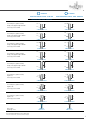
1
P R O ST HET IC MANUAL
CONOWELD
Shelta
P R O S T H E T I C M A N U A L TA B L E O F C O N T E N T S
Connection
platform
6
Use Protocols
18
Prosthetic
components
General indications
8
34
Bibliography
Instruments
41
14
Sweden & Martina develops and manufactures implant systems that offer excellent clinical performance hand in hand with perfect cosmetic results.
The prosthetic components available reflect the company’s updating and development capacity and cover all the requirements of prosthodontists and
laboratories. The same quality obtained for the production of implants is also guaranteed for the prosthesis: from abutments to screws, every single part is
milled with certified CNC machines and not moulded.
Training courses, continuous refresher courses and extensive assistance distinguish the service and reliability that have made Sweden & Martina a leader in
the Italian implant market.
KOHNO SWITCHING PLATFORM
Characterised by tapering
accentuated by the bevel for
the Switching Platform.
Premium SWITCHING PLATFORM
The morphology is the result
of clinical findings.
Premium
Kohno
Premium ø 3.30 mm
Dedicated to intraforaminal sectors and
useful with thin bone crests, or to replace upper
lateral incisors.
Premium
SHORTY implants
Intended for bone crests with reduced vertical
development.
4
KOHNO STRAIGHT
The same connection combined
with a conical morphology extends
the range of use of the family.
Premium Straight
The implant with 16 years
of clinical history.
Shelta
SHELTA SL
The large thread studied to obtain
the maximum primary stability.
Shelta standard
Three different diameters with the
same prosthetic connection.
5
Connection platform
COLLEX connection
The COLLEX connection, supported by 16 years of clinical studies, is characterised by a wide internal hexagon, synonym of high prosthetic stability,
guaranteed also by the collar that penetrates the posts, giving to the prosthetic structure an excellent and unique strength.
This interlocking solution gives stability and solidity to the implant-prosthesis complex, while also aiding the correct distribution of masticatory loads.
The COLLEX connection performs the same stabilising function regardless of the emergence of the implant, which may be straight in the case of Straight
implants or bevelled in the case of SP implants (Switching Platform).
The external collar of the COLLEX has the function of guide and engagement of the Easy Insert driver, the patented driver for the insertion of Premium
implants, which guarantees the total preservation of the angles of the internal connection during the surgical procedure, very important condition for a
correct prosthetic phase.
Collex connection
Premium implants
Ø 3.30 mm
Collex connection
Premium Kohno Straight implants
Ø 3.80, 4.25, 5.00 mm
Collex connection
Premium Kohno SP implants
Ø 3.80, 4.25, 5.00 mm
Collex connection
Shelta implants
Ø 3.80, 4.25, 5.00 mm
The strength properties of the COLLEX connection are also documented by a study carried out by the group of Prof. Covani, in which this connection was
compared with another internal hexagon connection, but without the external prosthetic collar; the results highlighted values 25% higher in terms of
robustness and stability of the prosthetic COLLEX compared to the connection without collar.
(Covani U., Ricci M., Tonelli P., Barone A. - An evaluation of new designs in implant-abutment connections: a finite element method assessment - Implant
Dentistry Volume 22, Number 3 2013).
IMPORTANT WARNING
With the same implant diameter, the implants with Straight emergence and with Switching Platform emergence use the same prosthetic components:
for this reason no distinction will be made in this manual between the two different emergences.
6
Switching Platform
The Switching Platform protocol, a prosthetic technique widely supported by scientific literature, aims to distance the implant-post junction from the
crestal bone. This result may be achieved either by designing ad-hoc an enlarged emergence at the level of the neck of the implant, or by using posts with a
diameter smaller than the implant platform, when the geometer of the connection is the same for all the sizes in the range. Premium Kohno SP implants are
specifically designed for use in prosthetic rehabilitations according to the Switching Platform protocol: the bevel around the connecting platform distances
the prosthetic junction both vertically and horizontally. The morphology of the neck of the implant is also very useful for obtaining an excellent primary
stability. The Switching Platform technique used in these implants is incorporated in the fixture morphology.
0.30 mm
Ground Section of Premium
Switching Platform implant 4
months after insertion.
0.50 mm
(Image by kind permission of
Dr D. Botticelli)
Maximising of soft tissues
The decision to simplify the range of Conoweld posts to two sizes is also supported by the excellent clinical results of the Switching Platform protocols
reported in the literature.
With the Premium Kohno system, the Conoweld posts that rest on the COLLEX collar result in mismatching being available for a larger amount of gingival
tissue, which is organised and stabilised in keratinised tissue around the prosthetic crown.
In addition, the implant-abutment joint is shifted not only horizontally but also vertically, maximising the distance from the soft tissues.
Conoweld post with
2.30 mm hexagon
Conoweld post with
2.50 mm hexagon
Conoweld post with
2.50 mm hexagon
Premium Implant SP Ø 3.80
Kohno Implant SP Ø 3.80
Premium Implant SP Ø 4.25
Kohno Implant SP Ø 4.25
Premium Implant SP Ø 5.00
Kohno Implant SP Ø 5.00
7
PROSTHETIC COMPONENTS
Conoweld conometric technique
The Conoweld technique embraces the advantages of two protocols already widely established in oral implantoprosthesis: intraoral welding and conometric
retention.
Intraoral welding for
the temporary stage
• stabilizes immediate loaded
implants
• provides an immediate temporary
prosthesis that is reinforced
• makes it possible to take a precise
impression
ADVANTAGES OF CONOMETRIC REHABILITATION:
• Extremely reliable hold: the connection between the prefabbricated conical cap incorporated
in the over-structure and the post offers total mechanical stability that can be relied upon.
• Fully reversible: the dentist can remove the prosthesis at any time applying the same degree
of force. Impossible for the prosthesis to move spontaneously: the consequent splinting of
the implants makes it immediate loading possible and secure.
• Extremely simple to use: the posts are standardised and all fit the same caps. • Low-maintenance for the patient, who has all the comfort of a fixed prosthesis, and easy to
access for the dental hygienist.
• Natural passivation of the posts.
• Primary stability ensured with intraoral welding.
8
Conometry for the temporary
and final restoration
• cement-free restoration
• removable, but only by the dentist
• low-maintenance for the patient at
home
The technique involving the intraoral welding of the temporary caps in titanium is carried out by using equipment specifically designed for the purpose and
which melt two pieces of titanium together, with a high intensity electric charge being passed through the point of contact, but for such a short time (a few
milliseconds) that the surrounding tissues are not heated at all.
The cap for the temporary stage is welded
to a titanium bar in order to create a structure
that is solid but can be removed without
having to unscrew the posts. In addition, being
passivated in the mouth, this can operate as a
reliable control key for accurately transferring
an impression to the laboratory.
The Conoweld post can operate as a
temporary as well as a permanent solution.
Here it has the advantage of eliminating the
biological stress linked to screwing, unscrewing
and replacing the prosthetic components,
protecting the intimate bond that is created
between the mucous tissues and the emergence
of the post.
The final cap works on the same operating principle
as the temporary cap; not needing to be of a thickness
that is sufficient for the intraoral welding, it takes up far
less space, making it easy to position in the prosthetic.
This system, currently available for Premium
Kohno and Shelta implants of all diameters,
includes posts with a conical body that are
straight as well as at 5°, 10° and 15° angles,
to be used together with the special titanium
caps for intraoral welding, at the temporary
stage, and together with the corresponding
permanent caps, to achieve rehabilitation with
conometric retention both partial and full arch.
Completing the range is a handy cap in PMMA,
which can be used for a snap-on impression as well as
for casting, and a laboratory analog that can be easily
repositioned not only on the impression but also on the
fixed structure.
9
PROSTHETIC COMPONENTS
Conoweld prosthetics for Premium, Kohno and
Shelta implants
Conoweld conical posts
The grade 5 titanium posts that form part of the Conoweld prosthetic range have been specifically designed to rest securely on the COLLEX collar.
This makes it possible to have only two posts for the Premium and Kohno platforms: one with a hexagon with a 2.30 mm key for platforms with implant
diameters of 3.30 mm and 3.80 mm, and one with a hexagon with a 2.50 mm key for platforms with implant diameters of 4.25 mm, 5.00 mm and 6.00
mm, without any difference between straight and SP implant emergences.
The Shelta implants, which share the same connection with a hexagon with a 2.30 key, use the same Conoweld post.
POSTS WITH A 2.30 MM HEXAGON
The same Conoweld straight and angled posts with a 2.30 mm hexagon are used on Premium Kohno implants with Ø 3.30 and 3.80 mm and on all
diameters of Shelta implants, that is 3.80, 4.25 and 5.00 mm.
Premium Ø 3.30
Premium Ø 3.80
Kohno Ø 3.80
Shelta Ø 3.80
Shelta Ø 4.25
Shelta Ø 5.00
POSTS WITH A 2.50 MM HEXAGON
The same Conoweld straight and angled posts with a 2.50
mm hexagon are used on Premium Kohno implants with
Ø 4.25 and 5.00 mm and for the Kohno implant Ø 6.00 mm.
Premium Ø 4.25
Kohno Ø 4.25
10
Premium Ø 5.00
Kohno Ø 5.00
Kohno Ø 6.00
2.30 mm
Premium Kohno Ø 3.30 - 3.80 mm
2.50 mm
Premium Kohno Ø 4.25 - 5.00 - 6.00 mm
Shelta Ø 3.80 - 4.25 - 5.00 mm
Conoweld post in grade 5 titanium,
straight, transgingival height 0.50 mm.
Connecting screw included
2.85
2.85
3.50
0.50
A-MD-TS-EX230-05
Conoweld post in grade 5 titanium,
straight, transgingival height 1.00 mm.
Connecting screw included
3.50
1.00
3.50
2.00
3.00
3.50
5.00
3.50
2.00
A-MD-TS-EX250-2
5.00
3.00
A-MD-TS-EX250-3
5.00
5.00
5.00
3.50
5.00
3.50
5.00
1.50
5.00
3.50
5.00
1.50
5.00
3.50
3.50
2.85
A-MA10-TS-EX230
Spare screws
In packages of 10
A-MD-TS-EX250-1
5.00
A-MA05-TS-EX230
Conoweld post in grade 5 titanium,
angled at 15°
Connecting screw included
1.00
2.85
2.85
A-MD-TS-EX230-5
Conoweld post in grade 5 titanium,
angled at 10°
Connecting screw included
5.00
3.50
5.00
2.85
Conoweld post in grade 5 titanium,
angled at 5°
Connecting screw included
A-MD-TS-EX250-05
2.85
2.85
A-MD-TS-EX230-3
Conoweld post in grade 5 titanium,
straight, transgingival height 5.00 mm.
Connecting screw included
0.50
5.00
A-MD-TS-EX230-2
Conoweld post in grade 5 titanium,
straight, transgingival height 3.00 mm.
Connecting screw included
3.50
2.85
2.85
A-MD-TS-EX230-1
Conoweld post in grade 5 titanium,
straight, transgingival height 2.00 mm.
Connecting screw included
5.00
5.00
5.00
1.50
5.00
3.50
A-MD-TS-EX250-5
5.00
3.50
5.00
1.50
A-MA05-TS-EX250
5.00
3.50
5.00
1.50
A-MA10-TS-EX250
5.00
3.50
5.00
1.50
A-MA15-TS-EX230
A-MA15-TS-EX250
VM2-180
VM2-180-10
VM2-200
VM2-200-10
Recommended tightening torque: 20-25 Ncm.
All measurements in mm, save where otherwise indicated.
11
PROSTHETIC COMPONENTS
Conoweld conometric caps
The Conoweld range includes three different caps, which are universal in relation to the diameters of the posts and the implant platforms: this is due to the
fact that conometric retention is in the most coronal portion of the post, which always has the same dimensions.
5°
10°
15°
The two titanium caps differ in thickness: the cap designed for the construction of a welded structure intraorally for the temporary stage is thicker in order
to withstand the welding with the titanium bars, without bonding with the underlying post, while the cap designed to anchor the final prosthesis glued on
is thinner, in order to reduce the impact on the anatomical morphology of the prosthesis; it should not, therefore, be used for the welding.
A cap in PMMA is also available, which allows a precise impression to be taken even when working without a intraoral welding machine and which can
be used for modelling and casting a structure entirely in cobalt chrome or other alloys, when the decision has been taken not to use gluing techniques for
assembly.
12
code
description
5.70
Temporary cap for intraoral welding
CAP-TS-PRO
5.70
Final cap for gluing
5.70
Cap for impression
CAP-TS-DEF
CAP-TS-IMP
17.00
Analog of the Conoweld post
ANA-TS
Bar in grade 2 titanium, L. 150 mm, Ø 1.20 mm, package of 5
DW-BARRA1.2
Bar in grade 2 titanium, L. 150 mm, Ø 1.50 mm, package of 5
DW-BARRA1.5
Bar in grade 2 titanium, L. 150 mm, Ø 1.80 mm, package of 5
DW-BARRA1.8
13
INSTRUMENTS
DENT WELD
Endoral welding machine
DENT WELD is a high-tech endoral welding machine for
fixing implants in the event of immediate loading and to
maintain maximum stability throughout the entire process of
rehabilitation up until osseointegration of the implants.
Welding is carried out by means of a high intensity electric
charge being passed through the point of contact between two
pieces of titanium, but for such a short time (a few milliseconds)
that the surrounding tissues are not heated at all.
General features
• Dent-weld is the endoral welding machine with 100% Italian technology and quality, and is a medical device manufactured by Swiss & Wegman S.r.l., via
Svezia 8, 35020 - Ponte San Nicolò (PD) ITALY and distributed in Italy exclusively by Sweden & Martina S.p.A..
• Safe and accurate, the welding machine is fitted with a microprocessor that guarantees complete safety in all applications. • The practical titanium bars (which come in various diameters) to weld to the posts mean that implants can be fixed in the space of only a few seconds,
with their stability being assured throughout the entire period of osseointegration.
• DENT WELD does not create sparks, it does not generate any risk for the patient because the forceps are automatically disconnected from the power
supply during the welding phase.
• The heat produced is dispersed through copper electrodes, which have greater thermal conductivity than titanium, without causing any discomfort to the
patient or to the doctor.
• DENT WELD also welds in the presence of saliva or any liquid.
• The pre-set programs make it easy to use.
• The forceps are easy to handle; fitted with a simple attachment, they are easy to sterilise.
Standard configuration
Control unit, forceps, cable, pedal, power cable, instruction leaflet.
Technical features:
Supply voltage:
Frequency:
Average power absorbed:
Weight:
Dimensions:
Classification:
230V AC
50 Hz
10W
7 kg
33x22x17 cm
I- BF, IPXO
See the instruction manuals for the equipment and the electrode forceps:
Preliminary instructions for welding
1.Coupling stage
The coupling stage involves bringing the two materials into direct contact with one another, avoiding any stress. The pressure applied by the tips must
bring the two shapes of the objects to be welded together so that they match perfectly, with absolutely no space between the two materials.
2.Welding stage
It is extremely important at this stage that pressure is maintained in order to keep the joints coupled, to act on contact resistance and to concentrate
maximum resistance at the centre. It is recommended at this stage not to apply any force that resists the action of the pre-load spring in the forceps.
3.Cooling stage
Having completed the welding, pressure must be maintained for a few seconds (around five) in order to allow the heat that has been generated to
dissipate via the electrodes
14
Drivers
Connecting screws for all Conoweld posts can be screwed using the following drivers, included within all surgical and prosthetic kits for Sweden & Martina
implants. They can also be ordered separately as spare parts.
code
description
Driver for tap screws and connecting screws, digital, extra-short
HSMXS-20-DG
Driver for tap screws and connecting screws, digital, short
HSM-20-DG
Driver for tap screws and connecting screws, digital, long
HSML-20-DG
Driver for connecting screws, with connector for dynamometric ratchet or
digital connector, short
HSM-20-EX
Driver for connecting screws, with connector for dynamometric ratchet or
digital connector, long
HSML-20-EX
Driver for connecting screws, with connector for dynamometric ratchet or
digital connector, extra long
HSMXL-20-EX
Driver for connecting screws, with right angle shank
HSM-20-CA
15
STRUMENTAZIONE
Dynamometric ratchet CRI5
The surgical kit of the implant system contains a special ratchet (CRI5), with its own adjustment key, for quickly screwing the torque adjustment ring nut, and
with gel lubricant for maintenance. The ratchet may be used with torque adjustment from 10 to 70 Ncm or in a blocked position without torque control. When
using as a prosthetic ratchet for fastening the screws, refer to the torque values given in the table on the previous page. The ratchet key CRI5 is a multi-purpose
instrument that can be disassembled, and is sold unsterile. It is included in the Screw Kit and in all surgical kits of Sweden & Martina Implant Systems. It is
also available as a replacement.
IMPORTANT WARNING
All components that are to be tightened at a torque of less than 10 Ncm must be tightened by hand.
For example: Transfer screws to be tightened at 8 Ncm.
Before each use, this instrument must be cleaned and sterilised according to the instructions on pages 38-39. Adequate maintenance, performed following
in detail all the step by step instructions for the disassembly and correct reassembly of the device during cleaning operations, is essential for the correct
functioning of the device and for its durability. Personnel who use this tool must be suitably trained, and they must have read the instructions in this manual
prior to handling the device.
After sterilisation, the key is ready for use. A test to verify the correct assembly and functioning of the key is necessary before any surgical or prosthetic
interventions.
Ratchet head cover
Pawl wheel
Wheel stop tooth
Cover fastening screw
Spring
Torque adjustment ring nut
Ratchet head
Handle
Guide pin
Hexagonal tip of the torque
adjustment screw
16
After sterilization the key is ready to use; before any surgical or prosthetic procedure a test to check the correct assembling and functioning of the key is
mandatory.
The torque is adjusted by aligning the marking of the desired torque in the circular opening of the handle. The “IN” arrow legible on the top of the head
indicates the screwing position of the key. The “OUT” arrow legible on the top of the head indicates the loosening or unscrewing position.
An unlimited torque position is obtained by positioning the torque adjustment device up to the line marked “R” on the handle of the ratchet body.
IMPORTANT WARNING
The torque is adjusted by screwing/unscrewing the ring nut located at the bottom of the instrument’s handle. The torque must always be adjusted on
the rise, starting screwing from a lower value until the desired torque is reached, or unscrewing the ring nut in a clockwise direction. To do this, if it is
necessary to set a torque lower than the last one used, you must unscrew the ring nut by two turns below the value of the desired new torque, and work
up to that value by rescrewing the ring nut in a clockwise direction.
The ring nut may be screwed and unscrewed by hand, but to speed up these
operations the kit also contains a driver that allows it to be turned quickly.
Any deterioration of the screwing, insertion and torque mechanisms must be
checked by personnel responsible for the use and maintenance of this dental
instrument.
The pieces of this mechanism are not interchangeable; one piece from one key
cannot be replaced by a piece from another key as each ratchet is calibrated
INDIVIDUALLY. If a piece is lost, please return the instrument to Sweden &
Martina for repair. No components for assembling the ratchet can be sold
individually.
Failure to follow the instructions provided may cause problems of maintenance
and stability of the prosthesis.
17
USE PROTOCOLS
Fixing stage - endoral welding on Conoweld caps
for immediate loading
Position the Conoweld posts on the implants, accurately assessing the transgingival height that is the most suitable and
the angle that is most appropriate in the case of disparallel
implants. Tighten the connecting screws using one of the
HSM drivers (see page 15 for lengths and types available) at
a torque of no more than 25-30 Ncm.
Position the temporary Conoweld caps on the abutments,
gently applying manual pressure. Take a titanium bar for intraoral welding of the most suitable thickness and curve it
manually in line with the arch to be rehabilitated.
Note: avoid cutting the bar to measure at the outset, as
the excess segment makes removal and repositioning easier to handle until the welding of the caps is
complete.
Line up the first segment of the bar with one of the two
distal caps and carry out welding with the special intraoral
welding machine, following the manufacturer’s instructions.
IMPORTANT WARNING
To set the operating parameters for the welding machine
and for the relevant information and warnings, read
and follow the instructions in the manual issued by the
manufacturer of the equipment.
18
In order to establish that the procedure has been carried out
correctly, it is advisable to remove the bar and caps that have
been fixed up until that point after each welding and before
the next. This helps to establish that the wall of the cap and
the underlying post have not bonded and to be sure that a
passive structure is being built.
Having positioned the first cap back on its post, guide the bar
round and, if necessary, remodel it, in order to weld it to the
next cap along.
Remove the structure made up of the bar and the first two
caps welded to it in order to establish that that the wall of this
second cap and the underlying post have not bonded and to
establish that the structure is passive.
19
USE PROTOCOLS
Having positioned the first and second caps back on their
posts, guide the bar round and, if necessary, remodel it, in
order to weld it to the next cap along.
Remove the structure made up of the bar and the three caps
welded to it in order to establish that that there is no casting
between the wall of this third cap and the underlying post
and to establish that the structure is passive.
Having positioned the three caps back on their posts, guide
the bar round and, if necessary, remodel it, in order to weld
it to the next cap along.
20
Remove the entire structure made up of the bar and the four
caps welded to it in order to establish that that the wall of
the last cap and the underlying post have not bonded and to
establish that the structure is passive.
In order to further increase the passive nature of the structure
and eliminate any residual stress, welding is advisable at a
halfway point along each segment of free titanium along the
bar.
21
USE PROTOCOLS
A
If a more rigid structure is required or if titanium bars with
a thin diameter are being used, an additional bar can be
welded onto the caps, following the exact same procedure as for the first bar, i.e. one cap at a time, removing the
structure after each welding. The preferable positioning is
the ‘gun barrel’ style (A), i.e. two parallel bars, one sitting
above the other. There are other solutions, however, such as
crossed bars (B) and bars forming a rail (C) i.e. parallel horizontally. This type of structure is particularly useful where
the caps descend 1.5 mm into the sulcus, leaving only 4 mm
of wall available for welding. This arrangement is, however,
only suggested in the distal sectors, where problems are not
usually encountered with thickness. Whilst the additional
bar should not be capable of generating traction, here too
the free segments can be passivated with a welding point.
Where the bars are positioned to form a rail, welding is carried out by inclining the forceps vertically in order that only
one segment is gripped between the two electrodes.
B
Note: whilst it is advisable for both bars to have the same
thickness (usually two bars of 1.50 mm), different
diameters can also be used.
IMPORTANT WARNING
Do not attempt to passivate both segments at the same
time. As the welded structure would in any event close
the circuit, the bars would soften and deform under the
pressure of the forceps, creating additional stress and
weakening the structure instead of passivating it.
C
22
Creation of the temporary prosthesis: direct protocol
The temporary prosthesis for immediate loading can be made
in the laboratory or chair-side, relining a ready-made moulded prosthesis. The resin will incorporate the welded structure
entirely, which also makes it easier for the patient to clean it
at home.
Once the base of the temporary prosthesis has been smoothed, it can be positioned onto the Conoweld posts immediately: the interaction via conometry between the posts and
the Conoweld caps will mean that, in terms of retention, the
dentist will be able to remove the temporary prosthesis at any
time, but the patient will not be able to remove it himselves.
This particular feature of conometric rehabilitation ensures
solid splinting throughout the entire osseointegration period,
limiting the micromovements of the implants, even where the
bone is only slightly mineralised.
23
USE PROTOCOLS
Indirect protocol: impression on the welded structure
The structure including the temporary Conoweld caps welded onto the titanium bars forms an accurate and reliable
impression key. The impression can therefore be taken incorporating the entire structure within the material inside
the tray. As the caps rub against the posts as a result of
conometry, it is advisable to use hard impression material for
edentulism (e.g. SKY IMPLANT ONEMIX- ED, code SKY08).
Push the impression tray onto the welded structure so that,
despite its fairly rigid consistency, the impression material
incorporates the bar and the caps welded onto it completely.
Lift the tray vertically: the entire welded structure will remain
incorporated in the impression material.
24
One by one, reposition the analogs in the metallic caps. Send
the impression to the laboratory. Cast the model in the usual
way.
Release the structure from the impression material, taking
care not to create distortions, and position it on the model in
order to check that it is completely passive.
On the structure, build a temporary prosthesis in resin, and
then return to the patient in order to position this in the mouth.
25
USE PROTOCOLS
Indirect protocol: impression with Conoweld transfer caps in PMMA
As an alternative, the impression can be taken using the special Conoweld caps in PMMA, inserting them on the Conoweld posts and gently applying manual pressure.
Fix the caps between each other using resin and wire, if desired.
Take a closed tray impression, ensuring that, vertically, the
caps and posts are included in the impression material in
their entirety. Leave to harden for the amount of time indicated by the manufacturer.
Lift the impression tray: the PMMA caps will remain securely
incorporated in the hardened material.
26
Reposition the Conoweld analogs in the caps, now incorporated into the impression material, and send the impression
to the laboratory together with the intraorally welded structure. Cast the model in the usual way.
IMPORTANT WARNING
As the emerging portion of all of the posts is equal, there
is one analog only (code ANA-TS) for all the straight and
angled posts of any diameter.
The resulting model can be used to build over it a reinforcement for the temporary prosthesis, which is welded in the
laboratory or obtained via traditional techniques using the
locker taping components of the system.
Test the passivity of the structure on the model, whether
created intraorally or in the laboratory.
On the structure, build a temporary prosthesis in resin, and
then return to the patient in order to position this in the mouth.
27
USE PROTOCOLS
Creation of final conometric prosthesis
Technique for gluing cast structure onto titanium caps
Take a precise impression in accordance with the protocol
set out on pages 26-27. Position the final titanium caps on
the posts, gently applying manual pressure.
Wax the structures, interposing a lab spacer in order to
passivate the final prosthesis and facilitate the gluing of the
caps that is to follow.
The caps will only be in direct contact with the structure
on the flat occlusal surface, making a precise reset of the
structure possible on the model in the laboratory as well as
during the various intraoral tests.
Cast only the structure made in wax, leaving the Conoweld
caps on the model.
28
Smooth the base of the structure and complete the coating
in ceramic.
Then glue the caps onto the base of the cast structure, each
one being housed within one of the respective spaces. It can
help here to spread small layer of primer before cementing.
IMPORTANT WARNING
To obtain the best results in terms of precision and passivity, we advise that the caps be glued intraorally. Gluing
cannot be carried out before ceramic is applied, as the
temperatures used in firing the ceramic are incompatible
with all cement types.
29
USE PROTOCOLS
Creation of final conometric prosthesis
Technique for complete casting with castable caps
Position the Conoweld caps in PMMA on the Conoweld
plaster analogs, gently applying manual pressure.
If necessary, reduce the castable caps appropriately to fit
within the silicone mask or with the spaces defined by the
articulator.
Complete a wax-up of the final structure that incorporates
the Conoweld castable caps.
30
Cast the structure made in wax that has the Conoweld castable caps inside it.
Finish the base of the structure and complete the coating
in ceramic.
IMPORTANT WARNING
Structures produced by bonding the castable caps may
require a minimal quantity of cement in order to be fixed
intraorally: given its own limitations, casting makes it difficult to achieve the same fit as with the standard caps
for gluing.
31
USE PROTOCOLS
Case report:
bridge on two elements using the Conoweld technique
Upon examination, we found that the patient required rehabilitation of the 3rd quadrant in region 35 and 36 following the avulsion of the severely damaged
element 36. At the patient’s request, the implants were not positioned immediately following extraction, the decision being taken to delay positioning.
Four months after extraction, using the flapless technique, two Shelta (Sweden & Martina) implants were positioned, being respectively 3.80 mm in
diameter and 11.5 in length in region 35 and 4.25 mm in diameter and 11.5 in length in region 36. An insertion torque of 65 Ncm was applied.
Two final conical posts of reduced diameter
for use with the Conoweld conometric system
were positioned on the implants.
32
In the same surgical phase, the Conoweld
preformed conometric caps were positioned.
The caps are fixed with intra oral welding
(DENT WELD, distributed by Sweden &
Martina) using a titanium bar with a 1.5 mm
diameter. The result is an extremely precise
prosthetic, that is removable, which means
that a temporary prosthesis can be made for
immediate loading.
The welded caps are removed in order to check for passivity and to see whether they can be
incorporated into the prefabricated temporary prosthesis.
Once the electro-welded structure has been
repositioned on the posts, direct relining
is carried out on the temporary prosthesis
in order to incorporate this structure and,
therefore, obtain a reinforced removable
prosthesis that will also have the advantage
of fixing the implants.
Removal of the relined temporary prosthesis, which will then be finished and polished.
Application of the temporary prosthesis
without cement, exploiting the properties of
the morse cone
X-rays which highlight the precision of the components and the switching platform achieved with this type of post (Sweden & Martina).
Conclusions
This case report shows how, using this technique, a prosthesis for immediate loading can be made that complies with the concepts of platform switching,
positioning, as early as the surgical stage, the final post that will now remain in place, given the availability of posts with various transgingival heights. Use
of the conometric caps built in advance means that a high level of precision can be obtained, with fixing via electro welding making it possible to stabilize
the positioned implants and, chair-side, to make a temporary prosthesis which, as well as being extremely resistant, can easily be removed by the dentist
at any time.
The fact that this method is standardized and can be adapted to suit the various Sweden & Martina implant systems makes it easy to use and cost-effective.
33
GENERAL INDICATIONS
Maximum allowed
values (%)
Tolerance
Nitrogen
0.03
+/- 0.02
Carbon
0.08
+/- 0.02
Hydrogen
0.015
+/- 0.002
Iron
0.30
+/- 0.10 (%<0.25)
GRADE 2 TITANIUM*
Chemical composition:
+/- 0.15 (%>0.25)
Oxygen
0.25
+/- 0.02 (%<0.20)
+/- 0.03 (%>0.20)
Titanium
remainder
-
Mechanical properties*
Tensile stress:
500 MPa (N/mm2)
Yield strength (0.2%):
275 MPa (N/mm2)
Elongation at yield:
20 %
Section reduction:
30 %
* This technical information complies with the express specifications of the regulations in force for the use of grade 2 titanium in implantology.
Maximum allowed
values (%)
Tolerance
Nitrogen
0.05
+/- 0.02
Carbon
0.08
+/- 0.02
Hydrogen
0.012
+/- 0.002
Iron
0.25
+/- 0.10
Oxygen
0.13
+/- 0.02
Aluminium
5.50÷6.50
+/- 0.40
Vanadium
3.50÷4.50
+/- 0.15
Titanium
remainder
-
GRADE 5 TITANIUM**
Chemical composition:
Mechanical properties**
Maximum allowed values (%)
Tensile stress (for bar diameters up to 44.45 mm):
860 MPa (N/mm2)
Yield strength (0.2%):
795 MPa (N/mm2)
Elongation at yield:
10 %
Section reduction
25 %
**This technical information complies with the express specifications of the regulations in force for the use of grade 5 titanium in implantology.
34
PMMA
Chemical designation:
Colour:
Polymethylmethacrylate
Transparent
Physical and mechanical properties
Density (DIN 53479):
1.18 g/cm3
Compressive yield strength (ISO 527, DIN 53454):
110 N/mm2
Elongation at Break (DIN 53455, ISO 527)
5.5 %
Flexural strength:
115 N/mm2
Modulus of elasticity (ISO 527, DIN 53457):
3300 N/mm2
Tangent modulus of elasticity at ca. Hz (DIN 53445)
1700 N/mm2
BRINELL HARDNESS BALL FALLING (DIN 53456)
200 N/mm2
Thermal properties
Linear expansion coefficient for 0…50° (DIN VDE 0304/01):
70-10 ∙ 1/°C
Thermal conductivity (DIN 52612):
0.19 W/m °C
Oven temperature
≈ 160 °C
Regaining temperature
>80 °C
Continuous max. exercise temperature:
78 °C
VICAT temperature proceeding B (DIN 53460):
115 °C
ISO 75 flection resistance 1.80 N/mm2 (DIN 53461):
105 °C
Heat resistance according Martens (DIN 53458):
95 °C
Various data
Water absorption in weight increase after 1 day immersion (DIN 53495):
0.3 %
35
GENERAL INDICATIONS
Clinical indications
The modern oral implantoprosthesis, with immediate or deferred loading, is a well-tested and reliable discipline able to solve virtually almost all edentulism
problems, both functional and cosmetic. Restorations can replace a single tooth (implant-supported crown), a group of neighbouring teeth (implantsupported bridge) or an entire arch. This manual deals with the use of Conoweld components for making temporary prostheses for immediate loading and
final prostheses of the conometric type.
Implant-prosthetic rehabilitation must meet certain fundamental criteria:
• the presence of a certain amount of bone;
• the primary stability of the implants after insertion;
• good periodontal (gingival) support; • no bruxism (teeth grinding) or serious malocclusion;
• the presence of good occlusal balance (correct masticatory occlusal plane).
Warnings and contraindications
When assessing the patient, in addition to his/her eligibility as regards implant-prosthetic rehabilitation, it is usually necessary to consider the contraindications
that apply to dental surgery procedures in general.
These include:
• clotting disorders, anticoagulant therapy;
• healing or bone regeneration disorders;
• decompensated diabetes mellitus;
• metabolic or systemic diseases that compromise tissue regeneration with a particular influence on healing and bone regeneration;
• alcohol abuse, smoking and use of drugs;
• immunosuppressive therapy, such as chemotherapy and radiotherapy;
• infections and inflammations, such as periodontitis and gingivitis;
• poor oral hygiene;
• inadequate motivation;
• occlusion and/or articulation disorders as well as an inadequate interocclusal space;
• inadequate alveolar process.
It is contraindicated to insert implants and prostheses in patients with poor general or oral health, those who are unable to monitor their general conditions
properly or those who have had organ transplants. Psychologically unstable patients and poorly motivated or uncooperative patients should also be
considered unsuitable for this kind of treatment. Patients with poor periodontal health should first be treated and allowed to recover. In the presence of a
lack of bone substance or poor quality of the receiving bone, such as to compromise the stability of the implant, suitable guided tissue regeneration must be
performed prior to implant treatment. Contraindications also include: bruxism, allergy to titanium (extremely rare), acute or chronic infectious diseases, subacute chronic maxillary osteitis, systemic diseases, endocrine disorders, diseases resulting in microvascular disorders, pregnancy, breastfeeding, previous
exposure to radiation, haemophilia, neutropenia, steroid use, diabetes mellitus, kidney failure and fibrous dysplasia. The normal contraindications common
to all oral surgery must also be observed. Surgery is not recommended for patients on anti-coagulant, anti-convulsant and immunosuppressant therapies,
with active inflammatory-infective processes of the oral cavity, and patients with BUN and creatinine values outside the norm. Patients with cardiovascular
disease, hypertension, thyroid or parathyroid diseases, malignant tumours found in the 5 years preceding the operation, or nodular swellings must also be
rejected. Chemotherapies reduce or eliminate the ability of osseointegration, therefore patients undergoing these treatments must be carefully screened
before being rehabilitated with oral implantoprostheses. Numerous cases of bisphosphonate-associated peri-implant osteonecrosis of the mandible have
been reported in the literature. This problem particularly applies to patients treated intravenously.
Restoration work must always be planned in advance. Restoration planning must be performed in concert with the dental technician. The restoration-guided
insertion of implants facilitates the prosthodontist’s work and provides better guarantees in terms of duration. It is recommended to collect and file all the
clinical, radiological and radiographic records.
Marked on each package are the code, a description of the content and the batch number. These same details, which are also indicated on the labels to be
attached to the patient’s records, must always be provided by the practitioner in any relevant correspondence. When handling the devices, both during use
and during cleaning and sterilisation, practitioners should at all times use surgical gloves for personal protection against bacterial contamination. Failure to
comply with these warnings may lead to cross-infection. The packaging conforms to European standards
Identification of the manufacturer
The manufacturer of the prosthetic components and of the instruments described in this manual is:
36
Sweden & Martina
Via Veneto 10
35020 Due Carrare (Padova) - Italy
Tel. +39 049.9124300 - Fax + 39 049.9124290
e-mail: [email protected] www.sweden-martina.com
Risk Classes
In accordance with Directive 93/42/EEC adopted in Italy with L.D. 46/97 of 26 March 1997, Annex IX, Sweden & Martina identifies the prosthetic components
and the surgical instruments described in this manual as medical devices and identifies the risk class as shown in the following table. Even though they can
be used in all patients who have the suitable therapeutic indications, all the devices listed must only be used by professional dentists or surgeons with the
necessary qualifications and training and by dental technicians in the preparation of the prostheses.
Device
Classification
Packaging
Rule annex IX
Risk Class
Caps for taking impressions on posts
Short-term, invasive, surgical components
Disposable, non sterile
7
2A
Conoweld posts and the respective locker
taping caps, both temporary and final
Long-term, invasive, surgical components for
use in the oral cavity.
Single use, non-sterile, complete with connecting screws.
8
2B
Connecting screws for posts
Long-term, surgical, invasive medical device
accessories for use in the oral cavity.
Disposable, non sterile Sold together with the
corresponding posts or individually, in single
or multiple packages
5
2A
Titanium bars for intraoral welding
Long-term, invasive, surgical components for
use in the oral cavity.
Disposable, non sterile
8
2B
Analogs of Conoweld posts
Non invasive medical device
Disposable, non sterile
1
1
Dynamometric ratchet
Reusable surgical instrument for temporary use (for less than 60 minutes at a
time).
Reusable, non sterile
6
1
Right-angle driver
Invasive surgical instrument for temporary
use (for less than 60 minutes at a time),
intended to be connected to an active
medical device.
Reusable, non sterile
6
2A
DRIVERS FOR RATCHET
Reusable invasive surgical instruments for
temporary use (for less than 60 minutes at
a time), not intended to be connected to
an active medical device.
Reusable, non sterile
6
1
Disposable devices
The prosthetic components are disposable. Disposable means that each individual device may be used just once, on a single patient. It is common practice
for prosthetic components to be tried in the patient’s mouth several times and then sent back to the dental technician for final restoration. This practice is
valid and does not alter the concept of ‘disposable’, provided the same prosthetic component is always used by the same patient and him/her alone. In the
case of multiple restorations, it is important that the same component is always used in the same position and connected to the same implant, i.e. that the
components are not switched within the same rehabilitation.
Failure to comply with these indications may compromise the precision of the work. Any reuse in other patients must be considered off-label use and, in such
cases, Sweden & Martina declines all responsibility.
37
GENERAL INDICATIONS
Special warnings
When tightening post screws, always use the tightening torques indicated below:
Passing screws for fastening posts to the implants
20-25 Ncm
Excessive tightening torques can weaken the screws’ mechanical structure and compromise restoration stability, with potential damage to the implant
connection.
Maintenance
Some implant restoration-related complications are reported in the literature. These complications may lead to a loss of osseointegration and implant
failure. Correct maintenance by the patient, good home dental care and regular sessions with a professional hygienist increase the device’s service life.
Complications, such as the pull-out of screws that fasten the restoration to the implants, can be easily prevented with regular check-ups. If post or prosthetic
connecting screws are needed, these operations must be performed by the practitioner using suitable devices with torque tightening control. The calibration
of these devices should be checked regularly.
In the event of complications of this kind, patients should contact their practitioner as soon as possible, so that the restoration can be repaired and
functionality restored. A delay in contacting the doctor may lead to a fracture of the connecting screw or of the prosthesis, in the first case, and to implant
failure in the second case, which could impair the rehabilitative result. Practitioners must make this clear to their patients.
Complications can be of a biological nature (loss of integration) or mechanical nature (fracture of a component due to overloading). If there are no
complications, duration depends on the devices and the whole restoration system depends on mechanical resistance in relation to the fatigue accumulated
by the device. Sweden & Martina has conducted 5,000,000-cycle fatigue resistance tests on its implant-post-connecting screw sets. The sets passed the
test. Fatigue tests are conducted according to applicable standards and further assessed by means of finite element calculations.
Cleaning / Sterilisation / Storage of prosthetic components and instruments
Attention!!! All the prosthetic components and the instruments for implants are sold NON-STERILE. Before use, such devices must be cleaned, disinfected
and sterilised according to the procedures validated by Sweden & Martina S.p.A. These processes must also be performed before intraoral use, i.e. before
each use for any test phases and in any case before final restoration loading. Repetition of the processes described in this paragraph does not alter the
characteristics of these devices. Failure to follow these instructions may lead to cross infections.
a.Cleaning: Containers and transport to be used for washing: there are no special requirements.
When cleaning automatically: use an ultrasound bath with a suitable detergent solution. Use neutral detergents only. Follow the manufacturer’s
instructions concerning concentrations and washing times. Use demineralised water to prevent the formation of stains and marks. When draining, check
the recesses of the devices, holes, etc. to make sure all residues have been completely removed. If necessary, repeat the cycle or clean manually.
When cleaning manually: use a suitable neutral detergent and follow the manufacturer’s user instructions. Brush the products with a soft-bristled brush
under plenty of running water. Use the brush to apply the detergent to all surfaces. Rinse with distilled water for at least four minutes. Make sure plenty
of running water passes through any holes. After rinsing, dry the devices thoroughly and place them inside suitable sterilisation bags. Do not exceed
120°C when performing a drying cycle in a washing and disinfection appliance.
b.Sterilisation: place in a vacuum autoclave and sterilise as follows:
- Temperature = 121 – 124°C, with autoclave cycle of at least 20 minutes and drying cycle of 15 minutes.
c.Storage: After sterilisation, the product must remain in the sterilisation bags. The bags should only be opened immediately prior to reuse. In normal
conditions, sterilisation bags maintain the sterility of the contents, unless the wrapping is damaged. Therefore, do not use components if the bags in
which they were kept are damaged, and resterilise in new bags before using them again. The storage time of products sterilised inside the bags should
not exceed that recommended by the manufacturer of the bags.
The product must be stored in a cool dry place, away from direct sunlight, water and heat sources.
38
Cleaning, sterilisation and storage of the dynamometric ratchet CRI5
The processes described below must be performed before use and before each subsequent operation. Repetition of the processes described in this paragraph
has minimal effect on the wear of the device.
Failure to follow these instructions may lead to cross infections.
Containers and transport to be used for washing: there are no special requirements.
As soon as possible after each use, the key must be placed in a container filled with a disinfecting/cleansing solution and covered with a cloth. This prevents
the desiccation of the contaminating agents coming from the patient, and dissolves them, thus making cleaning easier and more effective.
Completely disassemble the key as shown below:
Completely unscrew the torque adjustment screw
and remove the spring inside the handle of the
ratchet body. Do not separate the spring from the
pin that acts as a stop.
Use the hexagon tip at the bottom of the torque
adjustment screw to unscrew and completely remove
the connecting screw of the cover from the side
marked “OUT”. Exert a light pressure in order to
avoid damaging the hexagon tip.
After removing the cover, pull out the two
components contained inside the ratchet head:
the toothed pawl wheel and wheel stop tooth.
When cleaning manually, clean the outer and inner surfaces of the instrument mechanically under hot water with a soft bristled brush. Inject hot water using a
needleless syringe to wash the hard-to-access holes of the head and the area around the wheel pawl and wheel stop. If necessary, proceed in the same way for
the inside of the handle and of the torque adjustment device. Use a suitable neutral detergent and follow the manufacturer’s user instructions. Use the brush to
apply the detergent to all surfaces. Rinse with distilled water for at least four minutes. Make sure the running water passes abundantly through the passages.
When cleaning automatically: use an ultrasound bath with a suitable detergent solution. Use neutral detergents only. Follow the manufacturer’s instructions
concerning concentrations and washing times. Use demineralised water to prevent the formation of stains and marks. During this cycle, avoid contact between
the pieces because this causes the machined surfaces to deteriorate, and consequently, loss of precision of the torque measurement.
When draining, check the recesses of the devices, holes, etc. to make sure all residues have been completely removed. If necessary, repeat the cycle or clean
manually.
Note: Blood residues or other deposits reduce the efficacy of the sterilisation process, which is why it is important to clean thoroughly. During cleaning,
avoid sprays or jets of liquid and adopt adequate protections. Avoid contact between this instrument and other nickel-plated instruments. The pieces must
be reassembled prior to sterilisation. Dry the parts, lubricate the functional areas lightly and reassemble the key as shown in the figures below. Too much
lubrication may cause the surfaces of the instrument to resurface during sterilisation. Use only the lubricant supplied.
After lubricating the parts shown in
the figure, insert the two elements
of the ratchet head according to the
following sequence: the toothed pawl
wheel and then the wheel stop tooth.
Lubricate the contact areas between
the tooth of the wheel pawl and the
pin of the wheel stop tooth.
Once parts 2 and 3 have been
lubricated and inserted in the head
of the ratchet body, position the
cover and turn the ratchet body from
the “OUT” side. Tighten the screw
with the hexagon tip of the torque
adjustment screw.
Lubricate the spring inside the
ratchet handle as shown in the
figure. Assemble the torque
adjustment screw, making sure
the instrument functions properly.
Manually activate the wheel pawl.
Sterilisation: in a vacuum autoclave, proceeding as follows:
Temperature = 121 – 124°C, with autoclave cycle of at least 20 minutes and drying cycle of 15 minutes.
This procedure is important in order to preserve the precision of the instrument within a tolerance of ± 3.5Ncm. Operate the torque and insertion mechanism to
check their proper functioning. Remove any traces of lubricant from the outer surface of the key. Place the device in suitable sterilisation bags. It is recommended
to practise the disassembly and reassembly operations, following the instructions.
39
GENERAL INDICATIONS
Responsibility for defective products and warranty terms
Optimal patient care and attention to their needs are necessary conditions for the success of implantation procedures and, therefore, patients must be
carefully selected and informed of the associated risks and obligations connected with the treatment and encouraged to cooperate with the odontologist
in the interests of the success of the same treatment. The patient must, therefore, maintain good hygiene, which should be confirmed during check-up
appointments, guaranteed and recorded and the practitioner’s instructions and orders shall be observed. The warranty only covers manufacturing defects
as long as the faulty piece is identified by the article code and batch number and returned within the validity period of the warranty.
Warning
The prosthetic components manufactured by Sweden & Martina are designed for use with dental implants and prosthetic instruments also manufactured by
Sweden & Martina. Use of non-original components limits the responsibility of Sweden & Martina S.p.A. and renders the product warranty void.
Prosthetic components must be fastened to the implants using dedicated instruments. When tightening prosthetic components, use original instruments
manufactured by Sweden & Martina. Sweden & Martina declines all responsibility for use of any non-original instruments.
The instruments manufactured by Sweden & Martina are designed for use with dental implants and prosthetic components also manufactured by Sweden
& Martina. Use of the instruments for working with implants other than those manufactured by Sweden & Martina limits the responsibility of Sweden &
Martina and renders the product warranty void. Sweden & Martina declines all responsibility for use of any non-original instruments.
The devices in this user manual are designed and manufactured in accordance with the most recent directives and harmonised standards regarding the
materials used, production processes, sterilisation, information supplied and packaging.
Marked on each package are the code, a description of the content and the batch number. These same details, which are also indicated on the labels inside
the packages, must always be provided by the practitioner in any relevant correspondence.
The prosthetic components and instruments manufactured by Sweden & Martina do not contain any material of human or animal origin or phthalates.
Remember to ask patients whether they are allergic to any of the substances used.
Although very rare, titanium allergy is possible. Patients should therefore always be asked whether they are allergic to this material before use.
See pages 34-37 for detailed data sheets for all the materials used, to check the relative chemical compositions and the physical and mechanical properties.
Disposal
If removed from the oral cavity due to biological or mechanical failure, the prosthetic components must be disposed of as biological waste. The instruments
are made of small components, mostly metal. They may be disposed of as such. If dirty, they must be disposed of as biological waste. In general, the local
regulations apply.
Key of the symbols used on the packages:
symbol
description:
Attention!!! See instruction leaflet
Batch number
Code
Non sterile product (only prosthetic components and surgical instruments)
Disposable product, do not reuse
Manufacturer
Consult the instruction leaflet
CE conformity marking, class 1 products.
CE conformity marking, class 2a and 2b products.
American federal law restricts this device to sale by or on the order of a dental surgeon.
40
THE LATEST REVISION DATE OF THIS MANUAL IS MARCH 2014
BIBLIOGRAPHY ON SWEDEN & MARTINA IMPLANTS
-
Segura Andrés G., Martìnez Lage J. F., Ferreiroa A., Faus Lòpez J., Agustìn Panadero R.; Rehabilitación protésica en un maxilar atrófico a consecuencia de un trauma facial; Gaceta Dental, 2013;
244:112-118
-
Canullo L., Micarelli C., Clementini M.; Hard tissue response to argon plasma cleaning treatment on
titanium abutments: 2-year follow-up RCT; European Journal of Oral Implantology, 6 (Suppl. Spring),
2013: S21-S22
-Rossi F., Lang N. P., De Santis E., Morelli F., Favero G., Botticelli D.; Bone-healing pattern at the surface of titanium implants: an experimental study in the dog; Clinical Oral Implant Research, Early View,
First Published online on 2013, January 4th; DOI: 10.1902/jop.2010.100428
-
Clementini M., Canullo L., Micarelli C.; Fibroblast growth on titanium disks treated by argon plasma:
an in vitro triple-blinded study; European Journal of Oral Implantology, 6 (Suppl. Spring), 2013:
S29-S30
-
Canullo L., Micarelli C., Iannello G.; Microscopical and chemical surface characterization of the gingival portion and connection of an internal hexagon abutment before and after different technical
stages of preparation; Clinical Oral Implant Research, 2013, 24: 606-611; DOI: 10.1111/j.16000501.2012.02499.x
-
Canullo L., Heinemann F., Gedrange T., Biffar R., Kunert-Keil C.; Histological evaluation at different
times after augmentation of extraction sites grafted with a magnesium-enriched hydro xypatite:
double-blinded randomized controlled trial; Clinical Oral Implant Research, Early View, First Published online on 2013,January 4th; DOI: 10.1111/clr.12035
Canullo L., Micarelli C., Clementini M., Carinci F.; Cleaning procedures on customized abutments:
microscopical, microbiological and chemical analysis; Clinical Oral Implant Research, 2012; 23 Suppl
7: 55-56; DOI: 10.1111/clr.12019, 2012, 23(s7_128)
-
Canullo L., Micarelli C., Lembo-Fazio L., Iannello G., Clementini M.; Microscopical and microbiologic
characterization of customized titanium abutments after different cleaning procedures; Clinical
Oral Implant Research, Early View, First Published online on 2012, December 5th, DOI: 10.1111/
clr.12089
-
Wennstrom J. L., Derks I.; Is there a need for keratinized mucosa around implants to maintain
health and tissue stability?; Clinical Oral Implant Research, 2012; 23 Suppl 6: 136-146; DOI:
10.1111/j.1600-0501.2012.0254.x
-
Sisti A., Canullo L., Mottola M. P., Covani U., Barone A., Botticelli D.; Clinical evaluation of a
ridge augmentation procedure for the severely resorbed alveolar socket: multicenter randomized
controlled trial, preliminary results; Clinical Oral Implant Research, 2012; 23: 526–535; DOI:
10.1111/j.1600-0501.2011.02386.x
-
Sailer I., Muhlemann S., Zwahlen M., Hämmerle C. H. F., Schneider D.; Cemented and screw-retained
implant reconstructions: a systematic review of the survival and complication rates; Clinical Oral
Implant Research, 2012; 23 Suppl 6: 163-201; DOI: 10.1111/j.1600-0501.2012.02538.x
-
Barone A., Orlando B., Cingano L., Marconcini S., Derchi G., Covani U.; A randomized clinical trial to
evaluate and compare implants placed in augmented vs. non-augmented extraction sockets A 3-year
evaluation; Journal of Periodontology, 2012; 83: 836-846; DOI: 10.1902/jop.2011.110205
-
Sisti A., Canullo L., Mottola M. P., Iannello G.; Crestal minimally invasive sinus lift on severely
resorbed maxillary crest: prospective study; Biomedizinische Technik/Biomedical Engineering, 2012,
57, ISSN (Online) 1862-278X, ISSN (Print) 0013-5585; DOI: 10.1515/bmt-2011-0038
-
Crespi C., Capparè P., Gherlone E.; Sinus floor elevation by osteotome: hand mallet versus electric
mallet. A prospective clinical study; The International Journal of Oral & Maxillofacial Implants, 2012;
27: 1144-50; DOI: 10.1111/j.1708-8208.2012.00485.x
-
Al-Nsour M., Chan H. L., Wang H. L.; Effect of the platform-switching technique on preservation
of peri implant marginal bone: a systematic review; International Journal of Oral and Maxillofacial
Implants, 2012; 27: 138-145
-
Annibali S., Bignozzi I., Cristalli M. P., Graziani F., La Monaca G., Polimeni A.; Peri-implant marginal
bone level: a systematic rewiew and meta-analysis of studies comparing platform switching versus
conventionally restored implants; Journal of Clinical Periodontology, 2012; 39: 1097-1113; DOI:
10.1111/j.1600-051X.2012.01930.x
-
Csonka M.; Switching Platform chirurgico e protesico; Italian Dental Journal, 2012; 08: 24
-
Lang N. P., Pun L., Lau K. Y., Li K. Y., Wong M. C.; A systematic review on survival and success rates
of implants placed immediately into fresh extraction sockets after at least 1; Clinical Oral Implants
Research, 2012; 23 (Suppl 5): 39-66; DOI: 10.1111/j.1600-0501.2011.02372.x
-
Ortega Martinez J., Pérez Pascual T., Mereque Bueno S., Hernàndez Alfaro F., Ferrés Padrò E.;
Immediate Implants following tooth extraction - A systematic review; Medicina Oral Patología Oral y
Cirugía Bucal, 2012; 17: 251-261; DOI: 10.4317/medoral.17469
-
Vignoletti F., Morante Mudarra S., Lorenzo Vignau R., Oteo Pérez A., Rodrigo Gómez D.; ”Implantes
immediatos en alveolos posextracción. Factores críticos en la cicatrizaci ón de los tejidos duros y
blandos”; Maxillaris, Ciencia y Actualidad Profesional e Industrial del Sector Dental, 2012, XV, 155:
110-126
-
Bruschi G., Crespi R., Capparé P., Gherlone E.; Clinical study of flap design to increase the
keratinized gingiva around implants. 4-year, follow-up; Journal of Oral Implantology,Early View, First
published online in 2012, November; DOI: 10.1563/AAID-JOI-D-11-00236.1
-
Canullo L., Gotz W.; Peri-implant hard tissue response to glow-discharged abutments: Prospective
study. Preliminary radiological results; Annals of Anatomy, 2012; 194: 174-478; DOI: org/10.1016/j.
aanat.2012.03.006
-
Canullo L., Micarelli C., Clementini M.; Effect of titanium abutment plasma cleaning on peri-implant
bone level changes: randomized contolled trial, preliminary results; Poster, Winner, Prize Martignoni,
AIOP Congress Bologna, 22-24 Novembre 2012
-
Bastieri A.; espansione ossea controllata con Drill Bone Expander (D.B.e.): variabili e polifunzionalità;
Numeri Uno 15, 2013, 10-12
-
Scavia S.; Studio prospettico sul mantenimento del volume osseo verticale in impianti inseriti con
tecnica M.I.S.E.; Numeri Uno 14, 2012, 14-15
-
Crespi R., Bruschi G. B.; Vantaggi chirurgici nell’uso del Magnetic Mallet; Numeri Uno 13, 2012,
16-18.
-
Csonka M.; Postestrattivo a carico immediato con Tecnica Simple e Platform Switching; Numeri Uno
12, 2012, 8-9
-
Minenna F., De Leo L.; Riabilitazione implanto protesica con tecnica One Stage e protesi avvitata in
materiale composito; Numeri Uno, 12;2012, 12-13
-
Micarelli C., Canullo L., Baldissara A., Clementini M.; Abutment screw removal torque values before
and after plasma cleaning; Clinical Oral Implant Research, 2012; 23 Suppl 7: 72; DOI: 10.1111/
clr.12019, 2012, 23(s7_162)
-
Micarelli C., Canullo L., Baldissara P., Clementini M.; Abutment screw reverse torque values before
and after plasma cleaning; International Journal of Prosthodontics, Accepted and Overview Published online, on 2012, September, 12th
-
Peñarrocha Diago M., Pellicer Chover H., Peñarrocha Oltra D.; Rehabilitación con prótesis fija de
arco completo maxilary mandibular sobre implantes postextracción. A propósito de un caso; Numeri
Uno 11, 2012, 15-17
-
Galli C., Piemontese M., Meikle S. T., Santin M., Macaluso G. M., Passeri G.; Biomimetic coating
with phosphoserine-tethered poly(epsilon-lysine) dendrons on titanium surfaces enhances Wnt and
osteoblastic differentiation; Clinical Oral Implant Research, Early View, First Published online on
2012, December 5th; DOI: 10.1111/j.1600-0501.2011.02227.x
-
Bengazi F., Lang N. P., Canciani E., Viganò P., Urbizo Velez J., Botticelli D.; Osseointegration of
implants with dendrimers surface characteristics installed conventionally or with Piezosurgery®. A
comparative study in the dog; Clinical Oral Implant Research, Early View, First Published online on
2012, December 12th; DOI: 10.1111/clr.12082
-
Schweikert M., Baffone G., Botticelli D., Favero G., Lavia P. P., Lang N. P.; Influence of buccal bony
crest width on marginal dimensions of peri-implant tissues after implant installation in dogs; Clinical
Oral Implant Research, 2012; 23 Suppl 7: 77-78; DOI: 10.1111/clr.12019, 2012, 23(s7_171)
-
Baffone G. M., Botticelli D., Pereira F. P., Favero G., Schweikert M., Lang N. P.; Influence of buccal
bony crest width on marginal dimensions of peri-implant hard and soft tissues after implant
installation. An experimental study in dogs; Clinical Oral Implants Research, 2013; 24: 250-254;
DOI: 10.1111/j.1600-0501.2012.02512.x
-
Crespi R., Capparé P., Gherlone E.; A comparison of manual and electrical mallet in maxillary bone
condensing for immediately loaded implants: a randomized study; Clinical Implant Dentistry and
Related Research, Early View, First Published online on 2012, August, 15th, DOI: 10.1111/j.17088208.2012.00485.x
-
Calvo-Guirado J. L., Boquete-Castro A., Negri B., Delgado Ruiz R., Gomez-Moreno G., Iezzi G.;
Crestal bone reactions to immediate implants placed at different levels in relation to crestal bone.
A pilot study in foxhound dogs; Clinical Oral Implant Research, Early View, First Published online on
2013, January 25th; DOI: 10.1111/clr.12110
-
Scala A., Lang N. P., Schweikert M. T., de Oliveira J. A., Rangel- Garcia I. Jr, Botticelli D.; Sequential
healing of open extraction sockets. An experimental study in monkeys; Clinical Oral Implant Research, Early View, First Published online on 2013, April 1st;; DOI: 10.1111/clr.12148
-Rossi F., Lang N. P., Favero G., Pantani F., Tschon M., Botticelli D.; Bone healing pattern at the surface of titanium implants: an experimental study in the dog; Clinical Oral Implant Research, 2012; 23
Suppl 7: 76-77; DOI: 10.1111/clr.12019, 2012, 23(s7_171)
-
Sivolella S., Bressan E., Salata L. A., Urrutia Z. A., Lang N. P., Botticelli D.; Osteogenesis at implants
without primary bone contact - An experimental study in dogs; Clinical Oral Implant Research, 2012;
23: 542-549, DOI: 10.1111/j.1600-0501.2012.02423.x
-
Sivolella S., Bressan E., Salata L. A., Quiñones M. E., Urrutia Z. A., Lang N. P., Botticelli D.; Deproteinized bovine bone mineral particles and osseointegration of implants without primary bone contact:
an experimental study in dogs; Clinical Oral Implant Research, Early View, First Published online on
2013, April 8th; DOI: 10.1111/clr.12154
-
Caneva M., Botticelli D., Viganò P., Morelli F., Rea M., Lang N. P.; Connective tissue grafts in conjunction with implants installed immediately into extraction sockets. An experimental study in dogs;
Clinical Oral Implant Research, 2013; 24: 50-56; DOI: 10.1111/j.1600-0501.2012.02450.x
-
De Santis E., Lang N. P., Cesaretti G., Mainetti T., Beolchini M., Botticelli D.; Healing outcomes
at implants installed in sites augmented with particulate autologous bone and xenografts. An
experimental study in dogs; Clinical Oral Implants Research, 2013; 24: 77-86; DOI: 10.1111/j.16000501.2012.02456.x
-
Negri M., Lumetti S., Manfredi E., Galli C., Chiacci G., Macaluso G. M.; Marginal bone remodelling
of Sweden&Martina Premium implants: 2-years clinical results; Clinical Oral Implant Research, 2012;
23 Suppl 7: 98; DOI: 10.1111/clr.12019, 2012, 23(s7_218)
-
Cosyn J., Hooghe N., De Bruyn H.; A systematic review on the frequency of advanced recession
following single Immediate Implant treatment; Journal of Clinical Periodontology, 2012 Jun; 39:
582-589; DOI: 10.1111/j.1600-051X.2012.01888.x
-
Covani U., Chiappe G., Bosco M., Orlando B., Quaranta A., Barone A.; A 10-year evaluation of
implants placed in fresh extraction sockets: a prospective cohort study; Journal of Periodontology,
2012; 83: 1226-1234; DOI: 10.1902/jop.2012.110583
-
Covani U., Ricci M., D’Ambrosio N., Quaranta A., Barone A.; Changes in soft tissues around immediate full-arch rehabilitations: a prospective study; Clinical Oral Implant Research, Early View, First
Published online on 2012, January, 6th; DOI: 10.1111/j.1600-0501.2011.02394.x
-
Crespi R., Capparè P., Gherlone E., Romanos G.; Immediate provisionalization of dental implants
placed in fresh extraction sockets using a flapless technique; The International Journal of Periodontics & Restorative Dentistry, 2012; 32: 29-37
-
Morelli F.; Rigenerazione ossea orizzontale e verticale peri-implantare con mesh in titanio ed osso
autologo; Numeri Uno 11; 2011, 7-9
-
Crespi R., Capparè P., Gherlone E.; Electrical mallet provides essential advantages in maxillary bone
condensing. A prospective clinical study; Clinical Implant Dentistry and Related Research, Early View,
First Published online on 2012, January, 11th, DOI: 10.1111/j.1708-8208.2011.00432.x
-
Galli C., Macaluso G.M., Elezi E., Ravanetti F., Cacchioli A., Gualini G., Passeri G.; The Effects of
Er:YAG Laser Treatment on Titanium Surface Profile and Osteoblastic Cell Activity: An In Vitro Study;
Journal of Periodontology, 82 (8): 1169-1177, 2011; DOI: 10.1902/jop.2010.100428
-Ramaglia L., Postiglione L., Di Spigna G., Capece G., Salzano S., Rossi G.; Sandblasted-acid-etched
titanium surface influences in vitro the biological behavior of SaOS-2 human osteoblast-like cells;
Dental Material Journal, 30: 183-192, 2011; DOI:10.4012/dmj.2010-107
-
Scala A., Botticelli D., Faeda R.S., Rangel I.G. Jr., de Oliveira J.A., Lang N.P.; Lack of influence of the
Schneiderian membrane in forming new bone apical to implants simultaneously installed with sinus
floor elevation: an experimental study in monkeys; Clinical Oral Implant Research, Early View, First
Published online on 2011, June 13th; DOI: 10.1111/j.1600-0501.2011.02227.x
-Rossi F., Botticelli D., Pantani F., Pereira F.P., Salata L.A., Lang N.P.; Bone healing pattern in
surgically created circumferential defects around submerged implants: an experimental study in
dog; Clinical Oral Implant Research, Early View, First Published online on 2011, March 28th; DOI:
10.1111/j.1600-0501.2011.02170.x
-
Caneva M., Botticelli D., Pantani F., Baffone G.M., Rangel I.G. Jr., Lang N.P.; Deproteinized bovine
bone mineral in marginal defects at implants installed immediately into extraction sockets: an
experimental study in dogs; Clinical Oral Implant Research, Early View, First Published online on
2011, May 5th; DOI: 10.1111/j.1600-0501.2011.02202.x
-
De Santis E., Botticelli D., Pantani F., Pereira F.P., Beolchini M., Lang N.P.; Bone regeneration at
implants placed into extraction sockets of maxillary incisors in dogs; Clinical Oral Implant Research
22, 2011; 430-437; DOI: 10.1111/j.1600-0501.2010.02122.x
-
De Santis E., Lang N.P., Scala A., Viganò P., Salata L.A., Botticelli D.; Healing outcomes at implants
installed in grafted sites: an experimental study in dogs; Clinical Oral Implant Research, Early View,
First Published online on 2011, October 3rd; DOI: 10.1111/j.1600-0501.2011.02326.x
-
Caneva M., Botticelli D., Morelli F., Cesaretti G., Beolchini M., Lang N.P.; Alveolar process preservation at implants installed immediately into extraction sockets using deproteinized bovine bone
mineral - an experimental study in dogs; Clinical Oral Implant Research, Early View, First Published
online on 2011, October 21st; DOI: 10.1111/j.1600-0501.2011.02332.x
-
Caneva M., Botticelli D., Rossi F., Carvalho Cardoso L., Pantani F., Lang N.P.; Influence of implants
with different sizes and configurations installed immediately into extraction sockets on peri-implant
hard and soft tissues: an experimental study in dogs; Clinical Oral Implant Research, Early View, First
Published online on 2011, September 29th; DOI: 10.1111/j.1600-0501.2011.02310.x
-
Vignoletti F., De Sanctis M., Sanz M.; Impianti immediati post-estrattivi: fattori critici per la guarigione dei tessuti; Il Dentista Moderno, 9:94-114, 2011
-
Farronato D., Santoro G., Canullo L., Botticelli D., Maiorana C., Lang N.P.; Establishment of the
epithelial attachment and connective tissue adaptation to implants installed under the concept of
“platform switching”: a histologic study in minipigs; Clinical Oral Implant Research, Early View, First
Published online on 2011, April 15th; DOI: 10.1111/j.1600-0501.2011.02196.x
-
Baffone G.M., Botticelli D., Pantani F., Cardoso L.C., Schweikert M.T., Lang N.P.; Influence of various
41
BIBLIOGRAPHY ON SWEDEN & MARTINA IMPLANTS
implant platform configurations on peri-implant tissue dimensions: an experimental study in dog;
Clinical Oral Implant Research 22, 2011; 438-444; DOI: 10.1111/j.1600-0501.2010.02146.x
-
Canullo L., Pellegrini G., Allievi C., Trombelli L., Annibali S., Dellavia C.; Soft tissues around long-term
platform switching implant restorations: a histological human evaluation. Preliminary results; Journal
of Clinical Periodontology, 2011; 38: 86-94; DOI: 10.1111/j.1600-051X.2010.01641.x
-
Canullo L., Iannello G., Netuschil L., Jepsen S.; Platform switching and matrix metalloproteinase-8
levels in peri-implant sulcular fluid; Clinical Oral Implant Research, Early View, First Published online
on 2011, March 28th; DOI: 10.1111/j.1600-0501.2011.02175.x
-
Della Via C., Canullo L., Allievi C., Lang N.P., Pellegrini C.; Soft tissue surrounding switched platform
implants: an immunohistochemical evaluation; Clinical Oral Implant Research, Early View, First
Published online on 2011, September 29th; DOI: 10.1111/j.1600-0501.2011.02301.x
-
Baffone G.M., Botticelli D., Canullo L., Scala A., Beolchini M., Lang N.P.; Effect of mismatching
abutments on implants with wider platforms - an experimental study in dogs; Clinical Oral Implant
Research, Early View First Published online on 2011, November 2nd; DOI: 10.1111/j.16000501.2011.02320.x
-
Canullo L., Pace F., Coelho P., Sciubba E., Vozza I.; The Influence of Platform Switching on the Biomechanical Aspects of the Implant-Abutment System. A Three Dimensional Finite Element Study; Med
Oral Patol Oral Cir Bucal. 2011 Sep 1;16 (6):e852-6; DOI:10.4317/medoral.17243
-
Canullo L., Iannello G., Götz W.; The influence of individual bone patterns on peri-implant bone loss:
preliminary report from a 3-year randomized clinical and histologic trial in patients treated with implants restored with matching-diameter abutments or the platform-switching concept; International
Journal of Oral and Maxillofacial Implants, 2011 May-Jun;26(3):618-30
-
Serrano-Sánchez P., Calvo-Guirado J.L., Manzanera-Pastor E., Lorrio-Castro C., Bretones-López
P., Pérez-Llanes J.A.; The influence of platform switching in dental implants. A literature review;
Medicina Oral Patología Oral Cirugía Bucal. 2011 May 1;16 (3):e400-5; DOI: 10.4317/medoral.16.
e400
-
Bruschi G.B., Crespi R., Capparè P., Bravi F., Bruschi E., Gherlone E.; Localized Management of Sinus
Floor Technique for Implant Placement in Fresh Molar Sockets; Clinical Implant Dentistry and Related
Research, 2011 May 20. [Epub ahead of print]; DOI: 10.1111/j.1708-8208.2011.00348.x
-
Sisti A., Canullo L., Mottola M.P., Iannello G.; A case series on crestal sinus elevation with rotary
instruments; European Journal of Oral Implantology. 2011 Summer;4(2):145-52
-
Garcia-Fajardo Palacios C.; Sinus Lift. Ottimizzazione dei risultati; NumeriUno 10(06), 2011, 04-07
-
Canullo L.; RIGENERAZIONE DI UN SITO POST-ESTRATTIVO con grave deficit osseo vestibolare con
idrossiapatite arrichita di magnesio. CASE REPORT CON FOLLOW UP DEL RESTAURO IMPLANTOPROTESICO A 2 ANNI; Odontoiatria - Rivista degli Amici di Brugg, 2: 166-167, 2011
-
Becattelli A., Biscaro L., Ferlin P., Soattin M.; Riabilitazione implanto-protesica di entrambe le arcate
mediante Toronto Bridge su impianti tiltati a carico immediato; NumeriUno 10(06) 2011, 10-12
-
Marano G., Tomarelli F.; Carico implantare immediato e condizionamento dei tessuti molli con
provvisorio in ceramica; NumeriUno 10 (06) 2011, 18-21
-
Avvanzo P., Fabrocini L., Avvanzo A., Ciavarella D., Lo Muzio L., De Maio R.; Use of intra-oral
welding to stabilize dental implants in augmented sites for immediate provisionalization: a case
report; Journal of Oral Implantology, 2010 Oct 8. [Epub ahead of print]; DOI: 10.1563/AAID-JOID-10-00047
-
Dominci A.D.; Solidarizzazione elettronica su impianti a carico immediato: un caso clinico in chirurgia flapless; Dental Cadmos, 79(8):545-550, 2011; DOI: 1 0.10161j.cadmos.2011.03.002
-
De Paolis G., Quaranta A., Zappia S., Vozza l., Quaranta M.; Valutazione clinica e microbiologica
di impianti a connessione conometrica rispetto a impianti a connessione esagonale: caso clinico;
Dental Cadmos, 79(7):443-454, 2011; DOI: 10.1016/j.cadmos.2010.1 2.010
-
Canullo L., Bignozzi I., Cocchetto R., Cristalli M.P., Iannello G.; Immediate positioning of a
definitive abutment versus repeated abutment replacements in post-extractive implants: 3-year
follow-up of a randomised multicentre clinical trial; European Journal of Oral Implantology, 2010
Winter;3(4):285-96
-
Csonka M.; Carico Immediato di una Cresta Mandibolare Edentula con Tecnica SIMPLE; Italian
Dental Journal, 2011
-
Canullo L., Baffone G.M., Botticelli D., Pantani F., Beolchini M., Lang N.P; Effect of wider implant/
abutment mismatching: an histological study in dogs; Clinical Oral Implant Research, 22(9),
2011:910; DOI: 10.1111/j.1600-0501.2011.02271.x
-Rossi F., Botticelli D., Salata L.; Bone healing in animal surgically created circumferential defects
around submerged implants; Clinical Oral Implant Research, 22(9), 2011: 937; DOI: 10.1111/j.16000501.2011.02271.x
-Ricci M., Funel N., Orazio V., Bobbio A., Barone A., Covani U.; Analysis of osteoblastic gene dynamics
in the early human mesenchymal cell response to an implant support: an in vitro study; Clinical Oral
Implant Research, 22(9), 2011: 1071; DOI: 10.1111/j.1600-0501.2011.02271.x
-
Canullo L., Gotz W.; Cell growth on titanium disks treated by plasma of Argon: experimental study;
Clinical Oral Implant Research, 22(9), 2011: 1082-3; DOI: 10.1111/j.1600-0501.2011.02271.x
-
Bruschi G. B., Crespi R.; TECNICHE DI ESPANSIONE OSSEA IN CHIRURGIA IMPLANTARE; Quintessenza Edizioni S.r.l., 2011, Milano (Anteprima)
-
Avellino W., Milan U., Delle Rose D.; SOLUZIONI CLINICHE E TECNICHE PER LA REALIZZAZIONE DI
UN PROVVISORIO FULL-ARCH SU IMPIANTI CON FUNZIONE IMMEDIATA; NumeriUno, 7: 11-13,
2010
-
Branchi R., Vangi D., Virga A., Guertin G., Fazi G.; RESISTANCE TO WEAR OF FOUR MATRICES WITH
BALL ATTACHMENTS FOR IMPLANT OVERDENTURES: A FATIGUE STUDY; Journal of Prosthodontics,
19(8):614-619, 2010
-
Bruschi G.B., Crespi R., Capparè P., Gherlone E.; TRANSCRESTAL SINUS FLOOR ELEVATION: A
RETROSPECTIVE STUDY OF 46 PATIENTS UP TO 16 YEARS; Clinical Implant Dentistry and Related
Research, 2010 Oct 26
-
Caneva M., Salata L.A., Scombatti de Souza S., Baffone G., Lang N.P., Botticelli D.; INFLUENCE OF
IMPLANT POSITIONING IN EXTRACTION SOCKETS ON OSSEOINTEGRATION: HISTOMORPHOMETRIC ANALYSES IN DOGS; Clinical Oral Implant Research 21; 43-49, 2010
-
Caneva M., Salata L.A., Scombatti de Souza S., Bressan E., Botticelli D., Lang N.P.; HARD TISSUE
FORMATION ADJACENT TO IMPLANTS OF VARIOUS SIZE AND CONFIGURATION IMMEDIATELY
PLACED INTO EXTRACTION SOCKETS: AN EXPERIMENTAL STUDY IN DOGS; Clinical Oral Implant
Research, 21(9):885-90, 2010
-
Caneva M., Botticelli D., Stellini E., Souza S.L., Salata L.A., Lang N.P.; MAGNESIUM-ENRICHED
HYDROXYAPATITE AT IMMEDIATE IMPLANTS: A HISTOMORPHOMETRIC STUDY IN DOGS; Clinical
Oral Implant Research, Early View, first published online 2010 Dec 9
-
Caneva M., Botticelli D., Salata L.A., Scombatti de Souza S., Carvalho Cardoso L., Lang N.P.; COLLAGEN MEMBRANES AT IMMEDIATE IMPLANTS: A HISTOMORPHOMETRIC STUDY IN DOGS; Clinical
Oral Implant Research, 21(9):891-7, 2010
-
Caneva M., Botticelli D., Salata L.A., Scombatti de Souza S.L., Bressan E., Lang N.P.; FLAP VS.
“FLAPLESS” SURGICAL APPROACH AT IMMEDIATE IMPLANTS: A HISTOMORPHOMETRIC STUDY IN
DOGS; Clinical Oral Implant Research, 21 (12):1314-1319, 2010
-
Canullo L., Quaranta A., Teles R.P.; THE MICROBIOTA ASSOCIATED WITH IMPLANTS RESTORED WITH
PLATFORM SWITCHING: A PRELIMINARY REPORT; Journal of Periodontology, 81:403-411, 2010
-
Canullo L., Rossi Fedele G., Iannello G., Jepsen S.; PLATFORM SWITCHING AND MARGINAL BONELEVEL ALTERATIONS: THE RESULTS OF A RANDOMIZEDCONTROLLED TRIAL; Clinical Oral Implant
Research, 21:115-121, 2010
42
-
Canullo L., Bignozzi I., Cocchetto R.; “ONE ABUTMENT-ONE TIME”: OPTIMIZING PLATFORMSWITCHING CONCEPT. THREE-YEAR CONTROLLED PROSPECTIVE STUDY; Clinical Oral Implant
Research, 21 (10): 1085, 2010
-
Canullo L.; CASO CLINICO: AGENESIA DELL’INCISIVO LATERALE SUPERIORE DESTRO; Italian Dental
Journal, 4: 16, 2010
-
Canullo. L., Cocchetto R., Loi I.; PERI-IMPLANT TISSUES REMODELING: SCIENTIFIC BACKGROUND &
CLINICAL IMPLICATIONS; Quintessenza Edizioni S.r.l., 2010, Milano (Anteprima)
-
Canullo L., Sisti A.; EARLY IMPLANT LOADING AFTER VERTICAL RIDGE AUGMENTATION (VRA)
USING E-PTFE TITANIUMREINFORCED MEMBRANE AND NANOSTRUCTURED HYDROXYAPATITE:
2-YEAR PROSPECTIVE STUDY; European Journal Oral Implantology, 3(1):59-69, 2010
-
Canullo L., Patacchia O., Sisti A., Heinemann F.; IMPLANT RESTORATION 3 MONTHS AFTER ONE
STAGE SINUS LIFT SURGERY IN SEVERELY RESORBED MAXILLAE: 2-YEAR RESULTS ON A MULTICENTER PROSPECTIVE CLINICAL STUDY; Clinical Implant Dentistry and Related Research, Early view
- Published online in ahead of printing, 21-10-2010
-
Cicciù M., Risitano G., Maiorana C., Herford A., Oteri G., Cicciù D.; “TORONTO” SCREWED
MANDIBULAR OVERDENTURE ON DENTAL IMPLANTS: FEM AND VON MISES ANALYSIS OF STRESS
DISTRIBUTION; The Journal of Implants and Advanced Dentistry, 2(9): 41-58, 2010
-
Covani U., Marconcini S., Santini S., Cornelini R., Barone A.; IMMEDIATE RESTORATION OF SINGLE
IMPLANTS PLACED IMMEDIATELY AFTER IMPLANT REMOVAL. A CASE REPORT; International
Journal of Periodontics & Restorative Dentistry, 30:639-645, 2010
-
Crespi R., Capparè P., Gherlone E.; OSTEOTOME SINUS FLOOR ELEVATION AND SIMULTANEOUS
IMPLANT PLACEMENT IN GRAFTED BIOMATERIAL SOCKETS: 3 YEARS OF FOLLOW-UP; Journal of
Periodontology, 81:344-349, 2010
-
Crespi R., Capparè P., Gherlone E.; A 4-YEAR EVALUATION OF THE PERI-IMPLANT PARAMETERS OF
IMMEDIATE LOADED IMPLANTS PLACED IN FRESH EXTRACTION SOCKETS; Journal of Periodontology, 81 (11):1629-1634, 2010
-
Crespi R., Capparè P., Gherlone E.; IMMEDIATE LOADING OF DENTAL IMPLANTS PLACED IN
PERIODONTALLY INFECTED AND NON INFECTED SITES IN HUMANS: A FOUR YEARS FOLLOW-UP
CLINICAL STUDY; Journal of Periodontology, 81 (8):1140-1146, 2010
-
Crespi R., Capparè P. and Gherlone E.; FRESH-SOCKET IMPLANTS IN PERIAPICAL INFECTED SITES
IN HUMANS; Journal of Periodontology, 81:378-383, 2010
-
Galli C., Passeri G., Piemontese M., Lumetti S., Manfredi E., Carra M.C., Macaluso G.M.;
PHOSPHOSERINE-POLY (LYSINE) COATINGS PROMOTE OSTEOBLASTIC DIFFERENTIATION AND WNT
SIGNALING ON TITANIUM SUBSTRATES; Clinical Oral Implant Research, 21(10): 1172, 2010
-
Mantoan G.; LE CORONE PROVVISORIE IMMEDIATE SU IMPIANTI GLOBAL (METODICA CLINICA);
NumeriUno, 7: 17-18, 2010
-
Momen A. A., Hadeel M. I., Ahmad H. A.; PLATFORM SWITCHING FOR MARGINAL BONE PRESERVATION AROUND DENTAL IMPLANTS: A SYSTEMATIC REVIEW AND META-ANALYSIS; Journal of
Periodontology, 81 (10):1350-1366, 2010
-
Pantani F., Botticelli D., Rangel Garcia I. Jr., Salata L.A., Jayme Borges G., Lang N. P.; INFLUENCE OF
LATERAL PRESSURE TO THE IMPLANT BED ON OSSEOINTEGRATION: AN EXPERIMENTAL STUDY IN
DOGS; Clinical Oral Implant Research, 21(11): 1264-70, 2010
-
Passeri G., Cacchioli A., Ravanetti F., Galli C., Elezi E., Macaluso G.M.; ADHESION PATTERN AND
GROWTH OF PRIMARY HUMAN OSTEOBLASTIC CELLS ON FIVE COMMERCIALLY AVAILABLE
TITANIUM SURFACES; Clinical Oral Implant Research 21: 756-765, 2010
-Raddi F.; ANALISI COMPARATIVA TEST DI RESISTENZA A FATICA SPERIMENTALI E VIRTUALI; Relazione interna, Sweden & Martina, 2010
-
Scala A., Botticelli D., Oliveira J.A., Okamoto R., Garcia Rangel I. Jr., Lang N.P.; EARLY HEALING
AFTER ELEVATION OF THE MAXILLARY SINUS FLOOR APPLYING A LATERAL ACCESS - A HISTOLOGICAL STUDY IN MONKEYS; Clinical Oral Implant Research, 21 (12): 1320-6, 2010
-
Silvasan M.H.; TIMING OF DENTAL IMPLANT LOADING - A LITERATURE REVIEW; Implants - Oemus,
11 (3): 06-16, 2010
-
Sbordone L, Levin L, Guidetti F, Sbordone C, Glikman A, Schwartz-Arad D.; APICAL AND MARGINAL
BONE ALTERATIONS AROUND IMPLANTS IN MAXILLARY SINUS AUGMENTATION GRAFTED WITH
AUTOGENOUS BONE OR BOVINE BONE MATERIAL AND SIMULTANEOUS OR DELAYED DENTAL
IMPLANT POSITIONING; Clinical Oral Implants Research. , 2010 Nov 19. [Epub ahead of print]
-
Ballini D., Attini M., Giunta S., Mezzanotte E.; MINI IMPIANTI: UN CASE REPORT; NumeriUno, 5:
18-20, 2009
-
Biscaro L., Becattelli A., Soattin M.; RIABILITAZIONE IMPLANTO-PROTESICA DELLE DUE ARCATE
CON CARICO IMMEDIATO: PROTOCOLLO DI LAVORO CON L’UTILIZZO DELLA TECNICA DEL MODELLO UNICO E DELLA SISTEMATICA PAD; NumeriUno, 8, 04-05, 2009
-
Briguglio F., Briguglio E., Sidoti Pinto G.A., Lapi M., Zappia D., Briguglio R.; VALUTAZIONE CLINICA
COMPARATIVA SULL’UTILIZZO DI UN COPOLIMERO DELL’ACIDO POLIGLICOLICO E POLILATTICO
NEL SINUS LIFT; Implantologia.1:9-14, 2009
-
Bruschi G. B., Bravi F., Di Felice A.; RIABILITAZIONE PROTESICA SU DENTI E IMPIANTI MEDIANTE
TECNICHE CHIRURGICHE DI ESPANSIONE CRESTALE E SOLLEVAMENTO DEL SENO E CHIRURGIA
PROTESICAMENTE GUIDATA; NumeriUno, 5: 8-14, 2009
-
-
-
-
-
-
-
-
-
-
Calesini G., Micarelli C., Coppe S., Scipioni A.; EDENTOLOUS SITE ENHANCEMENT: A REGENERATIVE
APPROACH TO THE MANAGEMENT OF EDENTULOUS AREAS. PART 2- PERI-IMPLANT TISSUES;
International Journal of Periodontics & Restorative Dentistry, 29(1):49-57, 2009
Canullo L., Iurlaro G., Iannello G.; DOUBLE-BLIND RANDOMIZED CONTROLLED TRIAL STUDY
ON POSTEXTRACTION IMMEDIATELY RESTORED IMPLANTS USING THE SWITCHING PLATFORM
CONCEPT: SOFT TISSUE RESPONSE. PRELIMINARY REPORT; Clinical Oral Implant Research, 20
(4):414-420, 2009
Canullo L., Goglia G., Iurlaro G., and Iannello G.; SHORT-TERM BONE LEVEL OBSERVATIONS
ASSOCIATED WITH PLATFORM SWITCHING IN IMMEDIATELY PLACED AND RESTORED SINGLE
MAXILLARY IMPLANTS: A PRELIMINARY REPORT; International Journal of Prosthodontics, 22
(3):277-282, 2009
Canullo L., Iannello G., Jepsen S.; MATRIX-METALLOPROTEINASES AND BONE LOSS AT IMPLANTS
RESTORED ACCORDING TO THE PLATFORM SWITCHING CONCEPT: A RANDOMIZED CONTROLLED
TRIAL ON THE INFLUENCE OF DIFFERENT MISMATCHING; Clinical Oral Implant Research,
20(9):873-874, 2009
Canullo L., Vozza I., Caricato F., Dellavia C.; MAXILLARY SINUS FLOOR AUGMENTATION USING
A NANO-CRYSTALLINE HYDROXYAPATITE SILICA GEL. A PROSPECTIVE STUDY - HYSTOLOGICAL
RESULTS AFTER 3 MONTHS OF HEALING; Implants 2, 24-27, 2009
Cardelli P., Montani M., Gallio M., Biancolini M., Brutti C., Barlattani A.; ABUTMENTS ANGOLATI E
TENSIONI PERIMPLANTARI: ANALISI F.E.M.; Oral Implantology, 1:7-14, 2009
Carinci F., Guidi R., Franco M., Viscioni A., Rigo L., De Santis B., Tropina E.; IMPLANTS INSERTED IN
FRESH-FROZEN BONE: A RETROSPECTIVE ANALYSIS OF 88 IMPLANTS LOADED 4 MONTHS AFTER
INSERTION; Quintessence International, 40(5): 413-419, 2009
Carusi G., Sisti A., Mottola M.P., Matera G., Veruggio P., Gelmi L., Bailo A.; TECNICA DI RIALZO DI
SENO MINIMAMENTE INVASIVA NEL TRATTAMENTO IMPLANTARE DEL MASCELLARE EDENTULO;
Dental Cadmos, 77(10): 31-40, 2009
Ceccherini A., De Angelis L., Silvestrelli S.; CHIRURGIA SOFTWARE ASSISTITA CON LA TECNICA
MODEL GUIDE: PROGETTO 3D - POSA DELL’IMPIANTO GUIDATA; TeamWork, 11(6), 63:75, 2009
Cicciù M., Risitano G., Maiorana C., Franceschini G.; PARAMETRIC ANALYSIS OF THE STRENGTH IN
THE “TORONTO” OSSEOUS-PROSTHESIS SYSTEM; Minerva Stomatologica, 58(1-2):9-23, 2009
Covani U.; I VANTAGGI DELL’IMPIANTO PREMIUM/KOHNO NELLA CHIRURGIA SOSTITUIVA
DELL’ARCATA DENTARIA; Atti del Congresso, X Congresso Nazionale di Implantoprotesi Integrata
Premium Day 2009, 18-20 giugno 2009, Abano Terme, pp. 14-15
-
Crespi R., Capparè P., Gherlone E.; RADIOGRAPHIC EVALUATION OF MARGINAL BONE LEVELS
AROUND PLATFORM-SWITCHED AND NON -PLATFORM-SWITCHED IMPLANTS USED IN AN
IMMEDIATE LOADING PROTOCOL; The International Journal of Oral and Maxillofacial Implants,
24:920-926, 2009
-
Crespi R., Capparè P., Gherlone E.; DENTAL IMPLANTS PLACED IN EXTRACTION SITES GRAFTED
WITH DIFFERENT BONE SUBSTITUTES: RADIOGRAPHIC EVALUATION AT 24 MONTHS; Journal of
Periodontology, 80 (10):1616-1621, 2009
-
Figliuzzi M.; LA TECNICA FLAPLESS: INDICAZIONI E LIMITI; NumeriUno, 3 (12-3); 2009, 04-07
-
Lenzi C.; LA RIGENERAZIONE DEI DIFETTI OSSEI NEI SITI POSTESTRATTIVI MEDIANTE OSSO BOVINO DEPROTEINIZZATO. VALUTAZIONE DELLE DIFFERENTI TECNICHE CHIRURGICHE; Implantologia,
1: 51-59, 2009
-
Maiorana C., Cicciú M., Andreoni D., Beretta M.; CARICO IMMEDIATO DI DENTE SINGOLO: CASO
CLINICO E REVISIONE DELLA LETTERATURA; Journal of Osseointegration, 2(1): 1-10, 2009
-
Maiorana C., Cicciú M., Beretta M., Andreoni D.; RISULTATI DEL TRATTAMENTO CON CARICO
FUNZIONALE PRECOCE SU PROTESI TORONTO DOPO IL POSIZIONAMENTO DI IMPIANTI IN SITI
POSTESTRATTIVI; Journal of Osseointegration, 2(1): 95-100, 2009
-
Mazzella M., Prota V., Mazzella A.; IL PONTIC A CONFORMAZIONE OVOIDALE IN PROTESI IMPIANTARE; NumeriUno, 6: 6-7, 2009
-
Monguzzi R., Pozzi E., Franceschini F. G.; PROTESI IN ZIRCONIO SU IMPIANTI ED ELEMENTI NATURALI; NumeriUno, 6, 04-05, 2009
-
Paniz G.; L’UTILIZZO DELLA TECNOLOGIA CAD-CAM ECHO PER IL TRATTAMENTO PROTESICO DI
TIPO CEMENTATO DELLE EDENTULIE SINGOLE IN ZONA ESTETICA; NumeriUno, 4 (4-6):04-05, 2009
-
Quaranta A., Maida C., Scrascia A., Campus G., Quaranta A.; ER:YAG LASER APPLICATION ON
TITANIUM IMPLANT SURFACES CONTAMINATED BY PORPHYROMONAS GINGIVALIS: AN HISTOMORPHOMETRIC EVALUATION; Minerva Stomatologica, 58:317-30, 2009
-Ricci M., Tonelli P., Barone A., Covani U.; RUOLO DEL PLATFORM SWITCHING NEL MANTENIMENTO
DELL’OSSO PERIMPLANTARE; Dental Cadmos, 77(9): 31-39, 2009
-
Severi G.; CARICO PRECOCE DI IMPIANTI DENTALI CHE SOSTENGONO UNA PROTESI FISSA NELLA
MANDIBOLA POSTERIORE EDENTULA; NumeriUno, 4: 6-8, 2009
-
Barone A., Cornelini R., Ciaglia R., Covani U.; IMPLANT PLACEMENT IN FRESH EXTRACTION SOCKETS AND SIMULTANEOUS OSTEOTOME SINUS FLOOR ELEVATION: A CASE SERIES; International
Journal of Periodontics Restorative Dentistry, 28(3):283-9, 2008
-
Bosisio C.; CARICO IMMEDIATO MANDIBOLARE; NumeriUno, 0: 7-9, 2008
-
Canullo L., Malagnino G., Iurlaro G.; RIABILITAZIONE PROTESICA DI IMPIANTI SINGOLI A CARICO
IMMEDIATO: STUDIO PROSPETTICO; Dental Cadmos, 76(6): 1-8, 2008
-
Canullo L., Nuzzoli A., Marinotti F.; TECNICHE DI REALIZZAZIONE DI UN PROVVISORIO SU MONOIMPIANTO A CARICO IMMEDIATO; Implantologia, 1: 21-29, 2008
-
Canullo L.; PLATFORM SWITCHING E CARICO IMMEDIATO SUL DENTE SINGOLO IN ZONA ESTETICA; NumeriUno, 1: 6-7, 2008
-
Cicciú M., Beretta M., Risitano G., Maiorana C.; CEMENTED-RETAINED VS SCREW-RETAINED
IMPLANT RESTORATIONS: AN INVESTIGATION ON 1939 DENTAL IMPLANTS; Minerva Stomatol,
57(4):167-79, 2008
-
Covani U., Barone A., Cornelini R.; BUCCAL BONE AUGMENTATION AROUND IMMEDIATE IMPLANTS WITH AND WITHOUT FLAP ELEVATION: A MODIFIED APPROACH; International Journal Oral
and Maxillofacial Implants, 23:841-846, 2008
-
Crespi R.; CARICO IMMEDIATO IN IMPLANTOPROTESI. PROTOCOLLI SPERIMENTALI E APPLICAZIONI
CLINICHE; Quintessenza Edizioni S.r.l., 2008, Milano
-
Crespi R., Capparè P., Gherlone E., Romanos G.E.; IL CARICO IMMEDIATO IN IMPIANTI POSTESTRATTIVI IMMEDIATI; Implantologia, anno 6, marzo 2008
-
Crespi R., Capparè P., Gherlone E., Romanos G.E.; IMMEDIATE VERSUS DELAYED LOADING OF
DENTAL IMPLANTS PLACED IN FRESH EXTRACTION SOCKETS IN THE MAXILLARY ESTETHIC ZONE:
A CLINICAL COMPARATIVE STUDY; International Journal of Oral Maxillofacial Implants, 23:753-758,
2008
-
Lenzi C. C.; LA RIABILTAZIONE IMPLANTOPROTESICA FUNZIONALE ED ESTETICA CON SINUS LIFT
MONOLATERALE. CASE REPORT; Implantologia, 4:63-65, 2008
-Risitano G., Franceschini G., Cicciù M., Maiorana C.; ANALISI PARAMETRICA DELL’EFFICIENZA
DEL SISTEMA PROTESI TIPO “TORONTO”; XXXVII Convegno Nazionale, AIAS Associazione Italiana
per l’Analisi delle Sollecitazioni, 10-13 settembre 2008, Università di Roma la Sapienza, Atti del
Congresso
-
Scipioni A., Calesini G., Micarelli C., Coppe S., Scipioni L.; MORPHOGENIC BONE SPLITTING: DESCRIPTION OF AN ORIGINAL TECHNICQUE AND ITS APPLICATION IN AESTHETICALLY SIGNIFICANT
AREAS; The International Journal of Prosthodontics, 21(5):389-397, 2008
-
Tagliani M., Morandini E.; RIABILITAZIONE IMPLANTO-PROTESICA BIMASCELLARE DI UN PAZIENTE
CON GRAVE ATROFIA OSSEA MEDIANTE TECNICA DI ESPANSIONE CRESTALE ASSOCIATA A
CARICO IMMEDIATO NELL’ARCATA INFERIORE E A RIALZO DI SENO MASCELLARE LOCALIZZATO
CON APPROCCIO CRESTALE; NumeriUno, 2(10-12):04-06, 2008
-
Covani U., Marconcini S., Galassini G., Cornelini R., Santini S., Barone A.; CONNECTIVE TISSUE
GRAFT USED AS A BIOLOGIC BARRIER TO COVER AN IMMEDIATE IMPLANT; Journal of Periodontology, 78:1644-1649, 2007
-
Covani U., Barone A., Cornelini R.; VERTICAL CRESTAL BONE CHANGES AROUND IMPLANTS
PLACED INTO FRESH EXTRACTION SOCKETS; Journal of Periodontology, 78:810-815, 2007
-
Crespi R., Capparè P., Gherlone E., Romanos G.E.; IMMEDIATE OCCLUSAL LOADING OF IMPLANTS
PLACED IN FRESH SOCKETS AFTER TOOTH EXTRACTION; International Journal of Oral Maxillofacial
Implants, 22:955-962, 2007
-Elezi E., Galli C., Passeri G., Lumetti S., Manfredi E., Bonanini M., Macaluso G.M.; IL COMPORTAMENTO DEGLI OSTEOBLASTI UMANI SU SUPERFICI DI TITANIO TRATTATE IN MODO DIVERSO; IADR
General Session & Exhibition - Abstract Collection, 2007
-
Pappalardo S., Milazzo I., Nicoletti G., Baglio O., Blandino G., Scalini L., Mastrangelo F., Tete S.;
DENTAL IMPLANTS WITH LOCKING TAPER CONNECTION VERSUS SCREWED CONNECTION:
MICROBIOLOGIC AND SCANNING ELECTRON MICROSCOPE STUDY; International Journal of
Immunopathologic Pharmacology, 20 (Suppl1) Jan-Mar: 13-17, 2007
-Ravasini T., Malaguti G., Coppi C., Bortolini S., Consolo U.; APPLICAZIONE CLINICA DI UN NUOVO
IMPIANTO CORTO; Dental Cadmos, 6: 57-62, 2007
-
Barone A., Rispoli L., Vozza I., Quaranta A., Covani U.; IMMEDIATE RESTORATION OF SINGLE
IMPLANTS PLACED IMMEDIATELY AFTER TOOTH EXTRACTION; Journal of Periodontology, 77
(11):1914-1920, 2006
-
Barone A., Santini S., Sbordone L., Crespi R., Covani U.; A CLINICAL STUDY OF THE OUTCOMES AND
COMPLICATIONS ASSOCIATED WITH MAXILLARY SINUS AUGMENTATION; International Journal of
Oral Maxillofacial Implants, 21(1):81-5, 2006
-
Covani U., Barone A., Cornelini R., Crespi R.; CLINICAL OUTCOME OF IMPLANTS PLACED IMMEDIATELY AFTER IMPLANT REMOVAL; Journal of Periodontology, 77:722-727, 2006
-
Petrillo N.; CARICO IMMEDIATO POST-ESTRATTIVO SU MONOIMPIANTI MASCELLARI; Dental
Cadmos, 8:37-45, 2006
-
-
Sbordone L., Guidetti F., Menchini Fabris G.B., Sbordone C.; LANGERHANS’ CELL HISTIOCYTOSIS: A
CASE REPORT OF AN EOSINOPHILIC GRANULOMA OF THE MANDIBLE TREATED WITH BONE GRAFT
SURGERY AND ENDOSSEOUS TITANIUM IMPLANTS; International Journal of Oral and Maxillofacial
Implants, 21: 124-130, 2006
-
Seganti P.; TORONTO IN FIBRA, UN’ALTERNATIVA MODERNA; Quintessenza Odontotecnica, 3:3038, 2006
-
Barone A., Crespi R., Santini S., Cornelini R., Covani U.; RIALZO DEL SENO MASCELLARE IN CASI DI
SPLIT MOUTH: OSSERVAZIONI ISTOLOGICHE; Italian Oral Surgery, 2: 19-24, 2005
-
Conserva E., Tealdo I., Acquaviva A., Bevilacqua E., Volpara G., Pera P.; ANALISI MORFOLOGICA E
VALUTAZIONE DELLA PROLIFERAZIONE CELLULARE IN RELAZIONE A DIFFERENTI TRATTAMENTI DI
SUPERFICIE IMPLANTARE: STUDIO IN VITRO; Implantologia, 4: 303-317, 2005
-
Covani U., Barone A., Marconcini S., Cornelini R.; IMPIANTI POSTESTRATTIVI A PROTESIZZAZIONE
IMMEDIATA. STUDIO CLINICO PRELIMINARE; Implantologia Orale, 5 (11): 40-45, 2005
-
Pappalardo S., Baglio O.A., Carlino P., Grassi F.R.; INDAGINE AL MICROSCOPIO ELETTRONICO A
SCANSIONE DI DIVERSE SUPERFICI IMPLANTARI: STUDIO COMPARATIVO; European Journal of
Implant Prosthodontics, 1 (1): 25-34, 2005
-Riley D., Bavastrello V., Covani U., Barone A., Nicolini C.; AN IN-VITRO STUDY OF THE STERILIZATION
OF TITANIUM DENTAL IMPLANTS USING LOW INTENSITY UV-RADIATION; Dental materials; official
publication of the Academy of Dental Materials, 21 (8): 756-60, 2005
-
Barone A., Ameri S., Santini S., Covani U.; GUARIGIONE OSSEA MARGINALE IN IMPIANTI POSTESTRATTIVI IMMEDIATI; Implantologia Orale, 5 (11): 37-40; 2004
-
Barone A., Santini S., Covani U.; IMPIANTI POST-ESTRATTIVI IMMEDIATI. STUDIO LONGITUDINALE A
4 ANNI. RISULTATI PRELIMINARI; Il Circolo, Rivista Periodica di Odontostomatologia, 1: 7-11, 2004
-
Barone A., Ameri S., Santini S., Covani U.; IMPIANTI POST-ESTRATTIVI SENZA LEMBO CHIRURGICO:
GUARIGIONE DEI TESSUTI MOLLI; DoctorOs, 15 (3): 223-227, 2004
-
Bruschi G.B.; TECNICA DI ESPANSIONE VERTICALE DELLA CRESTA EDENTULA CON DISLOCAMENTO
DEL PAVIMENTO DEL SENO MASCELLARE; Implantologia Orale, 3 (5): 9-24; 2004
-
Calesini G., Scipioni A.; RITRATTAMENTO IMPLANTARE: RIFLESSIONI SU UN CASO CLINICO; Il
Circolo, Rivista Periodica di Odontostomatologia, 1: 21-26, 2004
-
Carinci F., Pezzetti F., Volinia S., Francioso F., Arcelli D., Marchesini J., Caramelli E., Piattelli A.;
ANALYSIS OF MG63 OSTEOBLASTIC-CELL RESPONSE TO A NEW NANOPOROUS IMPLANT SURFACE
BY MEANS OF A MICROARRAY TECHNOLOGY; Clinical Oral Implant Research, 15: 180-186, 2004
-
Cassetta M., Dell’Aquila D., Calasso S., Quaranta A.; LA MODULAZIONE DEI TEMPI DELLA TERAPIA
IMPLANTARE. ANALISI DELLA FREQUENZA DI RISONANZA (AFR); Dental Cadmos, 1: 1-30, 2004
-
Covani U., Barone A., Cornelini R., Crespi R.; SOFT TISSUE HEALING AROUND IMPLANTS PLACED
IMMEDIATELY AFTER TOOTH EXTRACTION WITHOUT INCISION: A CLINICAL REPORT; The International Journal Of Oral and Maxillofacial Implants, 19:(4) 549-553, 2004
-
Covani U., Crespi R., Cornelini R., Barone A.; IMMEDIATE IMPLANTS SUPPORTING SINGLE CROWN
RESTORATION: A 4-YEAR PROSPECTIVE STUDY; Journal of Periodontology, 75(7):982-8, 2004
-
Covani U., Bortolaia C., Barone A., Sbordone L.; BUCCO-LINGUAL CRESTAL BONE CHANGES AFTER
IMMEDIATE AND DELAYED IMPLANT PLACEMENT; Journal of Periodontology, 75(12):1605-12,
2004
-
Morra M., Cassinelli C., Crespi R., Covani U.; VALUTAZIONE IN VITRO DI UNA NUOVA SUPERFICIE
IMPLANTARE CON MORFOLOGIA NANO-STRUTTURATA; Il Circolo, Rivista Periodica di Odontostomatologia, 1: 27-34, 2004
-
Perrotti V., Scarano A., Iezzi G., Piattelli A.; RISPOSTA OSSEA AD IMPIANTI A SUPERFICIE ANODIZZATA; Il Circolo, Rivista Periodica di Odontostomatologia, 1: 13-20, 2004
-
Postiglione L., Di Domenico G., Ramaglia L., di Lauro A.E., Di Meglio F., Montagnani S.; DIFFERENT
TITANIUM SURFACES MODULATE THE BONE PHENOTYPE OF SAOS-2 OSTEOBLAST-LIKE CELLS;
European Journal of Histochemistry 49 (3): 213-222, 2004
-
Bellabona G.; IMPIANTO POST-ESTRATTIVO CON INNESTO DI OSSO AUTOLOGO E MEMBRANA
NON RIASSORBIBILE; Il Dentista Moderno, 7 (9): 121-126, 2003
-
Biancolini M., Brutti C.; STUDIO COMPARATIVO DEL COMPORTAMENTO MECCANICO DI QUATTRO
IMPIANTI; Relazione interna, Sweden & Martina, 15 gennaio 2003
-
Cassetta M., Vozzolo S.V., Stasolla A., Marini M.; LA RISONANZA MAGNETICA NELLA VALUTAZIONE
PREIMPLANTARE; Dental Cadmos, 3: 19-37, 2003
-
Covani U., Cornelini R., Barone A.; BUCCO-LINGUAL BONE REMODELING AROUND IMPLANTS
PLACED INTO IMMEDIATE EXTRACTION SOCKETS: A CASE SERIES; Journal of Periodontology,
74(2):268-73, 2003
-
Covani U., Barone A., Cornelini R., Gherlone E.; RADIOGRAPHIC BONE DENSITY AROUND IMMEDIATELY LOADED ORAL IMPLANTS; Clinical Oral Implant Research; nr. 14 (5); 610-615, 2003
-
Marzo G., Di Martino S., Marchetti E., Mummolo S.; VALUTAZIONE RETROSPETTIVA DELLA METODICA IMPLANTARE POSTESTRATTIVA: 10 ANNI DI OSSERVAZIONE; Doctor OS, 14 (4): 366-379, 2003
-
Masolini P.; IMPIANTO POSTESTRATTIVO IMMEDIATO; Il Dentista Moderno, 4:115-122, 2003
-
Postiglione L., Di Domenico G., Ramaglia L., Montagnani S., Salzano S., Di Meglio F., Sbordone L.,
Vitale M., Rossi G.; BEHAVIOR OF SAOS-2 CELLS CULTURED ON DIFFERENT TITANIUM SURFACES;
Journal of Dental Research, 82 (9): 692-696, 2003
-
Scarano A., Iezzi G., Petrone G., Quaranta A., Piattelli A.; VALUTAZIONE DELLA DENSITÀ OSSEA
PERIMPLANTARE: UNO STUDIO SPERIMENTALE SU CONIGLIO; Il Dentista Moderno, 5:95-107, 2003
-
Covani U., Barone A., Cornelini R.; RIALZO DEL SENO MASCELLARE UTILIZZANDO PLASMA RICCO
DI PIASTRINE E SOLFATO DI CALCIO; Doctor OS, 6: 725-730, 2002
-
Covani U., Barone A.; IMPIANTI IMMEDIATI E PLASMA RICCO DI PIASTRINE: CASO CLINICO; Dental
Cadmos, 10:79-85, 2002
-
Cilenti C., Grasso G., Pinco S., Pulvirenti G.; SISTEMA COMBINATO PER LA CARATTERIZZAZIONE
A FATICA DI MATERIALI IMPLANTOLOGICI E PROTESICI; Relazione interna, Università di Catania,
2001
-
Conforti A., Madini G.; MONO IMPIANTI: REALIZZAZIONI PROTESICHE CON L’AUSILIO DI VETROPOLIMERI OCCLUSO FUNZIONALI; Rassegna Odontotecnica, 9: 33-48, 2000
43
MP-IMP-CONOWELD-E
Rev. 03-14
Sweden & Martina Mediterranea S.L.
Sorolla Center, Oficina 801
Av.da Cortes Valencianas 58, 8pl
-46015-Valencia, España
Tel. +34.96.3525895
[email protected]
Numero gratuito 900993963
Contents up to date at time of publication.
Check with the company for any subsequent updates.
w w w. s w e d e n - m a r t i n a . c o m
Sweden & Martina S.p.A.
Via Veneto, 10
35020 Due Carrare (PD), Italy
Tel. +39.049.9124300
Fax +39.049.9124290
[email protected]