1
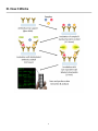
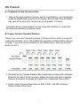
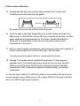
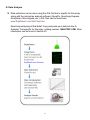
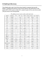
Quantibody® Human Periodontal
Disease Array 1
Quantitative measurement of 20 human periodontal disease associated
cytokines
Catalog #: QAH-PDD-1
User Manual
Last revised May, 2015
Caution:
Extraordinarily useful information enclosed
ISO 13485 Certified
3607 Parkway Lane, Suite 100
Norcross, GA 30092
Tel: 1-888-494-8555 (Toll Free) or 770-729-2992, Fax:770-206-2393
Web: www.RayBiotech.com, Email: [email protected]
1
Table of Contents
Section
Page #
I.
Overview
3
II.
Introduction
3
III.
How It Works
5
IV.
Materials Provided
6
V.
Storage
6
VI.
Additional Materials Required
6
VII.
General Considerations
A. Sample Preparation
B. Handling Glass Slides
C. Incubation
7
7
7
7
VIII.
Protocol
A. Completely Air Dry The Glass Slide
B. Prepare Cytokine Standard Dilutions
C. Blocking & Incubation
D. Incubation with Biotinylated Antibody Cocktail & Wash
E. Incubation with Cy3 Equivalent Dye-Streptavidin & Wash
F. Fluorescence Detection
G. Data Analysis
8
8
8
9
10
10
11
12
IX.
Array Map & Standard Curves
13
X.
Standard Concentrations
14
XI.
Spiking & Recovery
15
XII.
Q-Analyzer: Data Analysis Software
16
XIII.
Troubleshooting Guide
17
XIV.
Select Publications
18
XV.
Experiment Record Form
19
XVI.
How To Choose A Quantibody®
20
Please read the entire manual carefully before starting your experiment
2
I. Overview
Cytokines
Detected
(20)
CRP (C-Reactive Protein), IFN-gamma, IL-1 alpha (IL-1 F1),
IL-1 beta (IL-1 F2), IL-10, IL-12 p70, IL-17A, IL-2, IL-4, IL-6,
IL-8 (CXCL8), MIP-1 alpha (CCL3), MMP-13, MMP-9,
Osteoactivin (GPNMB), Osteopontin (SPP1),
Osteoprotegerin (TNFRSF11B), RANK (TNFRSF11A), TGF
beta 1, TNF alpha
See Section IX for Array Map
Format
One standard glass slide is spotted with 16 wells of identical
cytokine antibody arrays. Each antibody is arrayed in
quadruplicate.
Detection
Method
Fluorescence. Go to www.RayBiotech.com/Scanners for a
list of compatible laser scanners.
Sample Volume
50 - 100 µl per array
Reproducibility
CV <20%
Assay Duration
6 hours
II. Introduction
Periodontal disease is a gum disease. The symptom ranges from simple gum inflammation
(gingivitis) to periodontitis which results in major damage to the soft tissue and bone that
support the teeth. In the worst cases, teeth are lost. Because of the irreversible nature of
periodontitis, early diagnosis and treatment is critical. Clinical measurements include probing
pocket depth, bleeding on probing, clinical attachment loss, plaque index, and radiographs
etc. While such methods are useful for the staging of periodontal disease, they are only
indicators of previous disease status rather than the present disease activity. There is a need
for the development of new diagnostic tests that can reflect the active disease state, which
will be useful for disease diagnosis, prognosis, and monitoring the effectiveness of
periodontal therapy. Gingival crevicular fluid (GCF) is a tiny amount bodily fluid transuded
from periodontal tissues into the gingival crevice and periodontal pocket. The constituents of
GCF originate from serum, gingival tissues, and from both bacterial and host response cells,
which reflect the biology and physiology of the local tissues. Meanwhile, GCF could be easily
collected by noninvasive means such as paper strips, absorbent points and micropipettes. As
a result, proteins in GCF have been the ideal and hot targets pursued for candidate disease
3
specific biomarker research for the last several decades.
Most analyzed periodontal disease related proteins in GCF are inflammatory cytokines (eg. IL1b, IL-6, IL-8, IL-10, IL-12, IFNg, TNFa, and CRP); matrix metalloproteinases (eg. MMP-8,
MMP-9, and MMP-13) and their inhibitors (TIMPs); bone metabolism related cytokines (eg.
OPG, OPN, RANK, and RANKL); and enzymes (eg. alkaline phosphatase and aspartate
aminotransferase). Because of the limited availability of sample volumes, most of the
previous research only worked on one or several targets. The traditional method for cytokine
detection and quantification is through the use of an enzyme-linked immunosorbent array
(ELISA). While the traditional method works well for a single protein, the overall procedure is
time consuming and requires a lot of sample. Take the advantage of advancement in
microarray technology over the last decade; Raybiotech, has pioneered the development of
cytokine antibody arrays, which has now been widely applied in the research community with
hundreds of peer reviewed publications such as in Cell and Nature.The traditional method for
cytokine detection and quantification is through the use of an enzyme-linked immunosorbent
assay (ELISA). In this method, target protein is immobilized to a solid support. The
immobilized protein is then complexed with an antibody that is linked to an enzyme. Detection
of the enzyme complex can then be visualized through the use of a substrate that produces a
detectable signal. While this traditional method works well for a single protein, the overall
procedure is time consuming and requires a relatively high volume of sample. Thus,
conservation of precious small sample quantities becomes a challenging task. Innovations in
microarray technology over the last decade have addressed this problem. A long-standing
leader in the field, Raybiotech, has pioneered the development of cytokine antibody arrays,
which have now been widely applied in the research community with hundreds of peer
reviewed publications, including top-tier journals such as Cell and Nature.
The Quantibody® array, our multiplexed sandwich ELISA-based quantitative array platform,
enables researchers to accurately determine the concentration of multiple cytokines
simultaneously. It combines the advantages of the high detection sensitivity & specificity of
ELISA and the high throughput of arrays. Like a traditional sandwich-based ELISA, it uses a
pair of cytokine specific antibodies for detection. A capture antibody is first bound to the glass
surface. After incubation with the sample, the target cytokine is trapped on the solid surface.
A second biotin-labeled detection antibody is then added, which can recognize a different
epitope of the target cytokine. The cytokine-antibody-biotin complex can then be visualized
through the addition of the streptavidin-conjugated Cy3 equivalent dye, using a laser scanner.
Unlike the traditional ELISA, Quantibody products use an array format. By arraying multiple
cytokine specific capture antibodies onto a glass support, quantitative, multiplex detection of
cytokines in one experiment is made possible.
In detail, one standard glass slide is divided into 16 wells of identical cytokine antibody
arrays. Each antibody, together with the positive controls is arrayed in quadruplicate. The
slide comes with a 16-well removable gasket which allows for the process of 16 samples on
one slide. Four slides can be nested into a tray, which matches a standard microplate
footprint and allows for automated robotic high throughput process of 64 arrays
4
simultaneously. For cytokine quantification, the array specific cytokine standards, whose
concentration has been predetermined, are provided to generate a standard curve for each
cytokine. In a real experiment, standard cytokines and samples will be assayed in each array
simultaneously through a sandwich ELISA procedure. By comparing signals from unknown
samples to the standard curve, the cytokine concentration in the samples will be determined.
Quantibody® array kits have been confirmed to have similar detection sensitivity as traditional
ELISA. Our current high density Quantibody kits allow scientists to quantitatively determine
the concentration of 660 human, 200 mouse, and 67 rat cytokines in a single experiment.
This is not only one of the most efficient products on the market for cytokine quantification,
but makes it more affordable for quantification of large number of proteins. Simultaneous
detection of multiple cytokines undoubtedly provides a powerful tool for drug and biomarker
discovery.
5
III. How It Works
6
IV. Materials Provided
Catalog #
Component Name
1 Slide Box
2 Slide
Box*
1
2
1 QAH-PDD-1S
Human Periodontal Disease Array 1
Glass Slide
2 QA-SDB
Quantibody® Sample Diluent
3 AA-WB1-30ML
20X Wash Buffer I
4 AA-WB2-30ML
20X Wash Buffer II
30 ml
5 QAH-PDD-1-STD
Human Periodontal Disease Array 1
Lyophilized Standard Mix**
1 Vial
6 QAH-PDD-1B
Human Periodontal Disease Array 1
Biotinylated Antibody Cocktail
7 QA-CY3E
Cy3 equivalent dye-conjugated
Streptavidin
8 QA-SWD
Slide Washer/Dryer
1 x 30 ml Tube
9 QA-ADH
Adhesive Film
1
15 ml
2 x 30 ml
3 x 30 ml
1-25 µl
2 x 1-25 µl
5 µl
2 x 5 µl
2
* 4 slide kits are comprised of 2 separate 2 slide kits.
** See Section X for detailed cytokine concentrations after reconstitution.
V. Storage
Upon receipt, all components should be stored at -20°C. The kit will retain activity
for up to 6 months. Once thawed, the glass slide, standard mix, antibody cocktail
and dye-conjugated Streptavidin should be kept at -20°C. All other components
may be stored at 4°C. The entire kit should be used within 6 months of purchase.
VI. Additional Materials Required
Benchtop rocker or orbital rocker
Laser scanner for fluorescence detection
Aluminum foil
Distilled water
1.5 ml Polypropylene microcentrifuge tubes
7
VII. General Considerations
A. Preparation of Samples
Use serum-free conditioned media if possible.
If serum-containing conditioned media is required, it is highly recommended
that complete medium be used as a control since many types of sera contains
cytokines.
We recommend the following parameters for your samples: 50 to 100 µl of
original or diluted serum, plasma, cell culture media, or other body fluid, or 50500 µg/ml of protein for cell and tissue lysates.
If you experience high background or if the fluorescent signal intensities
exceed the detection range, further dilution of your sample is recommended.
B. Handling Glass Slides
Do not touch the surface of the slides, as the microarray slides are very
sensitive. Hold the slides by the edges only.
Handle all buffers and slides with powder free gloves.
Handle glass slide/s in clean environment.
The Quantibody slides do not have bar codes. To help distinguish one slide
from another, transcribe the slide serial number from the slide bag to the back
of the slide with a fine point permanent marker. Please write the number on the
very bottom edge of the slide, taking care to avoid writing on the array well
areas.
C. Incubation
Completely cover array area with sample or buffer during incubation.
Avoid foaming during incubation steps.
Perform all incubation and wash steps under gentle rocking or rotation.
Cover the incubation chamber with adhesive film during incubation, particularly
when incubation is more than 2 hours or <70 µl of sample or reagent is used.
Several incubation steps such as step 6 (blocking), step 7 (sample incubation),
step 10 (detection antibody incubation), or step 13 (Cy3 equivalent dyestreptavidin incubation) may be done overnight at 4°C. Please make sure to
cover the incubation chamber tightly to prevent evaporation.
8
VIII. Protocol
A. Completely Air Dry The Glass Slide
1. Take out the glass slide from the box, and let it equilibrate to room temperature
inside the sealed plastic bag for 20-30 minutes. Remove slide from the plastic
bag, peel off the cover film, and let it air dry for another 1-2 hours.
Incomplete drying of slides before use may cause the formation of "comet tails,"
thin directional smearing of antibody spots.
B. Prepare Cytokine Standard Dilutions
There is only one vial of standard provided in the two-slide kit, which is enough for
making two standard curves. Reconstitute the lyophilized standard within one hour
of usage. If you must use the standard for two different days, store only the Std1
dilution at -80°C.
2. Reconstitute the Cytokine Standard Mix (lyophilized) by adding 500 µl Sample
Diluent to the tube. For best recovery, always quick-spin vial prior to opening.
Dissolve the powder thoroughly by a gentle mix. Labeled the tube as Std1.
3. Label 6 clean microcentrifuge tubes as Std2 to Std7. Add 200 µl Sample
Diluent to each of the tubes.
9
4.
Pipette 100 µl Std1 into tube Std2 and mix gently. Perform 5 more serial
dilutions by adding 100 µl Std2 to tube Std3 and so on.
5.
Add 100 µl Sample Diluent to another tube labeled as CNTRL. Do not add
standard cytokines or samples to the CNTRL tube, which will be used as
negative control. For best results, include a set of standards in each slide.
Since the starting concentration of each cytokine is different, the serial
concentrations from Std1 to Std7 for each cytokine are varied which can be found
in Section X.
C. Blocking & Incubation
6.
Add 100 µl Sample Diluent into each well and incubate at room temperature
for 30 minutes to block slides.
7.
Decant buffer from each well. Add 100 µl standard cytokines or samples to
each well. Incubate arrays at room temperature for 1-2 hour.
Longer incubation time is preferable for higher signals. This step may be done
overnight at 4°C.
We recommend using 50 to 100 µl of original or diluted serum, plasma,
conditioned media, or other body fluid, or 50-500 µg/ml of protein for cell and
tissue lysates. Cover the incubation chamber with adhesive film during incubation,
especially if less than 70 ul of sample or reagent is used.
8.
Wash:
Decant the samples from each well, and wash 5 times (5 min each) with 150 µl
of 1X Wash Buffer I at room temperature with gentle rocking. Completely
remove wash buffer in each wash step. Dilute 20x Wash Buffer I with H2O.
(Optional for Cell and Tissue Lysates) Put the glass slide with frame into a box
with 1X Wash Buffer I (cover the whole glass slide and frame with Wash Buffer
I), and wash at room temperature with gentle rocking for 20 min.
10
Decant the 1x Wash Buffer I from each well, wash 2 times (5 min each) with
150 µl of 1X Wash Buffer II at room temperature with gentle rocking.
Completely remove wash buffer in each wash step. Dilute 20X Wash Buffer II
with H2O.
Incomplete removal of the wash buffer in each wash step may cause "dark spots,"
the background signals higher than the spots.
D. Incubation with Biotinylated Antibody Cocktail & Wash
9.
Reconstitute the detection antibody by adding 1.4 ml of Sample Diluent to the
tube. Spin briefly.
10. Add 80 µl of the detection antibody cocktail to each well. Incubate at room
temperature for 1-2 hour.
Longer incubation time is preferable for higher signals and backgrounds
11. Decant the samples from each well, and wash 5 times (5 mins each) with 150
µl of 1X Wash Buffer I and then 2 times with 150 µl of 1x Wash Buffer II at
room temperature with gentle rocking. Completely remove wash buffer in
each wash step.
E. Incubation with Cy3 Equivalent Dye-Streptavidin & Wash
12. After briefly spinning down, add 1.4 ml of Sample Diluent to Cy3 equivalent
dye-conjugated streptavidin tube. Mix gently.
13. Add 80 µl of Cy3 equivalent dye-conjugated streptavidin to each well. Cover
the device with aluminum foil to avoid exposure to light or incubate in dark
room. Incubate at room temperature for 1 hour.
14. Decant the samples from each well, and wash 5 times (5 mins each) with 150
µl of 1X Wash Buffer I at room temperature with gentle rocking. Completely
remove wash buffer in each wash step.
11
F. Fluorescence Detection
15. Disassemble the device by pushing clips outward from the slide side.
Carefully remove the slide from the gasket.
Be careful not to touch the surface of the array side.
16. Place the slide in the Slide Washer/Dryer (a 4-slide holder/centrifuge tube),
add enough 1x Wash Buffer I (about 30 ml) to cover the whole slide, and then
gently shake at room temperature for 15 minutes. Decant Wash Buffer I.
Wash with 1x Wash Buffer II (about 30 ml) and gently shake at room
temperature for 5 minutes.
17. Remove water droplets completely by gently applying suction with a pipette to
remove water droplets. Do not touch the array, only the sides.
You may also dry the glass slide by a compressed N 2 stream.
18. Imaging: The signals can be visualized through use of a laser scanner
equipped with a Cy3 wavelength (green channel) such as Axon GenePix.
Make sure that the signal from the well containing the highest standard
concentration (Std1) receives the highest possible reading, yet remains
unsaturated.
In case the signal intensity for different cytokine varies greatly in the same array,
we recommend using multiple scans, with a higher PMT for low signal cytokines,
and a low PMT for high signal cytokines.
12
G. Data Analysis
19. Data extraction can be done using the GAL file that is specific for this array
along with the microarray analysis software (GenePix, ScanArray Express,
ArrayVision, MicroVigene, etc.). GAL files can be found here:
www.RayBiotech.com/Gal-Files.html.
Need help analyzing all that data? Copy and paste your data into the QAnalyzer Tool specific for this array, catalog number: QAH-PDD-1-SW. More
information can be found in Section XII.
13
IX. Array Map & Standard Curves
14
X. Standard Concentrations
After reconstitution, the lyophilized cytokine standard mix contains the following
concentrations for each antigen included.
15
XI. Spiking & Recovery
The antibody pairs used in the kit have been tested to recognize their specific
antigen. The spiking and recovery rates of each cytokine in 2x diluted serum (SR),
plasma EDTA (PLE), plasma citrate (PLC), plasma heparin (PLH), and culture media
(CM) are listed in the following tables.
16
XII. Quantibody® Q-Analyzer
The Q-Analyzer is an array specific, Excel-based program. It is much more than a
simple calculation macro; it performs sophisticated data analysis (see below for
description).
The Q-Analyzer Tool specific for this array is catalog number: QAH-PDD-1-SW.
Key features:
Simplicity: Easy to operate and requires no professional training. With a simple
copy and paste process, the cytokine concentration is determined.
Outlier Marking & Removing: The software can automatically mark and remove
the outlier spots for more accurate data analysis
Normalization: The program allows for intra- and inter-slide normalization for
large numbers of samples.
Two Positive Controls: The program utilizes the two positive controls in each
array for normalization.
Two Analytical Algorithms: Users can choose either linear regression or log-log
algorithms to meet their analytical needs.
Two Data Outputs: standard curves and digital concentration.
User Intervention: The program allows for user manual handling of outliers and
other analytical data.
Lower and Upper Limits Determination: The program automatically marks out
the values below or above the detection range.
Standard Deviation: The program outputs the standard deviations of the
quadruplicate spots for data accuracy.
Analytical Tips: Q-Analyzer analysis tips are included in the program.
17
XIII. Troubleshooting Guide
Problem
Weak Signal
Uneven signal
Poor standard
curve
High
background
Cause
Recommendation
Inadequate detection
Increase laser power and PMT parameters
Inadequate reagent
volumes or improper
dilution
Check pipettes and ensure correct preparation
Short incubation time
Increase incubation time or change sample
incubation step to overnight
Too low protein
concentration in sample
Lessen dilution or do not dilute sample.
Concentrate sample if necessary.
Improper storage of kit
Store kit as suggested temperature. Don't
freeze/thaw the slide.
Bubble formed during
incubation
Decrease amount of rocking during incubations.
check for bubble formation and remove bubbles.
Arrays are not
completed covered by
reagent
Completely cover arrays with solution for all
required steps.
Reagent evaporation
Cover the incubation chamber with adhesive film
during incubation
Cross-contamination
from neighboring wells
Avoid overflowing wash buffer and other solutions
into neighboring wells.
Comet tail formation
Air dry the slide for at least 1 hour before usage
Inadequate standard
reconstitution or
Improper dilution
Reconstitute the lyophilized standard well at the
room temperature before making serial dilutions.
Check pipettes and ensure proper serial dilutions.
Inadequate detection
Increase laser power so the highest standard
concentration for each cytokine receives the
highest possible reading yet remains unsaturated.
Use freeze-thawed
cytokine standards
Always use new cytokine standard vial for new set
of experiment. Discard any leftover.
Overexposure
Lower the PMT or sigmal gain.
Dark spots
Completely remove wash buffer in each wash step.
Insufficient wash
Increase wash time and use more wash buffer
Dust
Work in clean environment
Slide is allowed to dry
out
Don't dry out slides during experiment.
18
XIV. Publications Citing This Product
1. Kinney JS, Morelli T, Oh M, Braun TM, Ramseier CA, Sugai JV, Giannobile WV.
Crevicular fluid biomarkers and periodontal disease progression. J Clin Periodontol 2014;
41: 113-120. doi: 10.1111/jcpe.12194
Species: Human
Sample Type: Gingival Crevicular Fluid
2. Filho-Nogueira G., Pesun I., Ploegman C., Wijegunasinghe M., McCulloch C. ongitudinal
Comparison of Cytokines in Peri-Implant Fluid and Gingival Crevicular Fluid in Healthy
Mouths. Journal of Periodontology (2014)
Species: Human
Sample Type: Gingival Crevicular Fluid
More citations for this product may be available.
Contact [email protected].
19
XV. Experiment Record Form
Date:_______________________
File Name:___________________
Laser Power:_________________
PMT:________________________
Well No.
Sample Name
1
CNTRL
2
Std7
3
Std6
4
Std5
5
Std4
6
Std3
7
Std2
8
Std1
Dilution factor
9
10
11
12
13
14
15
16
20
XVI. How to Choose a Quantibody® Array?
Species-based selection:
Human (QAH-)
Mouse (QAM-)
Rat (QAR-)
Bovine (QAB-)
Canine (QAC-)
Equine (QAE-)
Feline (QAF-)
Primates (QAN-)
Porcine (QAP-)
Rabbit (QAL-)
Function-based selection:
Adhesion Molecule
Arrays
Angiogenesis Arrays
Bone Metabolism
Arrays
Chemokine Arrays
Custom Arrays
Cytokine Arrays
Growth Factor Arrays
IGF Signaling
Arrays
IL-1 Family Arrays
Immune Response
Arrays
Inflammation Arrays
Interleukin Arrays
Isotyping Arrays
MMP Arrays
Obesity Arrays
Ophthalmic Arrays
Periodontal Disease
Arrays
Receptor Arrays
Th1/Th2/Th17 Arrays
Cytokine Number-based selection:
Arrays are available in the Quantibody ® platform to detect 660 human, 200 mouse, or
67 rat proteins. GLP-Compliant testing services are also available.
To learn more about the Quantibody ® Antibody Array, visit
www.RayBiotech.com/Quantibody-Multiplex-Elisa-Array.html
Quantibody® is the trademark of RayBiotech, Inc.
This product is for research use only.
©2015 RayBiotech, Inc
21