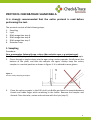
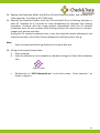
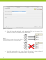
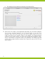
1
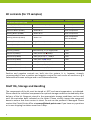
User manual Check&Trace Salmonella Routine Salmonella Serotype Identification 10-0010 72 Version 9.1 Issued December 16, 2011 Validated and certified by the OIE as fit for the purposes defined in this user manual. Registration number: 20110106 Previously marketed under the name of Premi® Test Salmonella. Key to symbols used Batch code Manufacturer Contains sufficient for < n > tests Consult instructions for use Use by YYYY-MM Catalog number Temperature limitation INTENDED USE 1 PRINCIPLE OF THE METHOD 1 CERTIFICATIONS 1 OIE certification AOAC-RI certification 1 2 KIT CONTENTS 3 SHELF LIFE, STORAGE AND HANDLING 3 MATERIALS REQUIRED BUT NOT SUPPLIED WITH THE KIT: 4 PRECAUTIONS AND RECOMMENDATIONS FOR BEST RESULTS 4 Prevention of contamination 5 PROTOCOL CHECK&TRACE SALMONELLA 6 1. Sampling 2. Lysis 3. DNA recognition step A 4. DNA recognition step B 5. DNA recognition step C 6. Detection step 6 7 7 8 8 9 FREQUENTLY ASKED QUESTIONS (FAQ) 19 LIMITATIONS OF THE TEST 22 APPENDIX 1 23 APPENDIX 2 24 APPENDIX 3 28 APPENDIX 4 29 Intended use Check&Trace Salmonella (10-0010) is designed for the rapid molecular confirmation and serotyping of presumptive Salmonella spp. The test employs highly specific DNA markers to allow accurate identification of the Salmonella serotype when Salmonella spp. is present. Principle of the method The principle of the Check&Trace Salmonella system is based on specific molecular recognition of DNA target sequences and subsequent amplification with universal primers. Each individual DNA target is recognized by a specific probe that contains a unique ZIP code corresponding to a unique position (address) on a microarray. These ZIP codes are used for detection on the microarray after amplification. Probe ends are joined by a DNA ligase when they match perfectly with the target DNA. Only ligated probes will result in amplification products. Probes that differ from the target DNA will not give amplification products, even in the case of a single nucleotide difference. Amplification products are hybridized to the microarray and visualized by colorimetric detection. The microarrays are contained in so called CP Array Tubes, which are inserted in the Check-Points Tube Reader upon completion of the detection reaction. This generates an array image that is analyzed by dedicated software to yield a definitive and objective assay result. Certifications The Check&Trace Salmonella is OIE certified for 22 Salmonella serotypes and AOAC-RI certified for 102 serotypes (see appendix 1). For a complete list of the Salmonella serotypes currently detected by Check&Trace Salmonella visit: www.checkandtrace.com OIE certification The validation data for this kit have been certified by the OIE, based on expert review, as fit for the following purposes: Rapid (molecular) confirmation and serotyping of presumptive Salmonella spp. of the following 22 serotypes: 1 Agona, Anatum, Bredeney, Derby, Dublin, Enteritidis, Hadar, Heidelberg, Indiana, Infantis, Kottbus, Mbandaka, Montevideo, Newport, Paratyphi B, Paratyphi B v Java, Saintpaul, Senftenberg, Tennessee, Typhimurium (and its monophasic variant 1,4,[5],12:i:-) and Virchow. For a summary of the OIE validation studies, see appendix 2. AOAC-RI certification See appendix 1 for an overview of the Salmonella serotypes for which the Check&Trace Salmonella is AOAC-RI certified. 2 Kit contents (for 72 samples) Components (Mat. No.) Description Storage conditions Colony samplers (9-0020) 1 vial sterile colony samplers Room temperature Lysis Buffer (9-0015) 1 bottle 10 ml Room temperature Detection Buffer (9-0007) 1 bottle 80 ml Room temperature Blocking Buffer (9-0008) 1 bottle 20 ml Room temperature Staining Solution (9-0014) 1 bottle 5 ml Room temperature store in the dark CP Array Tubes (10-0003) 6 bags of 4 each (total 24) Room temperature Manual (9-0010) Leaflet Not critical Blue tray (9-0022) 24x3-tube strip, 2.5 μl reagent/tube - 20°C Solution A (9-0021) 1 tube (brown cap ) 600 μl - 20°C Solution B1 (9-0003) 1 tube (purple cap ) 1300 µl - 20°C Solution B2 (9-0004) 1 tube (blue cap ) 120 µl - 20°C Solution C1 (9-0005) 1 tube (yellow cap ) 1300 μl - 20°C Solution C2 (9-0006) 1 tube (red cap ) 120 μl - 20°C Conjugate Solution (9-0027) 1 black tube & cap () 120 μl - 20°C Box Room Temperature (RT) Box -20°C Positive and negative controls are built into the system. It is, however, strongly recommended to use a positive and negative control for each series of reactions e.g a Salmonella of known serotype and a non-Salmonella strain. Shelf life, Storage and Handling The components of the kit must be stored at -20°C and room temperature, as indicated. Please check the individual components for optimal storage conditions immediately after delivery of the kit. Reagents stored in the appropriate storage conditions can be used until the expiration date indicated on the boxes. Please visually inspect the unopened boxes to ensure that their content is intact. Do not use the products if damaged. Please contact the Check-Points office at [email protected] if you have any questions or in case shipping has taken more than 5 days. 3 Materials required but not supplied with the kit: Pre-PCR Post-PCR Equipment Heating Block Flow cabinet (optional) Thermocycler Vortex Mixer Mini-centrifuge Supplies Disposable laboratory (powder-free) gloves Pipettes & disposable (preferably filter-) tips for volumes of 1 to 1000 µl 1.5 ml tubes (“Eppendorf tubes”) 10 ml tubes Thermocycler (optional) Vortex Mixer Mini-centrifuge Thermomixer with active cooling Check-Points Tube Reader with software Computer with USB port and internet connection Barcode Reader (optional) Disposable laboratory (powder-free) gloves Pipettes & disposable (preferably filter-) tips for volumes of 1 to 1000 µl 1.5 ml tubes (“Eppendorf tubes”) 10 ml tubes Please contact [email protected] if you have any questions regarding laboratory setup or equipment specifications. Precautions and recommendations for best results The test must be performed by adequately trained personnel. Food samples and enrichment cultures must be treated (and discarded) as potentially infectious material. Spinning down for a few seconds is necessary in the various steps to ensure that all material is properly collected in the bottom of the tubes. 4 The quality of the results depends on strict compliance with the following good laboratory practice, especially concerning PCR: - Do not use reagents after their expiration date. - Before use, thaw frozen reagents gradually at room temperature and vortex briefly to obtain a homogeneous solution. After vortexing briefly, spin down the solution to avoid contamination when opening the lid. - Periodically, verify the accuracy and precision of pipettes, as well as correct functioning of the instruments. Prevention of contamination PCR produces a very large quantity of DNA amplification products (amplicons) even from minute quantities of starting material. Check&Trace Salmonella may therefore yield unreliable results if samples become contaminated with amplicons from previous amplification reactions prior to the PCR (step C of the protocol). Preventive measures to minimize the risks of amplicon contamination must be taken, and are outlined below. Caution: To prevent contamination we recommend using pipettes with hydrophobic filter tips. Please read carefully and follow these instructions: Keep the detection step separate from recognition steps A, B and C, and preferably in a different room (”post-PCR room”). After the completion of step C transfer the reaction products to the location where the detection step is carried out. Do not open the tubes in the same location where steps A, B and C are performed. Open and close tubes with amplified material very carefully. Use separate equipment, pipettes, sample holders, lab coats, gloves, disposables and reagents, for the detection step than those that are used for the previous steps A, B and C. Never transfer items from the detection step location to the location where steps A, B and C are carried out. Wear a clean lab coat and clean gloves during all steps of the test. Keep the tubes of all kit components and samples closed as much as possible. Clean the lab benches and all equipment regularly with a 0.5% sodium hypochlorite solution. 5 PROTOCOL CHECK&TRACE SALMONELLA It is strongly recommended that the entire protocol is read before performing the test. The protocol consists of the following steps: 1. Sampling 2. Lysis 3. DNA recognition step A 4. DNA recognition step B 5. DNA recognition step C 6. Detection step 1. Sampling Procedure: Pure, presumptive Salmonella spp. culture (Non-selective agar, e.g.nutrient agar) 1. Dispense 100 μl Lysis Buffer into a 1.5 ml tube. Use a separate tube for each sample. 2. Pierce through a single colony into the agar using a colony sampler. Briefly touch the bottom of the plate, and take the sampler out again. Always keep the colony sampler in a vertical position as shown in figure 1. It is advised to wear gloves. Figure 1: Correct colony sampling technique. 3. Place the colony sampler in the 100 μl of Lysis Buffer and twist the samplerbetween thumb and index finger while remaining in the buffer. Remove the sampler and discard. Close the tube, vortex and continue with the lysis (step 2). 6 2. Lysis Procedure: 1. Transfer the 1.5 ml tubes with the resuspended cells to a heating block and incubate at 99°C for 15 min. This heat treatment disrupts the cells and releases the DNA into the Lysis Buffer. 2. Vortex and cool down to room temperature by placing the tubes on the laboratory bench. (If the samples aren’t used directly, store the tubes at -20°C). 3. (Thaw and) vortex before continuing with step 3. 3. DNA recognition step A Procedure: 1. First add 5 μl of solution A (brown cap ) to every reaction tube of the strip (supplied with the kit). Next add 10 μl of DNA extract (step 2), per sample. Please write down the sample reference for each tube of the strip (see figure 5 for guidance). 2. Close the tubes and spin down briefly using the mini-centrifuge to collect both sample and solution A at the bottom of the tubes. Mix well by tapping against each strip; the solution should have a uniform blue color. Spin down again briefly using the mini-centrifuge. 3. Place the strip(s) in the thermocycler and run the CP step A program, outlined in appendix 3 (total sample volume 18 μl). The program will run for approximately 2.5 hours. Note: 7 Always use a new pipette tip when adding solutions or samples to a reaction tube to avoid contamination. The reaction tubes supplied with the kit are prefilled with a small amount of blue-colored reagent. Proper mixing of this reagent, solution A and the sample is crucial for optimal test performance. Solution A is frozen and should be thawed at room temperature and mixed properly before use. When the DNA extracts have been stored at -20°C, please thaw the sample properly and mix well. When closing the tubes of the strip(s), don’t use excessive pressure as the cap may distort and the sample may then evaporate during the subsequent steps of the protocol. 4. DNA recognition step B Procedure: 1. Prepare a B-mix in a 1.5 ml tube for step B, while step A is proceeding. Take solution B1 (purple cap ) and solution B2 (blue cap ) from the freezer. Solution B1 is frozen (B2 is not), and should be thawed properly at room temperature, mixed well, and spun down briefly before use. Use the pipetting scheme (appendix 4) at the back of this user manual to prepare the required amount of B-mix. First add the required amount of B1 solution to the tube. Then dispense solution B2 into solution B1 by pipetting up and down 3 times. Mix very well by vortexing and spin down briefly. 2. Store the B-mix in the refrigerator or on ice until step A has finished. 3. Briefly spin down the reaction tubes when step A has finished. 4. Add 15 μl of the freshly prepared B-mix to each sample in the strip(s). Close the tubes, mix by tapping each strip, and spin down briefly. 5. Place the strip(s) in the thermocycler and run the CP step B program, as outlined in appendix 3 (total sample volume 33 μl). The program will run for approximately 1 hour. Note: B2 is a glycerol-based solution and is therefore not frozen and ready for use immediately. 5. DNA recognition step C Procedure: 1. Approximately 10 minutes before the end of step B, take solution C1 (yellow cap ) from the freezer. Thaw properly at room temperature, mix well and spin down briefly. Take solution C2 (red cap ) from the freezer and prepare a C-mix, in a 1.5 ml tube, using the pipetting scheme (appendix 4) at the back of this user manual. First add the required amount of C1 solution to the tube. Then dispense solution C2 in solution C1 by pipetting up and down 3 times. Mix very well by vortexing and spin down briefly. 2. Briefly spin down the reaction tubes when step B has finished. 3. Add 15 μl of the freshly prepared C-mix to each sample in the strip(s). Close the tubes, mix by tapping each strip, and spin down briefly. 4. Place the strip(s) in the thermocycler and run the CP step C program, as outlined in appendix 3 (total sample volume 48 μl). The program will run for approximately 1.5 hours. 8 Note: 5. 6. C2 is a glycerol-based solution and is therefore not frozen but is immediately ready for use. Briefly spin down the reaction mixture after step C has been completed and transfer the reaction tubes to the area where the detection step is carried out. Alternatively, store the reaction mixtures at 4°C (+/- 1°C) when the detection step is carried out within 24 hours or at -20°C when the detection step is carried out within two weeks. 6. Detection step A B Figure 2: A) the CP Array Tube (AT) and B) the Check-Points Tube Reader. Note: TM The ArrayTube DNA microarray platform (see figure 2) is sold under licence from Alere Technologies GmbH. Procedure: 1. Start preparing the required number of Array Tubes (ATs) for detection approximately 10 minutes before the end of the step C program. Heat the thermomixer to 50°C. Note: 2. 3. 9 One AT is required for every 3-tube strip. Remove the ATs from their package(s) and place them in the thermomixer at 50°C. Add 300 μl of Detection Buffer to every AT and incubate the tubes for 2 minutes (400 rpm) in the thermomixer. It is not necessary to close the tubes. Caution: Be careful when removing or adding liquids with the pipette to or from the AT. Do not touch the microarray at the bottom of the tube at any time. Pipet all material in or out of the AT at the side of the bottom of the tube without touching the array as shown in figure 3. Figure 3: Correct AT pipetting technique. 4. 5. 6. 7. Remove the Detection Buffer from the ATs and repeat step 3. Replace the Detection Buffer by 300 μl of fresh Detection Buffer. Take the samples from step C. Samples stored longer than 2 hours after step C was completed should be heated in the thermocycler at 95°C for 2 minutes (see CP step melt in appendix 3). Briefly spin down the reaction mixture. Transfer 10 μl of reaction mixture from each tube of one strip to the corresponding AT (in total 30 μl per AT). The total volume of the AT will be 330 µl. The lid of the AT should be labeled for reference. Caution: Samples may contain a white-colored precipitate. This is due to denaturation of one of the reaction components, a protein stabilizer. The presence of this precipitate has no effect on the result of the detection step and may be ignored when adding the sample. When adding samples to the AT do not remove the AT from the thermomixer, to prevent the buffer from cooling down. Add the sample directly into the Detection Buffer of the AT by pipetting up and down. With the completion of step 7, three samples have been added to one AT. 8. Close the lids of the ATs and incubate the tubes for 30 minutes at 50°C (400 rpm). Caution: Close the lid of the AT properly, to prevent the AT from drying out. 10 9. After 30 minutes, replace the Detection Buffer with 300 µl of Blocking Buffer: do this with one AT at a time! Remove the AT from the thermomixer, discard the Detection Buffer using a pipette and immediately replace with 300 µl Blocking Buffer using a new pipette tip. Place the AT back in the thermomixer at 50°C and proceed with the next AT until all the AT solutions have been replaced with Blocking Buffer. Incubate for 5 minutes at 50°C (400 rpm). Caution: Collect the liquids removed from the ATs in a disposable tube, and dispose of it the same day together with the other laboratory waste. It is important to replace the Detection Buffer with Blocking Buffer one AT at a time. Empty ATs in the thermomixer at 50°C may become very dry, thereby increasing the risk of background noise. Optional: Evaporated water condensed on the lid of the AT may be removed with a pipette before removing the Detection Buffer containing the sample. 10. Replace the Blocking Buffer with 300 μl of fresh Blocking Buffer. Adjust the temperature of the thermomixer to 30°C and incubate for 10 minutes (400 rpm). During this incubation time the thermomixer can cool down from 50°C to 30°C. 11. In the mean time prepare a dilution of the Conjugate Solution (black 2 ml tube with black cap ) with Detection Buffer using the pipetting scheme in appendix 4 at the back of this user manual. For this purpose a 1.5 ml tube or a 10 ml tube may be used depending on the amount required. Dispense the Conjugate Solution in the Detection Buffer by pipetting up and down 3 times. Mix well by vortexing for 30 seconds. Note: The Conjugate Solution is stored at -20°C but is not frozen and can be used immediately. Conjugate dilutions have to be made fresh, and should be used on the day of preparation. 12. Remove the Blocking Buffer completely and add 150 μl of the conjugate dilution. Incubate for 15 minutes at 30°C (400 rpm). 13. Remove the conjugate dilution from the ATs and add 600 μl of Detection Buffer. Incubate the tubes for 2 minutes at 30°C (400 rpm). 11 14. Replace the Detection Buffer with 600 μl of fresh Detection Buffer, and incubate the tubes again for 2 minutes at 30°C (400 rpm). 15. Remove the Detection Buffer from the ATs and add 150 μl of Staining Solution to each AT. Incubate for 15 minutes at room temperature to complete the staining procedure. Continue with the image analysis immediately after the 15 minutes incubation time. Do not incubate with Staining Solution for more than 15 minutes: images may become too dark. During the 15 minutes incubation time, enter the required sample information and relevant test data in the Check-Points software as outlined in point 16 a-g. Note: Store the bottle with Staining Solution in the dark after use. 16. Filling out the experimental data: a. Start computer b. Start the software on the computer by double-clicking the Check-Points desktop icon: Check-Points c. Double-click on “C&T Salmonella.arr” in the first screen, “Array selection”, as shown in figure 4. 12 Figure 4: “Array Selection”. d. Enter the lot number of the kit in the appropriate field and the name of the operator, followed by clicking the “Next Step” button. 1 Figure 5: Example of filling out sample references in the software and assigning samples to the corresponding tubes. 1 e. 13 Insert the sample codes in the screen “Sample information” as shown in figures 6 and 7. (Correct analysis is not possible without sample codes) Figure 6: ”Sample information”. Note: Additional remarks may be added per sample (optional), when sample references have been filled out. Double click on one of the sample reference fields. A pop-up will appear (see figure 7) allowing remarks to be added to individual samples or to all samples, by checking the box(es) of the relevant sample(s). Relevant information of the samples must be added at this stage as the database cannot be edited once the results have been read. Figure 7: Pop-up for additional sample information. f. Proceed to the “Detection step” screen (see figure 8), by clicking the “Next Step” button. 14 g. The software will now indicate the samples to be analyzed first. Figure 8: “Detection step”. 17. Enter the AT lot number in the appropriate field when the 15 minutes incubation time has been completed (optional: use the Barcode Reader to scan the AT lot numbers). Next, insert the AT with open cap into the reader, close the lid of the reader, and click on the “Confirm” button in the software. The Staining Solution remains in the AT while reading. The results will be displayed immediately. Finally, click on “Save Results” to save the results in the database. The software will now indicate which sample should be analyzed next. Repeat this step until all ATs have been analyzed. 15 Figure 9: Presentation of the final result. Note: Clicking “Confirm” leads to immediate scanning of the AT. Only click “Confirm” after the full incubation period of 15 minutes is completed. If the AT is not in the reader, after clicking the “Confirm” button, simply put the AT in the reader and click ”Confirm” again. It is important to adhere to the 15 minutes incubation time with Staining Solution as much as possible (step 15). A shorter incubation time may lead to faint images; exceeding the 15 minutes incubation time may lead to overstaining. In both cases incorrect results may be obtained. 18. When all ATs are analyzed, a new window with the summary of the results will appear (see figure 10), which may also be printed (click on the “Print” button). The results summary will display the sample name, confirm the presence of any Salmonella, report the Salmonella serotype (in case an unknown pattern is detected a genovar code will be reported instead) and any validation(s) available for these. Click the Quit button to end this run of analyses. 16 Figure 10: Summary of the results. Note: For support concerning results please send the result image along with the desired information to [email protected]. To do this, double click on the result(s) in question in the result summary window (see figure 10). A pop-up will appear (see figure 11) in which your comments may be added to the file. Figure 11: “Send image to Check-Points” pop-up. 17 Click on the “Save (Send later)” button to store the file, which will have a .cpfe extension on the computer, and send it by e-mail. Alternatively, if the computer has internet access, check the box “Send picture directly over internet connection” and enter the reply e-mail address, followed by clicking “Send”. In both cases feedback should be expected within two working days. Figure 12: Check the box to send pictures directly over the internet. Support may also be requested at a later stage when the program has been closed. For this purpose the “Send image to Check-Points” pop-up (see figure 11) may also be accessed from the Database viewer (DBview shortcut that is located on the desktop). Open the database by clicking “File” followed by “Open” (the database is located in C:\Images by default) and scroll down to the results which require support. By clicking “Save”, the “Send image to Check-Points” pop-up will appear just as in the result summary window. 18 Frequently asked questions (FAQ) 1. The thermocycler states an error in step A, B or C. Please contact Technical Support: [email protected]. 2. During the different steps (step A, B or C) sample(s) have (partly) evaporated. Tubes may not have not been closed properly. Please restart the procedure from step A. 3. I have left Solutions A, B and/or C out of the -20° C (-4 F) storage. These reagents must be stored at -20°C (-4°F) for proper performance of the test. The performance of the product cannot be fully guaranteed if these solutions were left out of -20 °C (-4°F) for longer than 24 hours. 4. Staining Solution turned blue after adding it to the AT. Conjugate dilution was not properly removed by washing steps 13 and 14 (detection step). Continue incubation with Staining Solution for 15 minutes and take the image as described in the protocol. If the image is too dark, please refer to question 5. 5. The picture of the array is very dark. The conjugate dilution (detection step 12) was not properly removed by washing steps 13 and 14 (detection step). Please replace the Staining Solution with Detection Buffer and take image immediately. If the image is still too dark, please repeat the detection step with a new AT. 6. The software indicates: “Reference spots not found”. The software did not find reference spots on the AT. Causes may be: 6.1. An air bubble is interfering with the result. Tap the AT gently or pipet the liquid gently up and down and then try to take the image again. 6.2. The picture is very dark: conjugate dilution not removed properly. Please refer to question 5. 6.3. The picture is completely white. Staining Solution was not added. Please add Staining Solution again and proceed from detection step 15. 6.4. Some spots are missing due to scratching of the array with a pipet tip during adding or removing of solution from or to the AT. Please repeat the detection step with a new AT. If the results do not improve, staining has failed. Most likely no conjugate dilution was added, or the conjugate dilution was not prepared properly. Please repeat detection step with new AT. 7. 19 The software indicates: “Hybridisation not OK”. The software did not find the hybridisation control spots on the AT. The hybridisation control is used to check if the hybridisation (at 50°C) of the PCR product with the AT has been performed properly. Causes may be: 7.1. The picture is completely dark: conjugate dilution not removed properly. Please refer to question 5. 7.2. The reaction mixture after completion of step C was not added to the AT. Please repeat detection step with a new AT. 7.3. Hybridisation temperature too high. Please verify that the thermomixer temperature has been at 50°C when the ATs were hybridised. Please repeat detection step with a new AT. 7.4. The C-mix was not prepared properly or was not added to the assay: please repeat the test from step A. 8. The software indicates: “DNA recognition not OK,”. The software did not find the reaction control spots on the AT. These reaction controls are used to check the performance of the assay in steps A and C. Possible explanations are: 8.1. The sample DNA contains contaminants inhibiting the reactions. Please redo the Lysis and repeat the test. 8.2. The A mix was not added in step A. Please repeat the test. 8.3. The C mix was not prepared properly or was not added to the assay: please repeat the test. 8.4. Reaction mixtures from step C (step 7 of detection step) were not all added to the AT: 3 reaction mixtures should be added, one or two have been omitted. 9. The software indicates: “Salmonella, genovar”. The sample is positive for Salmonella, but the (unique) serovar array pattern (genovar code) is not defined in the software. meaning the serotype most likely does not belong to one of the serotypes outlined in appendix 1. Check-Points has an extensive database containing results of more than 300 different serotypes. This Database is continuously growing so it is possible that the genovar code (array pattern) is in the CP database but not yet in the software. Please send the result image containing the genovar code to [email protected] as outlined on page 17 and 18 of the manual. The image is then compared with a database and feedback should be expected within two working days. 10. The software indicates: “Salmonella suspected”. The software did not find sufficient spots to give a conclusive result. Please inspect the picture visually: an air bubble or dust particles may interfere with the result. Tap the AT gently or pipet the liquid gently up and down, and try to take the image again. Please repeat the test if the results do not change. 11. The software indicates:“Salmonella ssp. (Serovar cannot be determined)”. The sample is positive for Salmonella but the software cannot determine a clear pattern and is therefore unable to specify the serotype. Causes may be: 11.1 An air bubble or dust particle may interfere with the result. Tap the AT gently and try to take the image again. 20 11.2 The sample DNA contains contaminates inhibiting the reactions. Please redo the lysis and repeat the test. In some specific cases the serotype can be determined manually. Please send the result image to [email protected] as outlined on page 17 and 18. 12. The software indicates: “Picture not found” or “Image capture error”. Check if the Check-Points Tube Reader is properly installed. Please contact Technical Support at [email protected] if reinstallation of the reader does not solve this problem. 13. The AT image is covered with small spots. There may be various causes for this phenomenon. Most likely the array dried out during the detection step. Please be sure that the AT always contains sufficient amounts of reagents (Detection Buffer, Blocking Buffer, conjugate dilution or Staining Solution). This is particularly critical during the incubations steps at 50°C. In most cases the software will be able to handle these small spots. If not, repeat the detection step with a new AT. 14. The AT was incubated for more than 15 minutes with Staining Solution before taking the image. Results may be unreliable due to overstaining. Inspect image: if spots are very dark, please repeat detection step with a new AT. 15. Duplex sample does not display identical result. 15.1 Inspect images for dust particles. Tap or gently shake the tubes to try to remove the particles from the array and rescan the images. 15.2 In case of extra spots repeat the test to confirm result. 16. I wish to stop the assay and continue at a later stage. What is the best point to stop the assay? You may stop the reactions after steps B or C. Store reaction mixtures at 4°C if they are to be used within 24 hours, or at -20°C for a maximum period of two weeks. 17. The AT image contains dust particles. The software will correct this in most cases. To prevent any interference with the results, please take the AT out of the reader and shake it gently until the dust particles have moved to the side of the AT. 21 Limitations of the test Check&Trace Salmonella uses highly specific DNA markers to identify the Salmonella serotype. The presence or absence of a range of these markers determines the serotype. The correlation between this DNA marker profile (expressed as genovar score) and the serotype has been tested and validated extensively for the serotypes listed in appendix 1 [1, 2, unpublished results]. Additional work with a wide range of serotypes has indicated that the vast majority of serotypes yield a unique genovar score. Check&Trace Salmonella has been tested against many, but not all, serotypes. It cannot be excluded that certain exotic serotypes generate an overlapping genovar score with another serotype as more than 2600 Salmonella serotypes have been described in literature. Therefore, Check&Trace Salmonella cannot and does not make any representation or warranty that it is capable of detecting every species, subspecies or serotype of the Salmonella genus in any sample source. Results may need to be confirmed by traditional methodology in specific cases (e.g. for regulatory samples). Check&Trace Salmonella has been developed to serotype Salmonella enterica subsp. enterica bacteria. The test also detects the other Salmonella species and subspecies, but does not discriminate all species. Consequently, the test indicates the presence of Salmonella in these cases as a genovar score without a specific serotype, except for Salmonella bongori. Other (biochemical) tests are required to identify these strains. Specific Salmonella serotypes originating from certain feed and food matrices may give suboptimal results with Check&Trace Salmonella in case of sampling from MSRV. Not all Salmonella serotypes have been extensively validated within all food and feed matrices with an MSRV pretreatment. Therefore an internal validation with the most occurring serotypes in the common food or feed matrices is recommended. Check&Trace Salmonella is able to fully serotype non-motile or monophasic Salmonella. So, for example a monophasic S. Typhimurium strain will be typed as S. Typhimurium. A specific exception is the S. 1,4,[5],12:i:-. When the specific DNA marker for the FljA gene is absent the score will be the S. 1,4,[5],12:i:-. In other cases S. 1,4,[5],12:i:- strains are identified as S. Typhimurium. For known overlapping scores the probability score of the dominant serotype is based on the prevalence of the respective serotypes according to the database of the Global Salmonella Survey of the World Health Organization (WHO): www.who.int/salmsurv/en/ In some cases Check&Trace Salmonella may yield two serovar names due to a known overlap in genovar score or due to historically given names to serovars containing the same global antigenic formula. As an example of the latter, S. Paratyphi C and S. Choleraesuis are considered to be part of the same group 6,7:c:1,5. This group also contains Choleraesuis var. Kunzendorf, var. Decatur and Typhisuis, which serotypes have not yet been validated with Check&Trace Salmonella. Therefore the result for this group will now read S. Paratyphi C or S. Choleraesuis. More details about the overlap in genovar scores as well as frequent updates of the Serotype list (appendix 1) can be found on the Check&Trace website: www.checkandtrace.com The Check-Points software is continuously improving thanks to the expanding global database of available results. In order to further improve the software, original result images (including specific data*) are shared with this database through a default setting in the software by means of an encrypted file. This data will be used solely for software improvements. All information will be kept strictly confidential and will not be shared with any third parties. *The specific data included are: the number identifying the CP Array Tube, the kit lot number, the software version used, the operator and the customer name. All patient data and sample information is removed in the encrypted file. 1. Wattiau, P. et al. Evaluation of the Premi®Test Salmonella, a commercial low-density DNA microarray system intended for routine identification and typing of Salmonella enterica. International Journal of Food Microbiology (2008). Vol 123. p 293 -298. 2. Wattiau, P et al. Comparison of Classical Serotyping and PremiTest Assay for Routine Identification of Common Salmonella enterica Serovars. J. Clin. Microbiol (2008), Vol. 46, p 4037–4040. 22 Appendix 1 Check&Trace Salmonella AOAC-RI and OIE certified serotypes For a complete list of the Salmonella serotypes currently detected by Check&Trace Salmonella visit: www.checkandtrace.com Serotypes not included in this test may yield a genovar score. 23 Appendix 2 OIE certification: summary of validation studies Analytical characteristics Calibration: The PTS [Check&Trace Salmonella, formerly Premi®Test Salmonella] is based on the presence/absence pattern of a number of specific DNA markers. The pattern is translated into a numerical code (genovar), which may be associated with known serotypes. Because the genovar is linked with DNA markers which are not always coding antigens used for the serotyping, it is possible to have different genovar (corresponding into different combination of DNA markers) linked with the same serotype. , This expresses the genetic variability inside a serotype. The initial choice of DNA markers potentially allows having a big spectrum of different combinations. In order to design the PTS, the selection of the targeted DNA sequences was done to have the possibility to differentiate the maximum of serotypes (in other words the targeted DNA sequences are the best to improve the chance to see the difference between two serotypes). To develop the software and to build the PTS database, the main serotypes (most often encountered in the laboratories) were tested. For these serotypes, the genovar (combination of positive spots) became known and were included in the PTS software (and became the first PTS-list). The “PTS database” can be expanded each time a new serotype can be linked with a specific (unique) genovar-code (combination of DNA markers). Repeatability: As regards serotype identification the repeatability was: - 91% in trial 1 (3 strains of 6 different serotypes tested twice by 2 different technicians); - 96% in trial 4 (5 strains of 5 major serotypes tested twice by 5 different laboratories). For spot reaction: - Trial 1 was designed in order to test all the spots involved in serotype identification; - For 17 spots 100% of the results were satisfactory and for 3 spots higher than 95% of the results were satisfactory; - Finally 390 interpretable spots (for chosen serotypes) were tested twice and 387 repetitions were satisfactory: 99.2% of repeatability (lower Confidence Limit = 98.5%). Analytical specificity and sensitivity of the kit The serotypes tested were serotypes with more than 20 positives strains and were the following ones: - The 9 regulatory serotypes; - The 10 serotypes of the other top 20; - 2 other serotypes. Globally, it means that, for each serotypes, at least 20 “positive” strains and around 1700 “negative” strains (other serotypes) were tested with both methods (Kaufman-White [KW] and PTS). 24 Analytical specificity The hypothesis tested was: H0: The percentage of same results between PTS and KW method is q=99%. The following table summarizes the results: For the False Positives, the following comments were made by the applicant: - For Dublin, the false positives concern 2 Salmonella Rostock and 2 Salmonella Kiel. These two serotypes have never been identified by CDC during ten years. - For Enteritidis, the false positives also includes very rare serotypes: 2 Salmonella Blegdam and 2 Salmonella Gallinarum gallinarum, with a frequency of 0,008% and 0,003% in the CDC database. - For Hadar, the FP is a confusion with Istanbul serotype (0,06% of the Salmonella database of CDC). - For Newport, the FP is a confusion with an Hadar serotype (1,02% of the Salmonella in the CDC database). - For Schwarzengrund, the FP is a confusion with a Panama serotype (0,43% of the Salmonella in the CDC database). - The mistakes mainly concern rare serotypes. So, the frequency of False Positives would be lower in a routine laboratory. Analytical sensitivity In a first step, the hypothesis tested was H0: a minimum q0=99% PTS in accordance with KW with a risk α of 1% only with the mistakes (wrong serotype) in the “false negatives.” The following table summarizes the results: The table gives the results for the 22 serotypes. - Hypothesis H0 is confirmed for all serotypes; - The risk β is always less than 6,5%, and less than 5% for 18/22 serotypes. 25 In a second step, the hypothesis tested was H1: a minimum q0=95% PTS in accordance with KW with a risk α of 1% with the mistakes (wrong serotypes) and genovar scores included in the “false negatives”. The following table summarizes the results: The table gives the results for the 22 serotypes. - Hypothesis H1 is confirmed for all serotypes; - The risk β is less than 36%, and less than 16% for 7/22 serotypes. The detailed results for the “false negatives” are given as follow by the applicant: For Derby: - There is one mistake. The PTS gives the serotype Livingstone. - In the two other cases, the PTS has given a genovar score without any identification. For Enteritidis: - The PTS has given a genovar score for the 4 False negatives. For Newport: - 4 strains have been identified with the genovar score. Three of them have the same genovar score. Reproducibility 5 European laboratories were involved in the trial (Belgium, Germany, Netherlands, Spain, United Kingdom). Each laboratory tested 26 samples (5 strains of Salmonella regulatory serotypes tested twice, 15 strains of other serotypes of Salmonella tested once and one Non Salmonella tested once) with two methods: reference method (KW) and PTS. In average 97% of strains were well identified with the PTS (Salmonella + good serotype) and 86% with KW (because one of the laboratories had bad results). Applications The kit is currently used in different laboratories world-wide and in the food industry. References Publications: - Wattiau P., Weijers T., Andreoli P., Schliker C., Veken H.V., Maas H.M.E., Verbruggen A.J., Heckc M.E.O.C, Wannetc W.J., Imberechts H., Vos P. Evaluation of the Premi®Test Salmonella, a commercial low-density DNA microarray system intended for routine identification and typing of Salmonella enterica. International Journal of Food Microbiology, 2008, 123 (3), pp. 293-298. - Wattiau P., Van Hessche M., Schlicker C., Vander Veken H., Imberechts H. Comparison of Classical Serotyping and PremiTest Assay for Routine Identification of Common Salmonella enterica Serovars. J. Clin. Microbiol., 2008; 46, pp. 4037 - 4040. Posters: 26 - Rapid detection of salmonella serotypes in feed and food using SST. Poster presented at the meeting “UW-RiverFalls Food Microbiology symposium” in 2006. - Salmonella Molecular Serotyping with a DNA microarray: an approach for non agglutinable Salmonella enterica serotypes. Congrès ICEID, Atlanta, 17-19 mars 2008. - The Effect of the Culture Medium on the Performance of “SST”: A Multiplex Molecular Serotyping Test using a DNA Microarray System. MedVetNet, June 08. - A multiplex molecular serotyping test using a DNA microarray system as an alternative Salmonella serotyping method. Rapid Methods, January 2009. - DNA Microarray evaluation for Salmonella serotyping. MedVetNet, June 09 - Short Evaluation of the Premi®Test Salmonella Method, Food Micro 2010, Copenhagen 30 August – 3 September 2010. - Comparison of Two Molecular Methods for Serotyping Salmonella, abstract presented at Fern 2010 by Junia Jean-Gilles Beaubrun, Ph.D. Microbiologist, Food and Drug Administration. - Interlaboratory study of a DNA Microarray for Salmonella Molecular Serotyping in comparison to the conventional serotyping method. i3S, International Symposium Salmonella and Salmonellosis, 28, 29 and 30th of June 2010 Saint-Malo – France by Anne Brisabois, AFSSA. 27 Appendix 3 CP Step A Cycle 1 (1x): 95°C 3 min. Cycle 2 (24x): 95°C 30 sec. 65°C 5 min. Cycle 3 (1x): 98°C 2 min. Holding at 4°C CP Step B Cycle 1 (1x): 37°C 45 min. 95°C 10 min. Holding at 4°C CP Step C Cycle 1 (1x): 95°C 10 min. Cycle 2 (35x): 95°C 5 sec. 55°C 30 sec. 72°C 30 sec. Cycle 3 (1x): 98°C 2 min. Holding at 4°C CP Step Melt Cycle 1 (1x): 98°C 2 min Holding at 4°C 28 Appendix 4 Pipetting scheme for B or C mix: samples 1–3 4–6 7–9 10 - 12 13 - 15 16 - 18 19 - 21 22 - 24 25 - 27 28 - 30 31 - 33 34 - 36 37- 39 40 - 42 43 - 45 46 - 48 49 - 51 52 - 54 55 - 57 58 - 60 61 - 63 64 - 66 67 - 69 70 - 72 μl B1 or C1 45 90 135 180 225 270 315 360 405 450 495 540 585 630 675 720 765 810 855 900 945 990 1035 1080 μl B2 or C2 3 6 9 12 15 18 21 24 27 30 33 36 39 42 45 48 51 54 57 60 63 66 69 72 Pipetting scheme for conjugate dilution: ATs 1 2 3 4 5 6 7 8 9 10 11 12 13 14 15 16 17 18 19 20 21 22 23 24 µl conjugate solution 5 5 5 10 10 10 15 15 15 20 20 20 25 25 25 30 30 30 35 35 35 40 40 40 μl Det. Buf. 495 495 495 990 990 990 1485 1485 1485 1980 1980 1980 2475 2475 2475 2970 2970 2970 3465 3465 3465 3960 3960 3960 Despite the utmost care in the development and preparation of the protocol Check-Points cannot take any responsibility for any errors, omissions and/or future changes herein. Check-Points BV Binnenhaven 5 6709 PD Wageningen The Netherlands 29 Tel: +31 317 453 908 Fax: +31 317 210 147 [email protected] www.checkandtrace.com