1
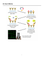
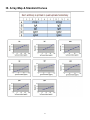
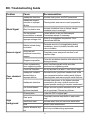
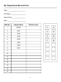
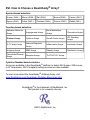
Quantibody® Human Ig Isotype Array 1
Quantitative measurement of 8 human Immnunoglobulins
Catalog #: QAH-ISO-1
User Manual
Last revised March, 2015
Caution:
Extraordinarily useful information enclosed
ISO 13485 Certified
3607 Parkway Lane, Suite 100
Norcross, GA 30092
Tel: 1-888-494-8555 (Toll Free) or 770-729-2992, Fax:770-206-2393
Web: www.RayBiotech.com, Email: [email protected]
1
Table of Contents
Section
Page #
I.
Overview
3
II.
Introduction
3
III.
How It Works
5
IV.
Materials Provided
6
V.
Storage
6
VI.
Additional Materials Required
6
VII.
General Considerations
A. Sample Preparation
B. Handling Glass Slides
C. Incubation
7
7
7
7
VIII.
Protocol
A. Completely Air Dry The Glass Slide
B. Prepare Cytokine Standard Dilutions
C. Blocking & Incubation
D. Incubation with Biotinylated Antibody Cocktail & Wash
E. Incubation with Cy3 Equivalent Dye-Streptavidin & Wash
F. Fluorescence Detection
G. Data Analysis
8
8
8
9
10
10
11
12
IX.
Array Map & Standard Curves
13
X.
Standard Concentrations
14
XI.
Notes Page
15
XII.
Q-Analyzer: Data Analysis Software
16
XIII.
Troubleshooting Guide
17
XIV.
Select Publications
18
XV.
Experiment Record Form
19
XVI.
How To Choose A Quantibody®
20
Please read the entire manual carefully before starting your experiment
2
I. Overview
Cytokines
Detected
(8)
Format
IgA, IgD, IgE, IgG1, IgG2, IgG3, IgG4, IgM
See Section IX for Array Map
One standard glass slide is spotted with 16 wells of identical
cytokine antibody arrays. Each antibody is arrayed in
quadruplicate.
Detection
Method
Fluorescence. Go to www.RayBiotech.com/Scanners for a
list of compatible laser scanners.
Sample Volume
50 - 100 µl per array (Sample Dilution: 40,000X - 200,000X)
Reproducibility
CV <20%
Assay Duration
6 hours
II. Introduction
The human immune system consists of two functional components classified as the innate
system (the physical, biochemical and cellular barriers), and the adaptive immune system
(including lymphocytes and immunoglobulins). Immunoglobulins are the key elements of the
humoral immune response in vertebrate against parasitic invasion. The polypeptide chains of
immunoglobulins composed of two identical heavy (H) chains and two identical light (L)
chains linked together by inter-chain disulfide bonds. While the amino-terminal portions that
exhibits highly variable amino-acid composition are involved in antigen binding, the C terminal
constant parts are involved in complement binding, placental passage and binding to cell
membranes. Based upon the variation of the constant region of the heavy chain, nine
immunoglobulin heavy chain isotypes are found in humans: IgA (with subclasses IgA1 and
IgA2), IgD, IgE, IgM, and IgG (with subclasses IgG1, IgG2, IgG3, and IgG4).
IgG is the predominant immunoglobulin in the serum (about 12 mg/ml), which accounts for
75% of the total serum antibody of healthy individuals. IgG has a molecular weight of about
150 kDa. Four distinct subgroups of human IgG (IgG1, IgG2, IgG3, and IgG4) were first
demonstrated in the 1960's by using polyclonal antisera prepared in animals immunized with
human myeloma proteins. Quantitatively, the relative abundance of the four subclasses in
adult human serum follows IgG1 > IgG2 > IgG3 = IgG4, which accounts for 6.98, 3.8, 0.56,
and 0.56 mg/ml respectively.
3
III. How It Works
4
IV. Materials Provided
Catalog #
Component Name
1 Slide Box
2 Slide
Box*
1
2
1 QAH-ISO-1S
Human Ig Isotype Array 1 Glass Slide
2 QA-SDB
Quantibody® Sample Diluent
3 AA-WB1-30ML
20X Wash Buffer I
4 AA-WB2-30ML
20X Wash Buffer II
5 QAH-ISO-1-STD
Human Ig Isotype Array 1 Lyophilized
Standard Mix**
1 Vial
2 Vials
6 QAH-ISO-1B
Human Ig Isotype Array 1 Biotinylated
Antibody Cocktail
1-25 µl
2 x 1-25 µl
7 QA-CY3E
Cy3 equivalent dye-conjugated
Streptavidin
5 µl
2 x 5 µl
8 QA-SWD
Slide Washer/Dryer
1 x 30 ml Tube
9 QA-ADH
Adhesive Film
1
15 ml
2 x 30 ml
3 x 30 ml
30 ml
2
* 4 slide kits are comprised of 2 separate 2 slide kits.
** See Section X for detailed cytokine concentrations after reconstitution.
V. Storage
Upon receipt, all components should be stored at -20°C. The kit will retain activity
for up to 6 months. Once thawed, the glass slide, standard mix, antibody cocktail
and dye-conjugated Streptavidin should be kept at -20°C. All other components
may be stored at 4°C. The entire kit should be used within 6 months of purchase.
VI. Additional Materials Required
Benchtop rocker or orbital shaker
Laser scanner for fluorescence detection
Aluminum foil
Distilled water
1.5 ml Polypropylene microcentrifuge tubes
5
VII. General Considerations
A. Preparation of Samples
Blood samples should be collected by venipuncture. Allow to clot naturally.
Undiluted samples may be stored at 2-8°C for up to 72 hours or in -20°C for
longer periods. Avoid repeated freezing and thawing.
Sample dilution: The suggested dilution for the patient sample is 1:40,000.
However, user may decide to use the optimum range for his own sample.
Dilute 1ul serum sample in 199ul sample diluent. Gently mix well, and then
proceed with another 1:200 dilution by adding 1ul of the diluted sample to
199ul sample diluent. The net dilution is 1:40,000.
B. Handling Glass Slides
Do not touch the surface of the slides, as the microarray slides are very
sensitive. Hold the slides by the edges only.
Handle all buffers and slides with powder free gloves.
Handle glass slide/s in clean environment.
Because there is no barcode on the slide, transcribe the slide serial number
from the slide bag to the back of the slide with a permanent marker before
discarding the slide bag. Once the slide is disassembled, you might not have
enough info to distinguish one slide from the other.
C. Incubation
Completely cover array area with sample or buffer during incubation.
Avoid foaming during incubation steps.
Perform all incubation and wash steps under gentle rocking or rotation.
Cover the incubation chamber with adhesive film during incubation, particularly
when incubation is more than 2 hours or <70 µl of sample or reagent is used.
Several incubation steps such as step 6 (blocking), step 7 (sample incubation),
step 10 (detection antibody incubation), or step 13 (Cy3 equivalent dyestreptavidin incubation) may be done overnight at 4°C. Please make sure to
cover the incubation chamber tightly to prevent evaporation.
6
VIII. Protocol
A. Completely Air Dry The Glass Slide
1. Take out the glass slide from the box, and let it equilibrate to room temperature
inside the sealed plastic bag for 20-30 minutes. Remove slide from the plastic
bag, peel off the cover film, and let it air dry for another 1-2 hours.
Incomplete drying of slides before use may cause the formation of "comet tails,"
thin directional smearing of antibody spots.
B. Prepare Cytokine Standard Dilutions
There is only one vial of standard provided in the two-slide kit, which is enough for
making two standard curves. Reconstitute the lyophilized standard within one hour
of usage. If you must use the standard for two different days, store only the Std1
dilution at -80°C.
2. Reconstitute the Cytokine Standard Mix (lyophilized) by adding 500 µl Sample
Diluent to the tube. For best recovery, always quick-spin vial prior to opening.
Dissolve the powder thoroughly by a gentle mix. Labeled the tube as Std1.
3. Label 6 clean microcentrifuge tubes as Std2 to Std7. Add 200 µl Sample
Diluent to each of the tubes.
7
4.
Pipette 100 µl Std1 into tube Std2 and mix gently. Perform 5 more serial
dilutions by adding 100 µl Std2 to tube Std3 and so on.
5.
Add 100 µl Sample Diluent to another tube labeled as CNTRL. Do not add
standard cytokines or samples to the CNTRL tube, which will be used as
negative control. For best results, include a set of standards in each slide.
Since the starting concentration of each cytokine is different, the serial
concentrations from Std1 to Std7 for each cytokine are varied which can be found
in Section X.
C. Blocking & Incubation
6.
Add 100 µl Sample Diluent into each well and incubate at room temperature
for 30 minutes to block slides.
7.
Decant buffer from each well. Add 100 µl standard cytokines or samples to
each well. Incubate arrays at room temperature for 1-2 hour.
Longer incubation time is preferable for higher signals. This step may be done
overnight at 4°C.
We recommend using 50 to 100 µl of original or diluted serum, plasma,
conditioned media, or other body fluid, or 50-500 µg/ml of protein for cell and
tissue lysates. Cover the incubation chamber with adhesive film during incubation,
especially if less than 70 ul of sample or reagent is used.
8.
Wash:
Decant the samples from each well, and wash 5 times (5 min each) with 150 µl
of 1X Wash Buffer I at room temperature with gentle shaking. Completely
remove wash buffer in each wash step. Dilute 20x Wash Buffer I with H2O.
(Optional for Cell and Tissue Lysates) Put the glass slide with frame into a box
with 1X Wash Buffer I (cover the whole glass slide and frame with Wash Buffer
I), and wash at room temperature with gentle shaking for 20 min.
8
Decant the 1x Wash Buffer I from each well, wash 2 times (5 min each) with
150 µl of 1X Wash Buffer II at room temperature with gentle shaking.
Completely remove wash buffer in each wash step. Dilute 20X Wash Buffer II
with H2O.
Incomplete removal of the wash buffer in each wash step may cause "dark spots,"
the background signals higher than the spots.
D. Incubation with Biotinylated Antibody Cocktail & Wash
9.
Reconstitute the detection antibody by adding 1.4 ml of Sample Diluent to the
tube. Spin briefly.
10. Add 80 µl of the detection antibody cocktail to each well. Incubate at room
temperature for 1-2 hour.
Longer incubation time is preferable for higher signals and backgrounds
11. Decant the samples from each well, and wash 5 times (5 mins each) with 150
µl of 1X Wash Buffer I and then 2 times with 150 µl of 1x Wash Buffer II at
room temperature with gentle shaking. Completely remove wash buffer in
each wash step.
E. Incubation with Cy3 Equivalent Dye-Streptavidin & Wash
12. After briefly spinning down, add 1.4 ml of Sample Diluent to Cy3 equivalent
dye-conjugated streptavidin tube. Mix gently.
13. Add 80 µl of Cy3 equivalent dye-conjugated streptavidin to each well. Cover
the device with aluminum foil to avoid exposure to light or incubate in dark
room. Incubate at room temperature for 1 hour.
14. Decant the samples from each well, and wash 5 times (5 mins each) with 150
µl of 1X Wash Buffer I at room temperature with gentle shaking. Completely
remove wash buffer in each wash step.
9
F. Fluorescence Detection
15. Disassemble the device by pushing clips outward from the slide side.
Carefully remove the slide from the gasket.
Be careful not to touch the surface of the array side.
16. Place the slide in the Slide Washer/Dryer (a 4-slide holder/centrifuge tube),
add enough 1x Wash Buffer I (about 30 ml) to cover the whole slide, and then
gently shake at room temperature for 15 minutes. Decant Wash Buffer I.
Wash with 1x Wash Buffer II (about 30 ml) and gently shake at room
temperature for 5 minutes.
17. Remove water droplets completely by gently applying suction with a pipette to
remove water droplets. Do not touch the array, only the sides.
You may dry the glass slide by a compressed N 2 stream.
18. Imaging: The signals can be visualized through use of a laser scanner
equipped with a Cy3 wavelength (green channel) such as Axon GenePix.
Make sure that the signal from the well containing the highest standard
concentration (Std1) receives the highest possible reading, yet remains
unsaturated.
In case the signal intensity for different cytokine varies greatly in the same array,
we recommend using multiple scans, with a higher PMT for low signal cytokines,
and a low PMT for high signal cytokines.
10
G. Data Analysis
19. Data extraction can be done using the GAL file that is specific for this array
along with the microarray analysis software (GenePix, ScanArray Express,
ArrayVision, MicroVigene, etc.). GAL files can be found here:
www.RayBiotech.com/Gal-Files.html.
Need help analyzing all that data? Copy and paste your data into the QAnalyzer Tool specific for this array, catalog number: QAH-ISO-1-SW. More
information can be found in Section XII.
11
IX. Array Map & Standard Curves
12
X. Standard Concentrations
After reconstitution, the lyophilized cytokine standard mix contains the following
concentrations for each antigen included.
13
Notes:
14
XII. Quantibody® Q-Analyzer
The Q-Analyzer is an array specific, Excel-based program. It is much more than a
simple calculation macro; it performs sophisticated data analysis (see below for
description).
The Q-Analyzer Tool specific for this array is catalog number: QAH-ISO-1-SW.
Key features:
Simplicity: Easy to operate and requires no professional training. With a simple
copy and paste process, the cytokine concentration is determined.
Outlier Marking & Removing: The software can automatically mark and remove
the outlier spots for more accurate data analysis
Normalization: The program allows for intra- and inter-slide normalization for
large numbers of samples.
Two Positive Controls: The program utilizes the two positive controls in each
array for normalization.
Two Analytical Algorithms: Users can choose either linear regression or log-log
algorithms to meet their analytical needs.
Two Data Outputs: standard curves and digital concentration.
User Intervention: The program allows for user manual handling of outliers and
other analytical data.
Lower and Upper Limits Determination: The program automatically marks out
the values below or above the detection range.
Standard Deviation: The program outputs the standard deviations of the
quadruplicate spots for data accuracy.
Analytical Tips: Q-Analyzer analysis tips are included in the program.
15
XIII. Troubleshooting Guide
Problem
Weak Signal
Cause
Recommendation
Inadequate detection
Increase laser power and PMT parameters
Inadequate reagent
volumes or improper
dilution
Check pipettes and ensure correct preparation
Short incubation time
Increase incubation time or change sample
incubation step to overnight
Too low protein
Lessen dilution or do not dilute sample.
concentration in sample Concentrate sample if necessary.
Uneven signal
Poor standard
curve
High
background
Improper storage of kit
Store kit as suggested temperature. Don't
freeze/thaw the slide.
Bubble formed during
incubation
Decrease amount of rocking/shaking during
incubations. check for bubble formation and
remove bubbles.
Arrays are not
completed covered by
reagent
Completely cover arrays with solution for all
required steps.
Reagent evaporation
Cover the incubation chamber with adhesive film
during incubation
Cross-contamination
from neighboring wells
Avoid overflowing wash buffer and other
solutions into neighboring wells.
Comet tail formation
Air dry the slide for at least 1 hour before usage
Inadequate standard
reconstitution or
Improper dilution
Reconstitute the lyophilized standard well at the
room temperature before making serial dilutions.
Check pipettes and ensure proper serial dilutions.
Inadequate detection
Increase laser power so the highest standard
concentration for each cytokine receives the
highest possible reading yet remains unsaturated.
Use freeze-thawed
cytokine standards
Always use new cytokine standard vial for new
set of experiment. Discard any leftover.
Overexposure
Lower the PMT or sigmal gain.
Dark spots
Completely remove wash buffer in each wash
step.
Insufficient wash
Increase wash time and use more wash buffer
Dust
Work in clean environment
Slide is allowed to dry
out
Don't dry out slides during experiment.
16
XIV. Publications Citing This Product
1. Du Y, Wei X, He Y, Wei G, Hampel H, et al. P2-380: Identification and characterization
of human autoantibodies that may be used for the treatment of prion diseases.
Alzheimer Dementia. 2008;4(4 Suppl):T484 (Abstract P2-380).
Species: Human
Sample Type: Plasma
2. Wei X, Roettger Y, Tan B, et al. Human Anti-prion Antibodies Block Prion Peptide Fibril
Formation and Neurotoxicity. The Journal of Biological Chemistry 2012;287(16):1285812866. doi:10.1074/jbc.M111.255836.
Species: Human
Sample Type: Conditioned Media
More citations for this product may be available.
Contact [email protected].
17
XV. Experiment Record Form
Date:_______________________
File Name:___________________
Laser Power:_________________
PMT:________________________
Well No.
Sample Name
1
CNTRL
2
Std7
3
Std6
4
Std5
5
Std4
6
Std3
7
Std2
8
Std1
Dilution factor
9
10
11
12
13
14
15
16
18
XVI. How to Choose a Quantibody® Array?
Species-based selection:
Human (QAH-)
Mouse (QAM-)
Rat (QAR-)
Bovine (QAB-)
Canine (QAC-)
Equine (QAE-)
Feline (QAF-)
Primates (QAN-)
Porcine (QAP-)
Rabbit (QAL-)
Function-based selection:
Adhesion Molecule
Arrays
Angiogenesis Arrays
Bone Metabolism
Arrays
Chemokine Arrays
Custom Arrays
Cytokine Arrays
Growth Factor Arrays
IGF Signaling
Arrays
IL-1 Family Arrays
Immune Response
Arrays
Inflammation Arrays
Interleukin Arrays
Isotyping Arrays
MMP Arrays
Obesity Arrays
Ophthalmic Arrays
Periodontal Disease
Arrays
Receptor Arrays
Th1/Th2/Th17 Arrays
Cytokine Number-based selection:
Arrays are available in the Quantibody ® platform to detect 440 human, 200 mouse,
or 67 rat proteins. GLP-Compliant testing services are also available.
To learn more about the Quantibody ® Antibody Array, visit
www.RayBiotech.com/Quantibody-Multiplex-Elisa-Array.html
Quantibody® is the trademark of RayBiotech, Inc.
This product is for research use only.
©2015 RayBiotech, Inc
19


![Progesterone Receptor (PR) [16]](http://vs1.manualzilla.com/store/data/005703733_1-5d4a6a4c070c4aacc906912b3410a27a-150x150.png)