1
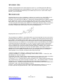
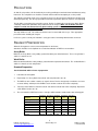
B-Bridge International, Inc.
Dehydroepiandrosterone sulfate (DHEA-S)
ELISA Kit
User Manual
Catalog # K3054-1
K3054-5
1 Plate
5 Plates
1
B-Bridge International, Inc.
[email protected]
www.b-bridge.com
DHEA-S 150902
+1-408-252-6200
TABLE OF CONTENTS
Intended Use
3
Background
3
Assay Principle
4
Kit Components
4
Materials Required
4
Precautions
5
Reagent Preparation
5
Sample Preparation
6
Assay Protocol
7
Calculations
7
Typical Data
8
Typical Standard Curve
8
Notice to Purchaser
This product is to be used for Research Purposes Only. It is not to be used for Drug or
Diagnostic Purposes, nor is it intended for Human Use. B-Bridge products may not be
resold, modified for resale, or used to manufacture commercial products without the
express written consent of B-Bridge International, Inc.
EXCEPT AS OTHERWISE EXPRESSLY SET FORTH IN THIS USER MANUAL, B-BRIDGE
DOES NOT MAKE ANY REPRESENTATION OR WARRANTIES OR CONDITIONS OF ANY
KIND, EITHER EXPRESSED OR IMPLIED, WITH RESPECT TO THE PRODUCTS, OR
INFORMATION DISCLOSED HEREUNDER, INCLUDING, BUT NOT LIMITED TO, THE IMPLIED
WARRANTIES OF MERCHANTABILITY, FIT FOR A PARTICULAR PURPOSE, OR
NONINFRINGEMENT OF THE INTELLECTUAL PROPERTY RIGHTS OF THIRD PARTIES.
B-Bridge International, Inc. All Rights Reserved.
2
B-Bridge International, Inc.
[email protected]
www.b-bridge.com
DHEA-S 150902
+1-408-252-6200
INTENDED USE
B-Bridge’s Dehydroepiandrosterone sulfate Immunoassay kit uses a specifically generated antibody to
measure Dehydroepiandrosterone sulfate (DHEA-S) in serum, plasma, urine, and saliva samples, and in
fecal extracts. The kit will also quantitatively measure DHEA-S present in tissue culture media samples.
DHEA-S is species independent.
BACKGROUND
Dehydroepiandrosterone sulfate, C19H28O5S, (5-androsten-3ß, 16a-diol-17-one sulfate, DHEA-S) is the
major C19 steroid secreted by the adrenal cortex, and is a precursor in testosterone and estrogen
biosynthesis. It is produced by the addition of a sulfate group to dehydroepiandrosterone (DHEA), catalyzed
by the sulfotransferase enzymes, SULT1A1 and SULT1E1, which also produce estrone sulfate from estrone.
DHEA sulfate can also be back-converted to DHEA through the action of steroid sulfatase. Due to the
17-ketone group rather than hydroxyl group, DHEA-S has relatively low androgenic activity (1). However the
bioactivity of DHEA-S may be high due to its high serum concentrations at 100-1,000-fold higher than
testosterone or DHEA and its weak affinity for sex-hormone binding globulin (2).
Dehydroepiandrosterone sulfate
The physiological role of DHEA-S is not well defined with serum levels being high in the fetus and neonates,
low during childhood and increased during puberty (3, 4). DHEA-S levels decline during the third decade of
life (5). DHEA-S, unlike DHEA and other steroids, does not show a significant diurnal or day-to-day variation.
DHEA-S levels are not increased due to ACTH administration and do not change significantly during the
normal menstrual cycle (2, 4). DHEA-S has a lower metabolic clearance rate than DHEA (6). Since DHEA-S
is primarily produced by the adrenal glands, it is useful as a marker for adrenal function. Adrenal tumors,
cancers, and hyperplasia can lead to the overproduction of DHEA-S. While elevated levels may not be
noticed in adult men, they can lead to amenorrhea and visible symptoms of virilization. These changes vary in
severity and may include: a deeper voice, hirsutism, male pattern baldness, muscularity, acne and
enlargement of the Adam’s apple. Women with polycystic ovary syndrome tend to have elevated levels of
DHEA-S. Excess levels of DHEA-S in children can cause precocious puberty in boys; and ambiguous
external genitalia, excess body hair, and abnormal menstrual periods in girls.
1. Dorfman RI, Shipley, RA., Androgens, J. Wiley and Sons, NY, 1956, 116-128.
2. Pang S. and Riddick, L, In: “Pediatric Endocrinology, A Clinical Guide, 2nd Ed.”. F. Lifshitz (Ed.) 1990,
Marcel Dekker, Inc. New York, 259-291.
3. de Peretti E, Forest MG., “Pattern of plasma dehydroepiandrosterone sulfate levels in humans from birth to
adulthood: evidence for testicular production”. 1978, J. Clin. Endocrinol. Metab. 47:572-577.
4. Lashansky, G., et. al., “Normative data for adrenal steroidogenesis in a healthy pediatric population: ageand sexrelated changes after adrenocorticotropin stimulation”. 1991, J. Clin. Endocrinol. Metab. 73:674-686.
5. Zumoff, B., et. al., “Sex differences in the twenty-four-hour mean plasma concentrations of
dehydroisoandroster one (DHA) and dehydroisoandrosterone sulfate (DHAS) and the DHA to DHAS ratio in
normal adults”. 1980, J. Clin. Endocrinol. Metab. 51:330-333.
6. Pang S., “Late-onset adrenal steroid 3 beta-hydroxysteroid dehydrogenase deficiency. I. A cause of
hirsutism in pubertal and postpubertal women”. 1985, J. Clin. Endocrinol. Metab. 60:428-39.
3
B-Bridge International, Inc.
[email protected]
www.b-bridge.com
DHEA-S 150902
+1-408-252-6200
ASSAY PRINCIPLE
A DHEAS standard is provided to generate a standard curve for the assay and all samples should be read off
the standard curve. Standards or diluted samples are pipetted into a clear microtiter plate coated with an
antibody to capture sheep antibodies. A DHEA-S-peroxidase conjugate is added to the standards and
samples in the wells. The binding reaction is initiated by the addition of a polyclonal antibody to DHEA-S to
each well. After 2 hours incubation, the plate is washed and substrate is added. The substrate reacts with the
bound DHEA-S-peroxidase conjugate. After a short incubation, the reaction is stopped and the intensity of
the generated color is detected in a microtiter plate reader capable of measuring 450nm wavelength. The
concentration of the Dehydroepiandrosterone sulfate in the sample is calculated, after making suitable
correction for the dilution of the sample, using software available with most plate readers.
KIT COMPONENTS
K3054-1
1 plate
K3054-5
5 plates
DHEA-S Standard (1,200 ng/ml) in stabilizing solution
70 µl
350 µl
DHEA-S Antibody
Sheep polyclonal antibody specific for DHEA-S
3 ml
13 ml
DHEA-S Conjugate
DHEA-S–peroxidase conjugate in stabilizing solution
3 ml
13 ml
5x Assay Buffer
28 ml
55 ml
20x Wash Buffer
30 ml
125 ml
TMB Substrate
11 ml
55 ml
Stop Solution
1M hydrochloric acid solution
5 ml
25 ml
Plate sealer
1 each
5 each
Clear 96 well Plate
Coated with donkey anti-sheep IgG
Store above components at 4°C
MATERIALS REQUIRED BUT NOT SUPPLIED
Distilled or deionized water
Colorimetric 96-well microplate reader capable of reading OD at 450 nm
Software for converting raw relative optical density readings from the plate reader and carrying out four
parameter logistic curve (4PLC) fitting.
Repeater or multi-channel pipet
Timer
ACS grade ethanol or ethyl acetate, glass test tubes, shaker, speedvac, centrifuge for extraction of dried
fecal samples
For saliva samples – 15 ml centrifuge tubes, centrifuge
4
B-Bridge International, Inc.
[email protected]
www.b-bridge.com
DHEA-S 150902
+1-408-252-6200
PRECAUTIONS
As with all such products, this kit should only be used by qualified personnel who have had laboratory safety
instruction. The complete insert should be read and understood before attempting to use the product.
The antibody coated plate needs to be stored desiccated. The silica gel pack included in the foil ziploc bag
will keep the plate dry. The silica gel pack will turn from blue to pink if the ziploc has not been closed properly.
This kit utilizes a peroxidase-based readout system. Buffers, including other manufacturers Wash Buffers,
containing sodium azide will inhibit color production from the enzyme. Make sure all buffers used for
samples are azide free. Ensure that any plate washing system is rinsed well with deionized water
prior to using the supplied Wash Buffer.
The Stop Solution is acid. The solution should not come in contact with skin or eyes. Take appropriate
precautions when handling this reagent.
In all cases, please consult your institution’s safety procedures for working with hazardous chemicals.
REAGENT PREPARATION
Allow the kit reagents to come to room temperature for 30 minutes.
Standards should be run in duplicate for accurate determination of DHEA-S concentrations.
Assay Buffer
Dilute the 5x Assay Buffer 1:5 by adding 1 part buffer with 4 parts deionized water. The 1x Assay Buffer is
stable for 3 months at 4°C.
Wash Buffer
Dilute the 20x Wash Buffer 1:20 by adding 1 part buffer with 19 parts deionized water. The 1x Wash Buffer is
stable for 3 months at room temperature.
Standard Preparation
Use all Standards within 2 hours of preparation.
1.
Label tubes #1 through #5.
2.
Pipet 380 µl of 1x Assay Buffer into tube #1 and 160 µl into tubes #2 - #5.
3.
The DHEA-S stock solution contains an organic solvent. Prerinse the pipet tip several times to ensure
accurate delivery. Add 20 µl of the DHEA-S stock solution to tube #1 and vortex completely.
4.
Take 40 µl of the DHEA-S solution in tube #1 and add it to tube #2 and vortex completely. Repeat the
serial dilution for tubes #3 - #5.
5.
The concentration of DHEA-S in tubes 1 through 5 will be 60,000, 12,000, 2,400, 480, and 96 pg/ml.
Reagent
1x Assay Buffer
DHEA-S Standard
Standard 1
Standard 2
Standard 3
Standard 4
Final Concentration
(pg/ml)
Standard Standard Standard Standard Standard
1
2
3
4
5
380 µl
160 µl
160 µl
160 µl
160 µl
20 µl
40 µl
40 µl
40 µl
40 µl
60,000
12,000
2,400
480
96
5
B-Bridge International, Inc.
[email protected]
www.b-bridge.com
DHEA-S 150902
+1-408-252-6200
SAMPLE PREPARATION
Samples should be run in duplicate for accurate measurements. Ensure that all samples have reached room
temperature and have been diluted appropriately prior to running the assay.
Samples containing visible particulates should be centrifuge prior to use.
Serum and Plasma Samples
The minimum Assay Buffer dilution for human serum and plasma samples is 1:2, but due to the high sample
concentration most samples will have to be diluted at least 1:100 with 1x Assay Buffer. For measurement of
DHEA-S in non-human samples it is recommended that the end user carry out a preliminary dilution series to
determine the correct dilution for their samples.
Urine Samples
Urine samples must be diluted at least 1:2 with 1x Assay Buffer, but due to the high sample concentration
most samples will have to be diluted at least 1:100 with 1x Assay Buffer. For measurement of DHEA-S in
non-human samples it is recommended that the end user carry out a preliminary dilution series to determine
the correct dilution for their samples.
For comparison to creatinine as a urine volume marker please see our NIST-calibrated 2 and 10 plate Urinary
Creatinine Detection kits, K3002-1 and K3002-5.
Saliva Samples
Whole saliva should be obtained at least 2 h after eating and rinsing mouth with water to avoid any food borne
antigens or materials from affecting the analysis. Methods of saliva collection vary widely, it is best to collect
saliva by allowing the saliva to passively flow into a 15 mL centrifuge tube.
Saliva samples must be diluted at least 1:2 times with 1x Assay Buffer.
A saliva clarification protocol, if needed.
Store samples frozen at -20°C. Upon thawing, the saliva is centrifuged at 2,500 x g for 20 minutes and the
clear supernatant is pipetted off any precipitated material. Samples should be kept on ice, analyze
immediately or aliquot and frozen at -20°C (-80°C long term storage). Minimize the number of freeze-thaws
cycles to prevent degradation of DHEA-S.
Dried Fecal Samples
Ensure that the sample is completely dry and powder the sample to improve extraction efficiency. Remove
any large particles, such as grass, if possible. We suggest checking the efficiency of extraction by preparing
a DHEA-S solution of known concentration in the 1x Assay Buffer. Spike one aliquot of your sample with a
volume of the DHEA-S solution in 1x Assay Buffer (Control Spike) and one aliquot of sample with the same
volume of only 1x Assay Buffer (Control Sample). Extract samples and both Controls with Ethanol or Ethyl
Acetate as described below.
1.
Weigh out ≥ 0.2 gm of dried fecal solid into a tube.
2.
Add 1 ml of Ethanol (or Ethyl Acetate) for every 0.1 gm of solid.
3.
Shake vigorously for at least 30 minutes.
4.
Centrifuge samples at 5,000 rpm for 15 minutes. Transfer measured volume of supernatant to a clean
tube for evaporation.
5.
Evaporate supernatant solution to dryness in a SpeedVac or under nitrogen. Keep dried extracted
samples frozen < -20°C in a desiccator.
6.
Dissolve extracted sample with 100µl ethanol, followed by at least 400µL 1x Assay Buffer. Vortex well
and allow to sit for 5 minutes at room temperature. Vortex and sit for 5 minutes twice more to ensure
complete DHEA-S solubility. For immunoassays ethanol content in the well should be below 5%. Dilute
the ethanol:Assay Buffer mixture ≥ 1:10 with 1x Assay Buffer.
7.
Run reconstituted diluted samples in assay immediately.
6
B-Bridge International, Inc.
[email protected]
www.b-bridge.com
DHEA-S 150902
+1-408-252-6200
8.
Determine the extraction efficiency by comparing the concentration of the DHEA-S measured in the
extracted Control (Control Spike - Control Sample) with the concentration of DHEA-S before extraction.
Note: In step 5 if only a portion of the organic solvent is being evaporated, ensure final amounts of measured
DHEA-S per gm solid accounts for volume of solution evaporated.
Tissue Culture Media (TCM) Samples
For measuring DHEA-S in tissue culture media (TCM), samples should be read off a standard curve
generated in TCM. Samples may need to be diluted further in TCM. The assay was validated using
RPMI-1640.
Use all samples within 2 hours of preparation.
ASSAY PROTOCOL
Standards and samples should be run in duplicate
1. Determine the number of wells needed and return the unused wells to the foil pouch with desiccant. Seal
the ziploc bag and store at 4°C.
2. Pipet 50 µl of samples or standards into wells in the plate.
3. Pipet 75 µl of 1x Assay Buffer into the non-specific binding (NSB) wells.
4. Pipet 50 µl of 1x Assay Buffer into wells to act as maximum binding wells (B0 or 0 ng/ml).
5. Add 25 µl of the DHEA-S Conjugate to each well using a repeater pipet.
6. Add 25 µl of the DHEA-S Antibody to each well, except the NSB wells, using a repeater pipet.
7. Gently tap the sides of the plate to ensure adequate mixing of the reagents. Cover the plate with the plate
sealer and shake at room temperature for 2 hour. If the plate is not shaken signals bound will be
approximately 20% lower.
8. Aspirate the plate and wash each well 4 times with 300 µl 1x Wash Buffer. Tap the plate dry on clean
absorbent towels.
9. Add 100 µl of the TMB Substrate to each well, using a repeater pipet.
10. Incubate the plate at room temperature for 30 minutes without shaking.
11. Add 50 µl of the Stop Solution to each well, using a repeater or a multichannel pipet.
12. Read the optical density generated from each well in a plate reader capable of reading at 450 nm.
13. Use the plate reader’s built-in 4PLC software capabilities to calculate DHEA-S concentration for each
sample.
CALCULATIONS
Average the duplicate OD readings for each standard and sample. Create a standard curve by reducing the
data using the 4PLC fitting routine on the plate reader, after subtracting the mean OD’s for the NSB. The
sample concentrations obtained, calculated from the %B/B0 curve, should be multiplied by the dilution factor
to obtain neat sample values.
7
B-Bridge International, Inc.
[email protected]
www.b-bridge.com
DHEA-S 150902
+1-408-252-6200
Typical Data
Always run your own standard curve for calculating results. Do not use this data.
Conversion Factor: 100 pg/ml of DHEA-S in equivalent to 256.1 pM
TYPICAL STANDARD CURVE
Standard curves vary with each assay. Always run your own standard curves for calculation of results; do not
use this data
8
B-Bridge International, Inc.
[email protected]
www.b-bridge.com
DHEA-S 150902
+1-408-252-6200