1
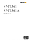
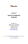
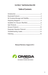
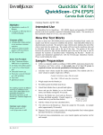
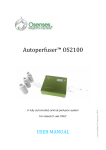
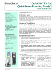
B-Bridge International, Inc. Urinary Retinol Binding Protein Immunoassay Kit User Manual Catalog # K3304-1 1 B-Bridge International, Inc. [email protected] www.b-bridge.com Retinol BP 062315 +1-408-252-6200 Table of Contents Intended Use...……………………………………………………………………..3 Background…………………………………………………………………………3 Assay Principle…..……………………………………….….…………………….3 Kit Components………………………………..…………………………………..4 Materials Required But Not Supplied…………………………………………....4 Precautions…………………………….…………………………………………..4 Reagent Preparation………………………………..……………………………..5 Sample Preparation…………………………..……………………………………5 Assay Protocol…………..………………………………………………………....6 Calculations…………….……………………………………………………….....6 Typical Standard Curve…………….…………………………………………......7 Typical Data…………...…………….…………………………………………......8 Notice to Purchaser This product is to be used for Research Purposes Only. It is not to be used for Drug or Diagnostic Purposes, nor is it intended for Human Use. B-Bridge products may not be resold, modified for resale, or used to manufacture commercial products without the express written consent of B-Bridge International, Inc. EXCEPT AS OTHERWISE EXPRESSLY SET FORTH IN THIS USER MANUAL, B-BRIDGE DOES NOT MAKE ANY REPRESENTATION OR WARRANTIES OR CONDITIONS OF ANY KIND, EITHER EXPRESSED OR IMPLIED, WITH RESPECT TO THE PRODUCTS, OR INFORMATION DISCLOSED HEREUNDER, INCLUDING, BUT NOT LIMITED TO, THE IMPLIED WARRANTIES OF MERCHANTABILITY, FIT FOR A PARTICULAR PURPOSE, OR NONINFRINGEMENT OF THE INTELLECTUAL PROPERTY RIGHTS OF THIRD PARTIES. B-Bridge International, Inc. All Rights Reserved. 2 B-Bridge International, Inc. [email protected] www.b-bridge.com Retinol BP 062315 +1-408-252-6200 Intended Use The B-Bridge Urinary Retinol Binding Protein Enzyme Immunoassay Kit (cat. # K3304-1) quantitatively measures Urinary Retinol Binding Protein (RBP) present in human, rat, dog, Rhesus monkey urine. RBP is a highly conserved protein and we have shown that this kit may measure RBP’s from sources other than human. The end user should evaluate recoveries of RBP in other urine samples being tested. For measuring retinol binding protein in serum or plasma samples, please refer to the Serum RBP Immunoassay kit, Catalog Number K3004-1. Background Retinol binding protein (RBP) is from a family of structurally related proteins that bind small hydrophobic molecules such as bile pigments, steroids, odorants, etc. RBP is a 21 kDa highly conserved, single-chain glycoprotein, consisting of 182 amino acids with 3 disulfide bonds that has a hydrophobic pocket which binds retinol (vitamin A). RBP binds retinol in a 1:1 stoichiometry, which serves to not only solubilize retinol but also protect it from oxidation. When in serum, the majority of RBP bound with retinol is reversibly complexed with transthyretin (prealbumin). This complex then transports retinol to specific receptors of various tissues in the body. Vitamin A status is reflected by serum concentration as it is hemostatically controlled and does not fall until stores are dramatically reduced. RBP has also been shown to be a useful marker for renal function as it is totally filtered by the glomeruli and reabsorbed by proximal tubules. This has made urinary RBP (uRBP) a tool to study renal function in heart or kidney transplant recipients, type 1 and 2 diabetics, and in people exposed to uranium from mining operations. Measurement of uRBP levels has also been useful in detection and characterization of diseases including hypertension and certain cancers. Assay Principle The Retinol Binding Protein (RBP) kit is designed to quantitatively measure RBP present in urine samples. Please read the complete kit insert before performing this assay. A RBP standard is provided to generate a standard curve for the assay and all samples should be read off the standard curve. Standards or diluted samples are pipetted into a clear microtiter plate coated with an antibody to capture rabbit antibodies. A RBP-peroxidase conjugate is added to the standards and samples in the wells. The binding reaction is initiated by the addition of the RBP polyclonal antibody to each well. After an hour incubation the plate is washed and substrate is added. The substrate reacts with the bound RBP-peroxidase conjugate. After a short incubation, the reaction is stopped and the intensity of the generated color is detected in a microtiter plate reader capable of measuring 450 nm wavelength. The concentration of the RBP in the sample is calculated, after making a suitable correction for the dilution of the sample, using software available with most plate readers. 3 B-Bridge International, Inc. [email protected] www.b-bridge.com Retinol BP 062315 +1-408-252-6200 Kit Components Clear 96-well plate with strip wells coated with goat anti-rabbit IgG 1 plate RBP Standard (stock solution of native human RBP at 20 µg/ml) 60 µL RBP Detection Antibody 3 mL RBP-Peroxidase Conjugate 5 mL 1X Assay Buffer 28 mL 20X Wash Buffer Concentrate 30 mL TMB Substrate 11 mL Stop Solution (IN HCl) Caution: Caustic 11 mL Plate Sealer 1 each Store all components at 4°C Materials Required But Not Supplied Deionized or distilled water Repeater pipet with disposable tips capable of dispensing 25 μL, 50 μL and 100 μL Microplate shaker and washer Colorimetric 96-well microplate reader capable of reading optical density at 450 nm Software for converting raw relative optical density readings from the plate reader and carrying out four parameter logistic curve (4PLC) fitting. Contact your plate reader manufacturer for details Precautions As with all such products, this kit should only be used by qualified personnel who have had laboratory safety instruction. The complete User Manual should be read and understood before attempting to use the product. Antibody coated plate needs to be stored desiccated. The silica pack wrapped with the plate will keep the plate dry. The silica gel pack will turn from blue to pink if the Ziploc has not been closed properly. The RBP Standard is purified from a human source and as such, should be treated as potentially hazardous. Proper safety procedures must be followed. This kit utilizes a peroxidase-based readout system. Buffers, including other manufacturers Wash Buffers, containing sodium azide will inhibit color production from the enzyme. Make sure all buffers used for samples are azide free. Ensure that any plate washing system is rinsed well with deionized water prior to using the supplied Wash Buffer. The Stop Solution is 1N hydrochloric acid. The solution should not come in contact with skin or eyes. Take appropriate precautions when handling this reagent and please consult your institution’s safety procedures for working with hazardous chemicals. 4 B-Bridge International, Inc. [email protected] www.b-bridge.com Retinol BP 062315 +1-408-252-6200 Reagent Preparation Allow the kit reagents to come to room temperature for 30 minutes. We recommend that all standards and samples be run in duplicate to allow the end user to accurately determine RBP concentrations. Wash Buffer Dilute 20x Wash Buffer 1:20 by adding one part of the concentrate to nineteen parts of deionized water. Once diluted this is stable at room temperature for 3 months at room temperature. Standard Preparation Label five tubes #1 through #5. Briefly spin vial of standard in a microcentrifuge to ensure contents are at bottom of vial. Pipet 475 µL of Assay Buffer into tube #1 and 300 µL into tubes #2 - #5. Carefully add 25 µL of the RBP Standard stock solution to tube #1 and vortex completely. Take 100 µL of the RBP solution in tube #1 and add it to tube #2 and vortex completely. Add 100 µL of tube #2 to tube #3 and vortex completely. Repeat these serial dilutions for tubes #4 and #5. The concentration of RBP in tubes 1 through 5 will be 1,000, 250, 62.5, 15.625, and 3.906 ng/mL. Use all standards within 2 hours of preparation. Reagents Std 1 Std 2 Std 3 Std 4 Std 5 Assay Buffer 475 µl 300 µl 300 µl 300 µl 300 µl RBP Standard 25 µl - - - - Standard 1 - 100 µl - - - Standard 2 - - 100 µl - - Standard 3 - - - 100 µl - Standard 4 - - - - 100 µl 1,000 ng/mL 250 ng/mL 62.5 ng/mL 15.625 ng/mL 3.906 ng/mL Concentration Sample Preparation Urine samples containing visible particulate should be centrifuged prior to using. We recommend that all standards and samples be run in duplicate to allow the end user to accurately determine RBP concentrations. Ensure that all samples have reached room temperature .and have been diluted as appropriate prior to running them in the kit. Samples must be diluted 1:2 by adding one part of urine to one part Assay Buffer prior to running in the kit. Any samples with RBP concentrations greater than the standard curve range should be diluted further with Assay Buffer to obtain readings within the standard curve. Samples that are too dilute to be measured should be concentrated prior to measuring in the assay. Use all samples within 2 hours of dilution. 5 B-Bridge International, Inc. [email protected] www.b-bridge.com Retinol BP 062315 +1-408-252-6200 Assay Protocol 1. Pipet 50 µl of samples or standards in duplicate into wells in the clear microplate. 2. Pipet 75 µl of Assay Buffer into the non-specific binding (NSB) wells. 3. Pipet 50 µl of Assay Buffer into wells to act as maximum binding wells (Bo or 0 pg/mL). 4. Add 25 µl of RBP-Peroxidase Conjugate to each well using a repeater pipet. 5. Add 25 µl of RBP Detection Antibody to each well except the NSB wells using a repeater pipet. 6. Gently tap the sides of the plate to ensure adequate mixing of the reagents. Cover the plate with the plate sealer and shake at room temperature for 1 hour. 7. Aspirate the plate and wash each well 4 times with 300 µl of Wash Buffer. Tap the plate dry on clean absorbent towels. 8. Add 100 µl of the TMB Substrate to each well using a repeater pipet. 9. Incubate at room temperature for 30 minutes without shaking. 10. Add 100 µl of the Stop Solution to each well using a repeater pipet. 11. Read the optical density (OD) of each well in a plate reader at 450 nm wavelength. 12. Use the plate reader’s built-in 4PLC software capabilities to calculate RBP concentration for each sample. Calculations Average the duplicate OD readings for each standard and sample. Create a standard curve by reducing the data using computer software capable of generating a four-parameter logistic curve (4PLC) fit, after subtracting the mean OD’s for the blank. The sample concentrations, calculated off the %B/B0 curve, should be multiplied by the dilution factor to obtain neat sample values. Sample RBP values should be normalized to creatinine levels by running the same samples in the Urinary Creatinine Detection Kit, K3002-1. 6 B-Bridge International, Inc. [email protected] www.b-bridge.com Retinol BP 062315 +1-408-252-6200 Typical Standard Curve 7 B-Bridge International, Inc. [email protected] www.b-bridge.com Retinol BP 062315 +1-408-252-6200 Typical Data Always run your own standard curve for calculation of results. Do not use this data. Conversion factor 1 µg/mL of human RBP is equivalent to 47.62 nM RBP 8 B-Bridge International, Inc. [email protected] www.b-bridge.com Retinol BP 062315 +1-408-252-6200