1
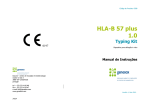
21001 Kittridge Street, Canoga Park, CA 91303-2801 USA Tel: +1 (818) 702-0042 Fax: +1 (818) 702-6904 P R O D U C T www.onelambda.com I N S E R T MICRO SSP™ HLA DNA TYPING TRAYS For In Vitro Diagnostic Use. Important: Refer to Micro SSP™ Product Reference Table (Document ID: MSSP_REF_TABLE_PI.DOC) for OLI product catalog numbers, specific product components and requirements. INTENDED USE DNA typing of Class I or Class II HLA alleles. SUMMARY AND EXPLANATION Historically, the established method for the determination of HLA antigens has been the lymphocytotoxicity test.1 However, with the advent of PCR technologies, DNA-based tissue typing techniques have become routine in the laboratory. For most DNA-based methodologies, the PCR process is used only as an amplification step to acquire the needed target DNA. The HLA typing process then requires a post-amplification step to discriminate between the different alleles (e.g., RFLP, SSOP, reverse dot blot). Unlike other PCR-based methods, the SSP methodology employed in the One Lambda, Inc., Micro SSP™ DNA typing test discriminates between the different alleles during the PCR process.2 This shortens the post-amplification processing time to a simple gel electrophoresis detection step. In contrast to the lymphocytotoxicity reaction scale (1 = negative to 8 = positive), Micro SSP™ test results are either positive or negative. This abolishes the need for complicated interpretation of results. In addition, single nucleotide changes can be discriminatory in PCR-SSP, while crossreacting groups (CREGs) provide major challenges to serological typing.3 Finally, due to the synthetic nature of DNA typing reagents (e.g., oligonucleotide primers), stability has been greatly improved and lot to lot variance has been reduced. PRINCIPLE(S) The PCR-SSP methodology is based on the principle that completely matched oligonucleotide primers are more efficiently used in amplifying a target sequence than a mismatched oligonucleotide primer by recombinant Taq polymerase. Primer pairs are designed to have perfect matches only with a single allele or group of alleles. Under strictly controlled PCR conditions, perfectly matched primer pairs result in the amplification of target sequences (i.e., a positive result) while mismatched primer pairs do not result in amplification (i.e., a negative result). After the PCR process, the amplified DNA fragments are separated by agarose gel electrophoresis and visualized by staining with ethidium bromide and exposure to ultraviolet light. Interpretation of PCR-SSP results is based on the presence or absence of a specific amplified DNA fragment. Since amplification during the PCR reaction may be adversely affected by various factors (pipetting errors, poor DNA quality, presence of inhibitors, etc.), an internal control primer pair is included in every PCR reaction. The control primer pair amplifies a conserved region of the Human -globin gene, which is present in all human DNA samples and is used to verify the integrity of the PCR reaction. In the presence of a positive typing band (specific amplification of an HLA allele), the product of the internal control primer pair may be weak or absent due to the differences in concentration and melting temperatures between the specific primer pairs and the internal control primer pair. The amplified DNA fragments of the specific HLA primer pairs are smaller than the product of the internal control primer pair, but larger than the diffuse, unincorporated primer band. Thus, a positive reaction for a specific HLA allele or allele group is visualized on the gel as an amplified DNA fragment between the internal control product band and the unincorporated primer band (see Expected Values/Gel Interpretation). REAGENTS A. Identification The Micro SSP™ DNA Typing Trays provide sequence-specific oligonucleotide primers for amplification of HLA alleles and the human -globin gene by the polymerase chain reaction (PCR). Pre-optimized primers are presented (dried) in different wells of a 96-well 0.2 ml thin-walled tube tray for PCR and are ready for the addition of DNA samples, recombinant Taq polymerase, and specially formulated dNTP-buffer mix (Micro SSP™ D-mix). Each typing tray includes a negative control reaction tube that detects the presence of the internal control PCR product generated by the Micro SSP™ DNA Typing Tray. The internal control PCR product amplified from the Human -globin gene is the most likely contaminating PCR product due to its amplification in every well. The amount of each primer is adjusted for optimal amplification of 100 ng of sample DNA when used in conjunction with the Micro SSP™ D-mix, the prescribed amount of recombinant Taq polymerase, and the PCR reaction profile detailed in this document (page 2). See the provided worksheet for specific alleles that can be amplified by each primer set under the specified PCR conditions of the assay. For lot specific primer site locations, refer to Primer Information document. SSP-DNAT-PI-EN-00, Rev. 16 Page 1 of 8 B. Warning or Caution 1. For In Vitro Diagnostic Use. 2. Pipettes used for post-PCR manipulations should not be used for pre-PCR manipulations. 3. Biohazard Warning: The ethidium bromide used for staining of DNA is a potential carcinogen. Always wear gloves when handling stained gels. 4. Biohazard Warning: All blood products should be treated as potentially infectious. Source material from which this product was derived was found negative when tested in accordance with current FDA required tests. No known test methods can offer assurance that products derived from human blood will not transmit infectious agents. 5. Caution: Wear UV-blocking eye protection, and do not view UV light source directly when viewing or photographing gels. 6. See Material Safety Data Sheet for detailed information. C. Instructions for Use See Directions for Use. D. Storage Instructions Store reagents at temperature indicated on package. Use before printed expiration date. E. Purification or Treatment Required for Use See Directions for Use. F. Instability Indications 1. Do not use primer set trays with cracks in the tubes or on the lips, cracks may cause evaporative losses. 2. If salts have precipitated out of the solution in the D-mix aliquots during shipping or storage, re-dissolve them by extended vortexing at room temperature (20 to 25C). 3. D-mix aliquots, upon thawing at room temperature (20 to 25C), should be pink to light purple in color. Any D-mix aliquot without the specified coloration should be considered unusable. INSTRUMENT REQUIREMENTS A. Programming the Thermal cycler 96-Well GeneAmp® PCR System 9600, 9700 or Veriti™ 96-Well Thermal Cycler (Applied Biosystems). Set ramp speed to 9600 for 96-Well GeneAmp® PCR System 9700. Set ramp speed to 9600 Emulation Mode for Veriti™ 96Well Thermal Cycler. Program your thermal cycler before starting the “Step-by-Step Procedure” below. # of Cycles 1 One Lambda PCR Program (OLI-1) Step Temp. (C) 1 96 2 63 Time (sec.) 130 60 9 1 2 96 63 10 60 20 1 2 3 96 59 72 10 50 30 End 1 4 --- For specific thermal cycler information, refer to the manufacturer's user manual. B. 2.5% Agarose Gel Preparation (For Micro SSP™ Gel System, OLI Cat. #MGS108) 1. To set up: Slide the locking pin on the base to the open position. Insert the gel box into the base matching the color-coded sides to assure proper orientation. Lock the gel box into the base by sliding the locking pin into the locked position. Use the leveling bubble and the three height adjustable legs to level the base. 2. 3. Orient and fully insert the 14 gel combs into the gel comb holder. To 100 ml of 1x Tris Borate EDTA buffer (1x TBE) with 0.5 g/ml ethidium bromide (in a 500 ml glass bottle), add 2.5 g electrophoresis grade agarose. Heat until a homogeneous solution is formed. SSP-DNAT-PI-EN-00, Rev. 16 Page 2 of 8 4. 5. Add 30 ml of the gel solution to the gel box. Make sure the agarose covers the entire surface evenly by tilting the gel box back and forth immediately after the gel solution is added. Quickly place the gel comb holder on the filled gel box by matching the color coding. Allow to set for 15 minutes. Remove the gel combs by lifting the gel comb holder while holding the base. Add 10 ml of 1xTBE containing 0.5 g/ml ethidium bromide evenly across the gel to fill every well. C. Gel Electrophoresis (For Micro SSP™ Gel System, OLI Cat. #MGS108) 1. After completing the PCR reaction: Orient the DNA primer set tray and gel box with the negative control well in the upper left hand corner. Gently remove the tray seal without splashing the samples. 2. Transfer each PCR reaction (10 l) in sequence to the 2.5% agarose gel. Make sure to transfer all samples in the proper sequence. (No addition of electrophoresis dye is necessary.) Use of an 8- or 12-channel Pipetman® is recommended. Note: The order of samples (to match the worksheet) is from left to right, top to bottom. 3. 4. 5. Cover the gel box with the gel box cover by matching the color-coded sides. Electrophorese the samples at 140 - 150 volts until the red tracking dye has migrated about 0.5 cm into the gel (approximately 3 - 5 minutes, depending on the agarose used.) Remove the cover. Slide the locking pin on the base to the open position and remove the gel box. Transfer the gel box to a UV transilluminator. Photograph the completed gel. Orient the photograph with the negative control reaction in the upper left corner, and mark the corresponding positive allele groups on the worksheet provided with the tray. SPECIMEN COLLECTION AND PREPARATION A. DNA can be purified from human leukocytes by any preferred method. B. The DNA sample to be used for PCR-SSP analysis should be re-suspended in sterile water or in 10 mM Tris-HCl, pH 8.0 - 9.0 at a concentration of 25 - 200 ng/l with the A260/A280 ratio of 1.65 - 1.80. C. Samples should not be re-suspended in solutions containing chelating agents, such as EDTA, above 0.5 mM in concentration. D. DNA samples may be used immediately after isolation or stored at -20 C for extended periods of time (over 1 year) with no adverse effects on results. E. DNA samples should be shipped at 4C or below to preserve their integrity during transport. PROCEDURE A. Materials Provided 1. Micro SSP™ DNA primer set trays 2. Pre-aliquoted D-mix tubes (appropriate number for test) 3. Tray seals (appropriate number for test) B. Materials Required, But Not Provided Recombinant Taq polymerase (5 units/l) Pipetting devices, such as: Gilson® P-20, Gilson® P200 Pipetman® Disposable pipette tips Vortex mixer Microcentrifuge PCR tray microtube storage rack and cover (Robbins Scientific, Cat. #1044-39-5) SSP-DNAT-PI-EN-00, Rev. 16 Page 3 of 8 Pressure pad for use with tray (OLI Cat. #SSPPAD, SSPPAD384, SSPPADGRY). Use only the pressure pad specifically for your thermal cycler. See Micro SSP™ Pressure Pad Protocol (PCR_PAD_INS.DOC.) Note: The pressure pad is good for a maximum of 300 PCR runs. Please order a new pad if the surface of the pad that contacts the tray seal is no longer smooth. Thermal cycler for PCR with 96-well format and tray/retainer for 0.2 ml thin-walled reaction tubes Hot plate or microwave oven for heating agarose solutions Electrophoresis apparatus/power supply (150V minimum capacity) UV transilluminator (Example: Fotodyne FOTO/UV21) Photographic or image documentation system 1x TBE buffer (89mM Tris-borate; 2 mM disodium EDTA, pH 8.0) with 0.5 g/ml ethidium bromide or 5XTBE Buffer with ethidium bromide (OLI Cat. # 5XTBE100) Electrophoresis grade agarose (e.g., FMC Seakem® LE) C. Step-by-step procedure. See Directions For Use below. DIRECTIONS FOR USE A. Sample Preparation 1. Purify genomic DNA from leukocyte sample by method of choice. Final DNA concentration should be 25 200ng/l (100ng/l is optimal) with the A260/A280 ratio between 1.65-1.80. 2. For specific information on sample preparation and storage, see Specimen Collection and Preparation above. 3. Perform PCR on the purified DNA sample using a Micro SSP HLA DNA Typing Tray, or store DNA sample at 20C or below until ready to type. B. Reagent/Equipment Preparation 1. Program a thermal cycler to run the One Lambda PCR program. (See “Instrument Requirements” above.) 2. Have available: recombinant Taq polymerase (5 units/l). Store at -20 C. 3. Prepare electrophoresis gel (2.5% agarose) using the Micro SSP™ Gel System (Cat. # MGS108) or with at least 96 sample wells (space rows of wells at least 1 cm apart). C. Instructions for Pipetting Taq Polymerase One Lambda's quality control tests indicate that the volume shown on the Taq polymerase label is more than sufficient for the number of tests indicated in this product. If additional Taq is required, it must be ordered directly from F. Hoffmann-La Roche or their representative. To avoid waste, please follow the simple instructions listed below for pipetting Taq polymerase. Note: Taq polymerase is very viscous and special care must be taken in the aliquoting process. Failure to follow the steps described below may result in reagent loss. 1. 2. Pipette slowly, using a calibrated Gilson® Pipetman®. (A P10 Pipetman® is recommended for increased accuracy.) The pipette tip should just pass the surface layer of the Taq. Warning: Do not immerse the pipette tip in the Taq. 3. Carefully wipe excess Taq from pipette tip on the rim of the vial. D. Stepwise Procedure 1. Remove from the indicated storage temperature: the volume (tube) of the Micro SSP™ D-Mix that matches the selected Micro SSP™ primer set tray, the primer set tray(s), and the appropriate number of DNA samples. Thaw at room temperature (20 to 25 C). Note: You may cut tray and tray seal into the number of tests needed for a single work session. Immediately return the unused portions to the appropriate storage temperature. 2. 3. 4. Vortex DNA samples to mix. Place the primer set tray in a PCR Tray Microtube Storage Rack (Robbins Scientific, Cat. #1044-39-5) and remove the tray label. Remove recombinant Taq polymerase from freezer, and keep on ice until ready to use. Using a Pipetman® (or equivalent), add 1 l of DNA diluent to the negative control reaction tube on the primer set tray. Using a Pipetman® (or equivalent), add recombinant Taq polymerase (5 units/l) to the Micro SSP™ D-mix tube. (See Micro SSP™ Product Reference Table for amount.) SSP-DNAT-PI-EN-00, Rev. 16 Page 4 of 8 5. 6. 7. 8. 9. Cap tube and vortex for 5 seconds. Pulse-spin the Micro SSP™ D-mix tube in a microcentrifuge to bring all liquid down from sides of the tube. Using a P20 Pipetman® (or equivalent), pipette 9 l of the Micro SSP™ D-mix to the negative control reaction tube. Using a Pipetman®(or equivalent), add the DNA sample to the Micro SSP™ D-mix tube. (See Micro SSP™ Product Reference Table for amount.) Cap tube and vortex for 5 seconds. Pulse-spin the Micro SSP™ D-mix tube in a microcentrifuge. Using a P20 Pipetman® or an electronic Pipetman®, aliquot 10 l of the sample-reaction mixture from the Micro SSP™ D-mix tube into each reaction tube, except the negative control reaction tube, of the Micro SSP™ primer set tray. Important: Be sure to apply the sample above the primers (dried at the bottom of each reaction tube) to avoid crosscontamination between tubes. Touch the inside wall of the tube with the pipette tip to allow the sample to slide down to the bottom of the tube. Check that all samples have dropped to the bottom of each tube. If not, tap the tray gently on the bench top so that all samples settle at the bottom of the tube before you begin PCR.. 10. Cover the reaction tubes with the tray seal provided. Check that all reaction tubes are completely covered by the tray seal to assure complete closure and to prevent evaporative loss during PCR. 11. Place the Micro SSP™ primer set tray in the thermal cycler. 12. Place a pressure pad on top of the tray with the textured side up before closing the thermal cycler lid. (See Micro SSP™ Pressure Pad Protocol for instructions on specific pad to be used with your thermal cycler.) 13. Enter your Micro SSP™ PCR program number. Specify a 10 l reaction volume option. 14. Start the PCR program. The program takes approximately 1 hour and 16 minutes. The last step will hold the sample at 4C until the run is stopped. 15. Remove the primer set tray from the thermal cycler. Gently remove the tray seal without splashing the samples. Or, store the samples in the primer set tray at -20 C or below for later gel electrophoresis. 16. Transfer each PCR reaction (10l) in sequence to a 2.5% agarose gel in the Micro SSP™ Gel System (OLI Cat. #MGS108). (Use of an 8- or 12-channel Pipetman® or of the 96-Well Transfer Device (OLI Cat. #TRNDV96) is highly recommended.) Note: Be sure to transfer all samples in the proper sequence, orienting the primer set tray with the negative control reaction well in the upper left corner. The order of samples (to match the worksheet) is from left to right, top to bottom. No addition of electrophoresis dye is necessary. 17. Electrophorese the samples at 140 - 150 volts until the red tracking dye has migrated about 0.5 cm into the gel (approximately 3 - 5 minutes, depending on the agarose used). 18. Photograph the gel on a UV transilluminator. 19. Interpret the typing results using the worksheet provided with the trays or with the assistance of the HLA software available from One Lambda, Inc. Note: When One Lambda’s DNA typing trays fill only a few rows of the electrophoresis gel, it is possible to place several samples on one gel. Care should be used to record sample positions on the gel. RESULTS Allele Groups DRB1*01 DRB1*15 DRB1*16 DRB1*0301, 0304 DRB1*0302, 0303 DRB1*04 DRB1*11 DRB1*12 DRB1*13 DRB1*14 DRB1*07 DRB1*08 DRB1*09 DRB1*10 DRB5* DRB3* DRB4* DQB1*05 DQB1*06 DQB1*02 DQB1*0301, 0304 DR 72 Serology 12 24 2 6 6 14 23a 5 17 11b 16 7 5 5 26 57c 32d 34 32 24 22 SSP-DNAT-PI-EN-00, Rev. 16 Micro SSP™ 12 24 2 6 6 14 24 5 17 10 16 7 5 5 26 56 33 34 32 24 22 a, b) The discrepancies on DRB1*11 and DRB1*14 are based on one sample in which serology assigned a DRB1*14 while DNA typing assigned DRB1*1117. The DRB1*14 serological reagents are monoclonal antibodies which are predicted to detect the epitope 31FHNQEEF37 based upon pattern analysis with DRB1*14 alleles. This amino acid sequence is also shared with DRB1*1117 (based on published amino acid sequences). c) The discord on DRB3 is based on one sample that was assigned a homozygous DRB1*08 by both serology and DNA typing, but was assigned an additional DRB3* by serology. DRB1*08 and DRB3* share amino acid sequences which under the double dose effects of DRB1*08 homozygosity may generate false positive serological reactions.6 d) The false negative discord on DRB4 is based on one sample in which only DNA typing assigned a DRB4. Assignments by both serology and DNA typing show that this sample contains the DR7-DQ9 haplotype, which is known to lack expression of the DRB4 allele at the protein level.7 Note: Lot specific performance data is available on request. Page 5 of 8 DQB1*0302, 0304 DQB1*0303 DQB1*04 11 3 11 11 3 11 LIMITATIONS OF THE PROCEDURE A. PCR-SSP is a dynamic process requiring highly controlled conditions to insure discriminatory amplification. The procedure provided in this product must be strictly followed. B. The extracted sample DNA provides the template for the specific amplification process and, thus, must have its concentration and purity within the ranges specified in the procedure. C. All instruments (e.g., PCR machine, pipetting devices) must be calibrated according to the manufacturer's recommendations. For lot specific information, refer to the Resolution Limitations document. D. For lot specific information, refer to the Resolution Limitations document. EXPECTED VALUES A. Data Analysis 1. An internal control band (slower-migrating) should always be visible in negative wells (except in the negative control well) as a control for successful amplification. Non-amplification of any well can void test results. 2. A faster-migrating, positive typing band will be observed on the electrophoretic gel if a specific HLA gene was amplified during PCR. This indicates a positive test result. 3. The internal control band may be weak or absent in positive wells. 4. Match the pattern of positive wells with the information on the Micro SSP™ worksheet to obtain the HLA typing of the sample DNA. 5. The presence of the internal control band and/or the positive typing band in the negative control well voids all test results. B. Gel Interpretation* Positive Reaction Negative Reaction NonAmplification Well Internal Control Band Positive Typing Band Primer Band *The internal control band and broad unincorporated primer band serve as size markers. Any visible band between the two size markers should be considered positive typing bands. SPECIFIC PERFORMANCE CHARACTERISTICS A comparison of Micro SSP™ and serological HLA typing methodologies was performed to assess the accuracy of the Micro SSP™ HLA typing system. In this comparison, eighty-one random whole blood samples were collected and analyzed for their HLA Class II typing using the One Lambda, Inc., HLA Class II Tissue Typing Tray DR72F and Micro SSP™ Generic HLA Class II DNA Typing Tray. The results show a 96% (78/81) concordance between the two methods. The table on the following page represents the total number of times each specificity was assigned by the two different methods. (Highlighted results indicate discordance between the two methods.) BIBLIOGRAPHY 1) Terasaki, P.I., Bernoco, F., Park, M.S., Ozturk, G., and Iwaki, Y. Microdroplet testing for HLA-A, -B, -C and -D antigens. American Journal of Clinical Pathology 69:103-120, 1978. 2) Newton, C.R., Graham, A., Heptinstall, E., Powell, S.J., Summers, C., Kalsheker, N., Smith, J.C., and Markham, A.F. Analysis of any point mutation in DNA. The amplification refractory mutation system (ARMS). Nucleic Acids Research 17: 2503-2516, 1989. 3) Slater, R.D. and Parham, P. Mutually exclusive public epitopes of HLA-A,B,C Molecules. Human Immunology 26: 85-89, 1989. 4) Bodmer J., Marsh S., Albert E., Bodmer W., Bontrop R., Charron D., Dupont B., Erlich H., Mach B., Mayr W., Parham P., Sasazuki T., Schreuder G., Strominger J., Svejgaard A. and Terasaki P. Nomenclature for factors of the HLA system, 1995. Human Immunology 43:149-164, 1995. 5) Terasaki, P.I. Histocompatibility Testing, 1980. UCLA Tissue Typing Laboratory, Los Angeles, California. SSP-DNAT-PI-EN-00, Rev 16 Page 6 of 8 6) Savage, D.A., Bidwell, J.L., Cullen, C., Bidwell, E.A., and Middleton, D. Identification of HLA-DRw52 associated antigens using HLA class II allogenotyping. Tissue Antigens 32: 278-285, 1988. 7) Sutton, V.R., Kienzle, B.K., and Knowles, R.W. An altered splice site is found in the DRB4 gene that is not expressed in HLA-DR7, Dw11 individuals. Immunogenetics 29: 317-322, 1989. TROUBLESHOOTING Problem Potential Causes No bands or faint bands DNA Solutions Not enough used Not pure enough (A260/A280 not within 1.65 1.80) PCR inhibitor present (e.g., EDTA above 0.5 mM) Repeat test; use amount indicated in ™ the Micro SSP Product Reference Table. Repeat DNA preparation; check for A260/A280 within 1.65 - 1.80, then repeat test. Repeat DNA preparation; re-suspend DNA in solution with EDTA < 0.5 mM, then repeat test. Recombinant Taq polymerase Not enough used Enzyme degraded Repeat test; use amount indicated in ™ the Micro SSP Product Reference Table. Repeat test; use fresh tube of enzyme. False bands EtBr Not enough used Remake 1 X TBE with EtBr @ 0.5 g/ml. Repeat test. Pressure Pad Pressure pad worn out Change pressure pad after 300 PCR runs. DNA Too much used Not pure enough (A260/A280 not within 1.65 1.80) Contaminated (other DNA or PCR product) Repeat test; use amount indicated in ™ the Micro SSP Product Reference Table. Repeat DNA preparation; check for A260/A280 within 1.65 - 1.80, then repeat test. Use new aliquots of all buffers/reagents for DNA extraction and PCR; repeat DNA extraction and test. Bands in negative control Recombinant Taq polymerase Too much used Repeat test; use amount indicated in ™ the Micro SSP Product Reference Table. EtBr Too much used Remake 1 X TBE with EtBr @ 0.5 g/ml. Repeat test. Reagents Contaminated (other DNA or PCR product) Use new aliquots of all buffers/reagent DNA extraction and PCR; repeat DNA extraction and test. SSP-DNAT-PI-EN-00, Rev 16 Page 7 of 8 TRADEMARKS USED IN THIS DOCUMENT/PRODUCT Micro SSP™ One Lambda Inc. FMC SeaKem FMC Corporation GeneAmp Applied Biosystems ARMS™ Zeneca Limited Fotodyne FOTO/UV21 FOTODYNE Incorporated Gilson® and Pipetman® Rainin Instrument Co., Inc. Veriti™ Applied Biosystems PATENTS USED IN THIS DOCUMENT Notice to purchaser of licensed product: The purchase price of this product includes limited, non-transferable rights under U.S. Patents 4,683,202, 4,683,195 and 4,965,188 and their foreign counterparts, owned by Roche Molecular Systems, Inc. and F. Hoffmann-LaRoche Ltd (“Roche”), to use only this amount of the product to practice the Polymerase Chain Reaction (“PCR”) Process described in said patents solely for the HLA Typing applications of the purchaser solely for organ, bone marrow, or tissue transplantation, and explicitly excludes analysis of forensic evidence or parentage determination. The right to use this product to perform and to offer commercial services for HLA Typing for organ or tissue transplantation using PCR, including reporting the results of the purchaser’s activities for a fee or other commercial consideration, is also hereby granted. Further information on purchasing licenses to practice PCR may be obtained by contacting, in the United States, the Director of Licensing at Roche Molecular Systems, Inc., 1145 Atlantic Avenue, Alameda, California 94501, and outside the United States, the PCR Licensing Manager, F. Hoffmann-La Roche Ltd, Grenzacherstr.124, CH-4070 Basel, Switzerland. SSP technology is licensed from Zeneca Limited, through its Zeneca Diagnostics business, Blacklands Way, Abingdon Business Park, Abingdon, Oxfordshire, England OX14 IDY and covered under the following patents held by Zeneca Corporation: European Patent No. 0 332 435 B1, United States Patent No. 5,595,890, entitled “Method of detecting nucleotide sequences,” and Canadian Patent No. 1323592, and corresponding patents and patent applications worldwide. The Micro SSP™ DNA typing reagents are manufactured and distributed by One Lambda, Inc., 21001 Kittridge Street, Canoga Park, CA 91303, U.S.A. Recombinant Taq polymerase is manufactured by F. Hoffmann-LaRoche. EUROPEAN AUTHORIZED REPRESENTATIVE MDSS GmbH, Schiffgraben 41, D-30175, Hannover, Germany REVISION HISTORY Revision Date 15 2009/02 16 2009/07 SSP-DNAT-PI-EN-00, Rev 16 Revision Description Added the Veriti™ Thermal Cycler in the Instrument Requirements Section; Separated “Thermal Cycler” into two words; Template Update-Inserted a Revision History Section Page 3, Materials Provided; remove reference to Micro SSP™ Reference Table. Move Taq Polymerase to Materials Required, But Not Provided. Page 8 of 8