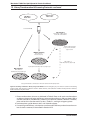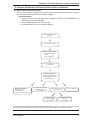
1
User Manual
Matchmaker™ GAL4 Two-Hybrid System 3
& Libraries User Manual
United States/Canada
800.662.2566
Asia Pacific
+1.650.919.7300
Europe
+33.(0)1.3904.6880
Japan
+81.(0)77.543.6116
Clontech Laboratories, Inc.
A Takara Bio Company
1290 Terra Bella Ave.
Mountain View, CA 94043
Technical Support (US)
E-mail: [email protected]
www.clontech.com
PT3247-1 (PR742219)
Published 3 April 2007
Matchmaker™ GAL4 Two-Hybrid System 3 & Libraries User Manual
Table of Contents
I. Introduction II. Overview: A Yeast Two-Hybrid Screen
4
8
III. Lists of Components
9
IV. Additional Materials Required
10
V. Yeast Strains & Phenotypes
A. Yeast Host Strains
B. Phenotypes
11
11
11
VI. Control Vectors
A. Positive Controls
B. Negative Control
13
13
13
II. Matchmaker™ GAL4 cDNA & Genomic Libraries
V
A. Library Construction
B. Library Quality Control Information
14
14
15
III.
V
Preparing for a Two-Hybrid Screen
A. Construct Fusion Genes
B. Obtain or Construct and AD Fusion Library
C. Verify that Constructs Do Not Activate Reporter Genes
D. Verify Protein Expression
16
16
16
17
17
IX.
Library Transformation & Screening Protocols
A. Transformation Scales
B. Yeast Transformation Protocols
C. Plating and Screening Transformation Mixtures
D. Calculations
18
18
19
21
23
X.
Analysis & Verification of Putative Positive Clones
A. Retest Phenotypes
B. Isolate Plasmid DNA from Yeast C. Sort Colonies to Eliminate Duplicates
D. Rescue AD/library Plasmids via transformation of E. coli
E. Retest Protein Interactions in Yeast
F. In vitro Analysis
G. Sequencing AD/Library Inserts
H. In vivo Analysis
I. Additional Two-Hybrid Tests 24
24
24
24
24
25
26
27
27
27
XI. Troubleshooting Guide
28
XII. References
30
XIII. Related Products
32
Appendix A: Media & Solution Recipes
33
Appendix B: Library Titering
34
Appendix C: Plasmid Library Amplification 35
Clontech Laboratories, Inc.
www.clontech.com
Protocol No. PT3247-1
Version No. PR742219
Matchmaker™ GAL4 Two-Hybrid System 3 & Libraries User Manual
List of Tables
Table I.
Table II.
Table III.
Table IV.
Table V.
Table VI.
List of Abbreviations
Matchmaker Yeast Strain Genotypes
Matchmaker Yeast Strain Phenotypes Matchmaker Two-Hybrid System 3 Vectors
Comparison of Two-Hybrid Transformation Methods
Set up for a Two-Hybrid Library Screen
7
11
11
13
18
21
List of Figures
Figure 1.
Figure 2.
Figure 3.
Figure 4.
Figure 5.
Figure 6.
Figure 7. Overview of Matchmaker Two-Hybrid System 3 advances and screening process 4
The two-hybrid principle
5
Reporter constructs in yeast strains AH109 and Y187
5
Overview of performing a yeast two-hybrid screen
8
Screening an AD fusion library using strain AH109
22
Strategies for analyzing and verifying putative positive clones 25
Yeast mating to verify protein interactions
26
Notice to Purchaser
Clontech products are to be used for research purposes only. They may not be used for any other purpose, including,
but not limited to, use in drugs, in vitro diagnostic purposes, therapeutics, or in humans. Clontech products may not be
transferred to third parties, resold, modified for resale, or used to manufacture commercial products or to provide a service
to third parties without written approval of Clontech Laboratories, Inc.
NucleoSpin® and NucleoBond are a registered trademarks of Macherey-Nagel GmbH & Co.
Parafilm® is a registered trademark of the American Can Co.
Clontech, the Clontech logo and all other trademarks are the property of Clontech Laboratories, Inc., unless noted
otherwise. Clontech is a Takara Bio Company. ©2007 Protocol No. PT3247-1
www.clontech.com
Clontech Laboratories, Inc.
Version No. PR742219
Matchmaker™ GAL4 Two-Hybrid System 3 & Libraries User Manual
I. Introduction
MatchmakerTwo-Hybrid System 3 is an advanced GAL4-based two-hybrid system that provides a
transcriptional assay for detecting protein interactions in vivo in yeast. You can use this system to
screen a library for novel proteins that interact with a known bait protein, or to test two previously
cloned proteins for an interaction. Matchmaker Two-Hybrid System 3 incorporates many features
that reduce the incidence of false positive results and allow you to quickly identify and confirm
protein interactions. Key features of System 3 are detailed in Figure 1.
Strain AH109
• Virtually eliminates
false positives
Library screening
Ade+, His+
Colonies
• Allows stringency of
selection to be varied
• Use simple α-gal
or β-gal assay
α-gal or β-gal assay
Improved vectors
• Increased transformation
efficiency
Verification of
true positives
• High-level protein expression
• c-Myc and HA tags facilitate
detection of fusion proteins
• Distinct bacterial selection
markers
AD—Ampr
DNA-BD—Kanr
• High copy vectors
Matchmaker™
Co-IP Kit
• T7 promoter allows in vitro
transcription and translation
Epitope-Tagged
Expression Vectors
or Mammalian
Matchmaker™ Kit
Figure 1. Overview of Matchmaker™ System 3 advances and screening process. Principle of the two-hybrid assay
In Matchmaker System 3 a bait gene is expressed as a fusion to the GAL4 DNA-binding domain
(DNA-BD), while another gene or cDNA is expressed as a fusion to the GAL4 activation domain
(AD; Fields & Song, 1989; Chien et al., 1991). When bait and library fusion proteins interact, the
DNA-BD and AD are brought into proximity, thus activating transcription of four reporter genes
(Figure 2). This technology can be used to identify novel protein interactions, confirm suspected
interactions, and define interacting domains. Moreover, two-hybrid technology provides immediate
access to the genes encoding the interacting proteins.
Sensitive in vivo assay
Yeast two-hybrid systems provide a sensitive method for detecting relatively weak and transient
protein interactions. Such interactions may not be biochemically detectable, but may be critical
for proper functioning of complex biological systems (Guarente, 1993; Estojak et al., 1995). The
sensitivity of Matchmaker GAL4 Two-Hybrid System 3 is primarily attributable to high-fold
amplification of positive signals in vivo (i.e., transcriptional, translational, and enzymatic). In
addition, because the two-hybrid assay is performed in vivo, the proteins are more likely to be in
their native conformations, which may lead to increased sensitivity and accuracy of detection.
Clontech Laboratories, Inc.
www.clontech.com
Protocol No. PT3247-1
Version No. PR742219
Matchmaker™ GAL4 Two-Hybrid System 3 & Libraries User Manual
I. Introduction continued
AD
Bait
protein
Library
protein
DNA-BD
GAL UAS
minimal promoter
transcription
Reporter gene
Figure 2. The two-hybrid principle. The DNA-BD is amino acids 1–147 of the yeast GAL4 protein, which binds to the GAL
UAS upstream of the reporter genes.The AD is amino acids 768–881 of the GAL4 protein and functions as a transcriptional
activator.
The sensitivity of the two-hybrid assay means that it can be used to pinpoint specific residues
critical for protein interactions and to evaluate protein variants for the relative strength of their
interactions (Yang, et al., 1995). The binding data reported by Yang et al. (ibid.) lead them to
suggest that protein interactions with dissociation constants (Kd) above ~70 µM can be detected
using a GAL4-based two-hybrid assay.
New yeast strain reduces false positives
System 3 features the yeast strain AH109, which virtually eliminates false positives by using
three reporters—ADE2, HIS3, and MEL1 (or lacZ)—under the control of distinct GAL4 upstream
activating sequences (UASs) andTATA boxes (Figure 3).These promoters yield strong and specific
responses to GAL4. As a result, two major classes of false positives are eliminated: those that
interact directly with the sequences flanking the GAL4 binding site and those that interact with
transcription factors bound to specific TATA boxes.
The ADE2 reporter alone provides strong nutritional selection: the option of using HIS3 selection
reduces the incidence of false positives and allows you to control the stringency of selection
(James et al., 1996). Furthermore, you have the option of using either MEL1 or lacZ, which encode
α-galactosidase and β-galactosidase, respectively. MEL1 is an endogenous gene found in several
yeast strains. Because α-galactosidase is a secreted enzyme, it can be assayed directly on X-α-Gal
(Cat. No. 630407) indicator plates, which employ blue/white screening.
AH109 Constructs
GAL1 UAS
GAL1 TATA
HIS3
GAL2 UAS
GAL2 TATA
ADE2
MEL1 UAS
MEL1 TATA
lacZ
MEL1 UAS
MEL1 TATA
MEL1
GAL1 TATA
lacZ
Y17 Constructs
GAL1 UAS
Figure 3. Reporter constructs in yeast strains AH109 and Y187. Strain AH109 is a derivative of strain PJ69-2A and includes
the ADE2 and HIS3 markers (James et al., 1996). MEL1 is an endogenous GAL4-responsive gene. The lacZ reporter gene
was introduced into PJ69-2A to create AH109 (A. Holtz, unpublished). The HIS3, ADE2, and MEL1/lacZ reporter genes are
under the control of three completely heterologous GAL4-responsive UAS and promoter elements—GAL1, GAL2, and
MEL1, respectively. Strain Y187 contains the lacZ reporter gene under control of the GAL1 UAS.
Protocol No. PT3247-1
www.clontech.com
Version No. PR742219
Clontech Laboratories, Inc.
Matchmaker™ GAL4 Two-Hybrid System 3 & Libraries User Manual
I. Introduction continued
Optimized vectors facilitate downstream confirmation
The Matchmaker System 3 DNA-BD and AD fusion vectors, pGBKT7 and pGADT7, were designed
for high-level protein expression and to facilitate confirmation of protein interactions. Bait and
library inserts are expressed as GAL4 fusions with c-Myc and hemagglutinin (HA) epitope tags,
respectively. The epitope tags eliminate the need to generate specific antibodies to each new
protein and allow convenient identification of the fusion proteins.
System 3 vectors also include T7 promoters downstream of the GAL4 coding sequences. These
promoters allow you totranscribe and translate the epitope-tagged fusion proteins in vitro. The
Matchmaker Co-IP Kit (Cat. No. 630449) allows you to confirm protein interactions through an in
vitro coimmunoprecipitation. The T7 promoter is also a priming site for DNA sequencing.
Finally, pGBKT7 and pGADT7 were designed to express different bacterial selection markers—
kanamycin and ampicillin, respectively—to simplify their independent isolation in E. coli. Purifying
the plasmids from E. coli allows the isolation of AD vectors after a library screening. Each vector also
contains thehigh-copy pUC origin of replication to ensure high yields from plasmid preparations.
These features provide the high quality and quantity of DNA necessary for sequencing and
characterizing inserts by PCR or restriction digests.
Increased transformation efficiency
Another benefit of System 3 is that yeast strains carrying pGBKT7 are transformed more efficiently
than strains containing other DNA-BD vectors (Louret et al., 1997; Clontechniques, January 1999).
This higher transformation efficiency facilitates the introduction of AD fusion libraries into yeast,
which maintains the complexity of the library and increases the probability of detecting novel
two-hybrid protein interactions.
Considerations
The Matchmaker Systems have been used to identify many different types of protein interactions,
including those from prokaryotes, yeast, plants, Drosophila, and mammals. However, all yeast
two-hybrid systems have potential limitations:
• Some bait proteins may have intrinsic DNA-binding and/or transcriptional activating properties;
hence, deletion of certain portions of bait proteins may be required to eliminate unwanted
activity before the proteins can be used in a two-hybrid screen (Bartel et al., 1993b).
• Some hybrid proteins may not be stably expressed in yeast or localized to the yeast nucleus.
For protein interactions that normally occur on the cell surface, a phage display system may
be a more appropriate choice. However, the two-hybrid system has been used to identify
extracellular protein interactions (Ozenberger & Young, 1995; Kuo et al., 1992).
• In some cases, the DNA-BD or AD fusion moiety may occlude the normal site of interaction,
or may impair the proper folding of the hybrid protein, and thus interfere with the ability of
the two hybrid proteins to interact (van Aelst et al., 1993).
• The conditions in yeast cells may not allow the proper folding or posttranslational modifications
(such as glycosylation) required for interaction of some mammalian proteins. Conversely, the
detection of a specific interaction between mammalian proteins in a yeast system does not
necessarily indicate that there is a corresponding interaction in the proteins’ native environment
(Fields & Sternglanz, 1994).
• Some protein interactions may not be detectable in a GAL4-based two-hybrid system, but may
be detectable using a LexA-based system such as the Matchmaker LexA Two-Hybrid System
(Cat. No. K1609-1; Gyuris et al., 1993; reviewed in Golemis et al., 1996; and Mendelsohn &
Brent, 1994), and vice versa. We cannot predict which system will give the best results for
particular protein interactions.
Clontech Laboratories, Inc.
www.clontech.com
Protocol No. PT3247-1
Version No. PR742219
Matchmaker™ GAL4 Two-Hybrid System 3 & Libraries User Manual
I. Introduction continued
Compatible Matchmaker™ products
• Pretransformed Matchmaker™ cDNA Libraries provide a high-quality library previously
transformed into Y187. Simply mate to AH109 that has been transformed with your bait.
• Matchmaker™ GAL4 cDNA Libraries provide a convenient means to quickly screen a highquality premade cDNA library.
• pBridgeTM Three-Hybrid Vector (Cat. No. 630404) allows you to investigate ternary protein
complexes (Tirode et al., 1997). pBridge allows expression of the DNA-BD/bait and a third
protein. The third protein is only expressed in the absence of methionine.
• The Matchmaker ™Co-IP Kit (Cat. No. 630449) provides reagents for quickly and independently
confirming protein interactions through an in vitro coimmunoprecipitation.
• Matchmaker™ Antibodies allow you to easily detect fusion proteins. See Related Products for
details.
• The pCMV-Myc & pCMV-HAVector Set (Cat. No. 631604) allows in vivo coimmunoprecipitations
in mammalian cells.
• The Mammalian Matchmaker™ Two-Hybrid Assay Kit (Cat. No. 630301) is ideal for confirming
protein interactions in mammalian cells.
• X-α-Gal (Cat. No. 630407) allows you to detect α-galactosidase activity.
table i. list of abbreviations
Two-Hybrid Terminology
AD/library
A fusion of the GAL4 AD with a library cDNA/protein.
DNA-BD/bait A fusion of the GAL4 DNA-BD with a bait cDNA/protein.
Yeast Phenotypes
Ade–, His–, Leu–, or Trp– Requires adenine (Ade), histidine (His), leucine (Leu), or tryptophan (Trp) in the medium to grow; is auxotrophic for at least one of these specific nutrients.
Ade+
Expresses the ADE2 reporter gene; i.e., does not require Ade in the medium to grow.
His+
Expresses the HIS3 reporter gene; i.e., does not require His in the medium to grow.
LacZ+
Expresses the lacZ reporter gene; i.e., is positive for β-galactosidase activity.
Mel1+
Expresses the MEL1 reporter gene; i.e., is positive for α-galactosi
dase activity.
Miscellaneous
Ade2p
Protein encoded by the yeast ADE2 gene.
3-AT
3-amino-1,2,4-triazole; a competitive inhibitor of the His3 protein.
CHX
Cycloheximide
DO
Dropout (supplement or solution); a mixture of specific amino acids and nucleosides used to supplement SD base to make SD medium; DO solutions are missing one or more of the nutrients required by untransformed yeast to grow on SD medium.
His3p
Protein encoded by the yeast HIS3 gene.
SD medium
Minimal Synthetic Dropout medium; comprised of a nitrogen base, a carbon source (glucose or galactose), and a DO supplement.
YPH
Yeast Protocols Handbook (PT3024-1) Protocol No. PT3247-1
www.clontech.com
Clontech Laboratories, Inc.
Version No. PR742219
Matchmaker™ GAL4 Two-Hybrid System 3 & Libraries User Manual
II. Overview: A Yeast Two-Hybrid Screen
Construct DNA/bait
Construct AD/target
Construct or Obtain AD/Library
Section VIII.A
Section VIII.A
Section VIII.B
(1 week)
(1–3 weeks)
Titer and Amplify Library
Test for Autonomous Activation
Appendix B & C
Section VIII.C
(3 days–1 week)
(3 days–1 week)
Transform AH109
Use sequential or simultaneous methods
Section IX.B
(3 hr)
Select Transformants
High Stringency: SD/–Ade/–His/–Leu/–Trp/X-α-Gal
Medium Stringency: SD/–His/–Leu/–Trp
Low Stringency: SD/–Leu/–Trp
Section IX.C
(5 days–2 week)
Verify Protein Interactions
Section X
(1 week–3 months)
Figure 4. Overview of performing a yeast two-hybrid screen. The appropriate User Manual sections are indicated.
Clontech Laboratories, Inc.
www.clontech.com
Protocol No. PT3247-1
Version No. PR742219
Matchmaker™ GAL4 Two-Hybrid System 3 & Libraries User Manual
III. Lists of Components
A. Matchmaker™ Two-Hybrid System 3
Store all yeast strains at –70°C.
Store sequencing primers and plasmid DNA at –20°C.
• 50
• 50
• 50
• 50
• 50
• 50
•0.5
•0.5 ml Y187 Saccharomyces cerevisiae in YPD medium/25% glycerol.
• 40 µl T7 Sequencing Primer (0.1 µg/µl)
5'–TAATACGACTCACTATAGGGC–3' (21-mer)
•
•
•
•
•
•
µl
µl
µl
µl
µl
µl
ml
40 µl 40 µl
10 g
10 g
1 ml
pGBKT7 Cloning Vector (0.1 µg/µl)
pGADT7 Cloning Vector (0.1 µg/µl)
pGBKT7-53 Control Vector (0.1 µg/µl)
pGBKT7-Lam Control Vector (0.1 µg/µl)
pGADT7-T Control Vector (0.1 µg/µl)
pCL1 Control Vector (0.1 µg/µl)
AH109 Saccharomyces cerevisiae in YPDA medium/25% glycerol.
3' DNA-BD Sequencing Primer (0.1 µg/µl)
5'–ATCATAAATCATAAGAAATTCGCC–3' (24-mer)
3' AD Sequencing Primer (0.1 µg/µl)
5'–AGATGGTGCACGATGCACAG–3' (20-mer)
–Leu/–Trp Dropout Supplement
–Ade/–His/–Leu/–Trp Dropout Supplement
Herring Testes Carrier DNA, denatured
Vector Information Packets (PT3248-5 & PT3249-5)
B. Matchmaker™ GAL4 cDNA & Genomic Libraries
Store all components at –70°C.
Divide the library cultures into 100 µl aliquots, and store at –70°C. Avoid multiple freeze/thaw cycles.
Retiter and amplify the library before use (Appendices B and C).
The appropriate vector information is provided in the accompanying vector information packet.
• 2 x 1.0 ml Plasmid Library Culture in LB broth/25% glycerol
• 0.5 ml AH109 Saccharomyces cerevisiae in YPDA medium/25% glycerol.
• 0.5 ml CG-1945 Saccharomyces cerevisiae in YPD medium/25% glycerol.
Protocol No. PT3247-1
www.clontech.com
Clontech Laboratories, Inc.
Version No. PR742219
Matchmaker™ GAL4 Two-Hybrid System 3 & Libraries User Manual
IV. Additional Materials Required
The following reagents and materials are not supplied but are required. Recipes for these materials are provided in Appendix A and the YPH.
•YPDA or the appropriate SD liquid medium
•Sterile 1X TE/LiAc (Prepare immediately prior to use from 10X stocks)
•TE buffer or sterile, distilled H2O
• Appropriate sterile tubes or flasks for transformations.
•Appropriate SD agar plates
Notes:
• Prepare the selection media and pour the required number of agar plates in advance.
• Allow SD agar plates to dry at room temperature for 2–3 days or at 30°C for 3 hr prior to plating any transformation mixtures. Moisture droplets on the agar surface can cause uneven spreading. • Yeastmaker™ Yeast Transformation System (Cat. No. 630439) provides all the necessary
reagents for yeast transformations.
Note:
• Boil the carrier DNA for 20 min and quickly cool it on ice just prior to use.
• Sterile PEG/LiAc solution (Prepare immediately prior to use from 10X stocks)
• 100% DMSO (Dimethyl sulfoxide; Sigma Cat. No. D-8779)
• 1X TE buffer
• Sterile glass rod, bent pasteur pipette, or 5-mm glass beads for spreading cells on
plates.
•65% Glycerol/MgSO4 solution for the low-stringency procedure.
•X-α-Gal (Cat. No. 630407)
Clontech Laboratories, Inc.
www.clontech.com
10 Protocol No. PT3247-1
Version No. PR742219
Matchmaker™ GAL4 Two-Hybrid System 3 & Libraries User Manual
V. Yeast Strains & Phenotypes
This section provides detailed phenotypes of the yeast strains included with Matchmaker System
3 and Matchmaker GAL4 cDNA and Genomic Libraries. For additional information on the growth
and maintenance of yeast, see the YPH, Chapter III. We also recommend Guthrie & Fink’s Guide
to Yeast Genetics and Molecular Biology (1991) and Heslot & Gailardin’s Molecular Biology and
Genetic Engineering of Yeasts (1992). A. Yeast Host Strains
The complete genotypes of AH109, Y187, and CG-1945 are provided in Table II. All strains are
gal4– and gal80–; this prevents interference of native regulatory proteins with the regulatory
elements in the two-hybrid system.
1.Use AH109 as the host strain if you plan to screen an AD/library using HIS3, ADE2, and
MEL1.
2.System 3 Users Only: Use Y187 as the host strain if you plan to test for an interaction
between two known proteins using the lacZ reporter only. In addition, use Y187 as a
mating partner to verify protein interactions.
3.Library Users Only: Use CG-1945 as the host strain if you plan to separate DNA/bait and AD/library plasmids by cycloheximide counterselection. Alternatively, use pGBKT7
to construct your bait. This DNA-BD vector contains a kanamycin resistance marker;
therefore, vectors can be separated in E. coli without cycloheximide counterselection.
table ii. matchmaker™ yeast strain genotypes
Strain
Genotype
References
AH109
MATa, trp1-901, leu2-3, 112, ura3-52, his3-200,
James et al., 1996;
gal4Δ, gal80Δ, LYS2 : : GAL1UAS-GAL1TATA-HIS3, A. Holtz, unpublished
GAL2UAS-GAL2TATA-ADE2, URA3 : : MEL1UAS-MEL1 TATA-lacZ
Y187 MATα, ura3-52, his3-200, ade2-101, trp1-901,
Harper et al., 1993
leu2-3, 112, gal4Δ, met–, gal80Δ, URA3 : : GAL1UAS-GAL1TATA-lacZ
MATa, ura3-52, his3-200, ade2-101, lys2-801, Feilotter et al., 1994;
trp1-901, leu2-3, 112, gal4-542, gal80-538, cyhr2,
C. Giroux, pers. comm.
LYS2 : : GAL1UAS-GAL1TATA-HIS3, URA3 : : GAL4 17-mers(x3)-CYC1TATA-lacZ
CG-1945
B. Phenotypes
1.Nutritional Requirements
To verify phenotypes and to become familiar with the yeast strains, test them for the
phenotypes shown in Table III.
a. Streak each strain onto adenine-supplemented YPD (YPDA) plates. Incubate at 30°C
for 3–5 days. Propagate additional cultures only from isolated colonies.
Note: The stock may be refrozen several times without significantly decreasing viability. b. Using a sterile loop or toothpick, streak 3–4 colonies onto the indicated SD selection
plates.
c. Incubate plates at 30°C for 4–6 days; yeast strains grow more slowly on SD selection
media than on YPDA.
d. Seal stock plate with Parafilm, and store at 4°C.
table iii. matchmaker™ yeast strain phenotypes
Strain
SD/–Ade
SD/–Met
SD/–Trp
SD/–Leu
SD/–His
SD/–Ura
AH109
–
+
–
–
– +
+
–
Y187
CG-1945
–
–
–
+
–
–
–
–
–
–
+
+
+
+
–
+
Protocol No. PT3247-1
www.clontech.com
Version No. PR742219
YPDA YPD/CHX
Clontech Laboratories, Inc.
11
Matchmaker™ GAL4 Two-Hybrid System 3 & Libraries User Manual
V. Yeast Strains & Phenotypes continued
2.Colony Color and Size
a. Y187 and CG-1945 carry the ade2-101 mutation. On medium with low amounts of
adenine, the colonies will turn pink after a few days and may turn darker as the colony
ages. These colonies grow to >2 mm in diameter. However, small (<1 mm) white
colonies will form at a rate of 1–2% due to spontaneous mutations that eliminate
mitochondrial function (Holm, 1993). Avoid these white colonies when inoculating
cultures.
In the absence of GAL4, AH109 also exhibits the ade2-101 phenotype. However, in
the presence of protein interactions, the ADE2 marker complements in cis the AH109 ade2-101 phenotype.
b. When transformed with pAS2-1, or any pAS2-derived plasmid, CG-1945 grows more
slowly and forms noticeably smaller colonies than untransformed CG-1945.
3.Antibiotic Resistance
CG-1945 is cycloheximide resistant. When making competent CG-1945 cells, use liquid
YPD medium without cycloheximide.
4.MEL1 and lacZ Reporter Gene Expression Levels
a. In response to GAL4 activation, AH109 and Y187 secrete α-galactosidase, which can
be detected on medium containing X-α-Gal (Aho et al., 1997).
b. In response to GAL4 activation,Y187 exhibits a higher level of induced β-galactosidase
activity than both AH109 and CG-1945.This is because of differences in the strengths of
the lacZ promoter constructs. In Y187, lacZ is under control of the intact GAL1 UAS; in
AH109 and CG-1945, lacZ is under control of the weaker MEL1 UAS and UASG 17-mer
consensus sequence, respectively.Therefore, use liquid cultures ofY187 for quantitative
β-galactosidase assays. For further information on β-galactosidase assays, see the
YPH.
5.Leaky HIS3 Expression
a. 3-AT is a competitive inhibitor of the yeast HIS3 protein (His3p). 3-AT is used to
inhibit low levels of His3p expression, and thus, to suppress background growth on
SD medium lacking His (Fields, 1993; Durfee et al., 1993).
b. CG-1945 transformants are suppressed by the addition of 5–15 mM 3-AT.
In general, AH109 does not require 3-AT. However, if your DNA/bait produces background
growth on SD/–His/–Trp plates, you will need to optimize the concentration of 3-AT.
To optimize the 3-AT concentration, plate cells transformed with your DNA-BD/bait
plasmid on SD/–His/–Trp plates containing 0, 2.5, 5, 7.5, 10, 12.5, and 15 mM 3-AT. Use
the lowest concentration of 3-AT which, after one week, allows only small (<1 mm)
colonies to grow.
c. A high concentration of 3-AT in the medium can kill freshly transformed cells. Thus,
if you wish to use excess 3-AT to select only very strong two-hybrid interactions, we
recommend using the low-stringency selection protocol.
6.Clumping
For unknown reasons, strain CG-1945 often clumps in liquid culture. Disperse clumps
by vortexing vigorously.
Clontech Laboratories, Inc.
www.clontech.com
12 Protocol No. PT3247-1
Version No. PR742219
Matchmaker™ GAL4 Two-Hybrid System 3 & Libraries User Manual
VI. Control Vectors
Matchmaker Two-Hybrid System 3 provides positive and negative control vectors. Vector information is
provided in Table IV. A. Positive Controls
pCL1 encodes the full-length, wild-type GAL4 protein and provides a positive control for
α-galactosidase and β-galactosidase assays.
pGBKT7-53 and pGADT7-T encode fusions between the GAL4 DNA-BD and AD and murine p53
and SV40 large T-antigen, respectively. p53 and large T-antigen interact in a yeast two-hybrid
assay (Li & Fields, 1993; Iwabuchi et al., 1993).
B. Negative Control
pGBKT7-Lam encodes a fusion of the DNA-BD with human lamin C and provides a control for
a fortuitous interaction between an unrelated protein and either the pGADT7-T control or your
AD/library plasmid. Lamin C neither forms complexes nor interacts with most other proteins
(Bartel et al., 1993b; S. Fields, pers. comm.; Ye & Worman, 1995).
table iv. matchmaker™ two-hybrid system 3 vectors
Fusion Epitopea
Cloning vectors
pGBKT7
pGADT7
DNA/bait
AD/library
c-Myc
HA
TRP1
LEU2
kanamycin
ampicillin
Control vectors
pCL1
pGADT7-T
pGBKT7-53
GAL4
AD/T-antigen
DNA-BD/p53
HA
c-Myc
LEU2
LEU2
TRP1
ampicillin
ampicillin
kanamycin
pGBKT7-Lam
DNA-BD/lamin C
c-Myc
TRP1
kanamycin
a HA
Yeast selection
Bacterial selection
= hemagglutinin
Protocol No. PT3247-1
www.clontech.com
Version No. PR742219
Clontech Laboratories, Inc.
13
Matchmaker™ GAL4 Two-Hybrid System 3 & Libraries User Manual
VII. Matchmaker™ GAL4 cDNA & Genomic Libraries
You can use premade Matchmaker GAL4 cDNA and Genomic Libraries with all Matchmaker GAL4based systems.
A. Library Construction
cDNA libraries are prepared using a modified Gubler & Hoffman procedure (1983).
cDNA Priming Methods
• Oligo(dT) priming eliminates the synthesis of lengthy poly (dT) regions and ensures that
full-length clones and 3' ends will be well-represented in the library (Chenchik et al., 1994;
Borson, et al., 1992; Moqadam & Siebert, 1994).
• Oligo(dT) + random-priming may lead to a greater representation of all portions of the
gene, including amino-terminal and internal domains, regardless of mRNA secondary
structure; random priming also generates a wider size-range of cDNA.
• Unidirectional libraries are made with oligo(dT) primers that have one vector-compatible
restriction enzyme site. The other site is added (with sticky ends) by the adaptor that is
ligated to the cDNA. Thus, digestion with one restriction enzyme ensures the cDNA's
proper orientation when ligated to a vector that has been digested with the appropriate
two enzymes.
Adaptors and Linkers
Please refer to the Product Analysis Certificate (PAC) for information on the specific adaptor
or linker used in the construction of your Matchmaker Library.
Notes:
• The open reading frame of the insert starts at the codon immediately following the C-terminal codon (a.a. 881)
of the GAL4 AD, not within the adaptor.
• If an EcoR I linker is used, the cDNA is methylated to protect any internal EcoR I sites.
• If an adaptor is used in the construction of nondirectionally cloned libraries, no methylation or restriction enzyme
digestion of the cDNA is required; therefore, any internal EcoR I sites present in the cDNA will not be cut.
• If an adaptor is used in the construction of unidirectionally cloned libraries, the cDNA is methylated to protect
the alternative site.
• If the library is synthesized using EcoR I/Not I/Sal I adaptors, you may excise the inserts from the vector using
sites within the adaptor.
cDNA Size Fractionation
The adaptor-ligated double-stranded cDNA is size-fractionated to remove unincorporated
primers, unligated adaptors, and adaptor dimers; this process also removes low-molecular
weight (<400 bp) incomplete cDNAs.
Insert Size Range and Average Insert Size
Sizes are determined by running the cDNA on a gel prior to cloning, and comparing the
profile to MW size markers.
Library Amplification
Unless otherwise stated on the PAC, all libraries are amplified once.
Clontech Laboratories, Inc.
www.clontech.com
14 Protocol No. PT3247-1
Version No. PR742219
Matchmaker™ GAL4 Two-Hybrid System 3 & Libraries User Manual
VII. Matchmaker™ GAL4 cDNA & Genomic Libraries continued
B. Library Quality Control Information
The following information is provided on the PAC. These data were obtained at the time of
library construction.
Number of Independent Clones
The number of independent clones is estimated before amplification. Most libraries are
guaranteed to have at least 1 x 10⁶ independent clones.
Library Titer
Library titer is determined after amplification and must be >108 cfu/ml for plasmid libraries.
Insert Size Analysis
The insert size of 15 randomly selected clones is determined by PCR amplification using
insert screening primers.
Sequence Representation
Sequence representation is evaluated by colony hybridization using a gene-specific probe. All Human Matchmaker cDNA Libraries must show a minimum β-actin frequency of 0.1%, and
all other mammalian Matchmaker cDNA Libraries must show a minimum β-actin frequency of
0.05%. Nonmammalian cDNA libraries are screened with a ubiquitously expressed speciesspecific probe.
Note: The frequency of β-actin positive clones varies among libraries made with RNA from different tissues and
species. A frequency of >0.1% in a human cDNA library suggests a reasonably high probability of finding a rare
transcript (Hagen et al., 1988). For nonhuman mammalian cDNA libraries, a frequency of 0.05% suggests a reasonably
high probability of finding a rare message (Clontech observations, unpublished).
Presence of Genomic DNA or rRNA Sequences in cDNA Libraries
The purified poly A+ RNA used to construct Matchmaker cDNA libraries is not treated with
DNase, due to potential degradation by contaminating RNase activity. Therefore, the poly
A+ preparation may have <1% of genomic DNA and <5% of rRNA.
PCR-based Sequence Screening
• Human cDNA libraries
A representative sample (up to 107 cfu or >107 pfu) of the total library is used as a template
in a PCR reaction with human β-actin PCR primers.These primers amplify a 1.1-kb fragment
located at the 5' end of the gene. A sample of the library may also be used as a template
for PCR amplification of G3PDH and transferrin receptor cDNA fragments.
• Nonhuman mammalian cDNA libraries
A representative sample (up to 107 cfu or >107 pfu) of the total library is used as a template
in a PCR reaction with β-actin PCR primers. Species-specific PCR primers are used for
Mouse and Rat Matchmaker cDNA Libraries; human primers are used for other nonhuman
mammalian libraries. Mouse and rat libraries may also be used as templates in PCR
reactions using species-specific G3PDH primers.
• Nonmammalian cDNA libraries
A representative sample of the total library (containing up to 107 cfu or >107 pfu) is used
as a template in a PCR reaction using the appropriate species-specific primers for a
ubiquitously expressed gene.
Protocol No. PT3247-1
www.clontech.com
Version No. PR742219
Clontech Laboratories, Inc.
15
Matchmaker™ GAL4 Two-Hybrid System 3 & Libraries User Manual
VIII.Preparing for a Yeast Two-Hybrid Screen
A. Construct Fusion Genes
The following is a brief protocol on constructing gene fusions. For more detailed information,
see Sambrook et al. (1989). The orientation and reading frame of each fusion must be
maintained in order to express fusion proteins.
• You can generate a fusion gene if compatible restriction sites are present in the test genes
and the corresponding vector. If not, generate the gene fragment by PCR with useful
restriction sites incorporated into the primers (Scharf, 1990). A restriction site at the end
of a gene can often be changed into a different site or put into a different reading frame
using a PCR primer that incorporates the desired mutation.
• If you are investigating two known genes, use either vector—unless one has an activation or
DNA-binding activity that would interfere with the proper functioning of the two-hybrid system.
1.Purify the gene fragment, whether generated by PCR or cut out of a plasmid.
Note: We recommend the NucleoSpin Extraction Kit (Cat. No. 635961) for rapid isolation of DNA fragments.
2.Digest the DNA-BD or AD vector with the appropriate restriction enzyme(s), treat with
phosphatase, and purify.
3.Ligate the appropriate vector and insert. Transform ligation mixtures into E. coli.
4.Identify insert-containing plasmids by restriction analysis or PCR using the Matchmaker
Insert Screening Amplimer Set (Cat. No. 630433).
5.Use the Sequencing Primers included with Matchmaker System 3 to check the orientation
and reading frame of the junctions.
B. Obtain or Construct an AD Fusion Library
Premade Matchmaker cDNA Libraries and Pretransformed cDNA Libraries from a variety of
tissues and species are available from Clontech. Alternatively, construct an AD fusion library
in pGADT7 using either intronless genomic DNA or cDNA such that at least 106 different
hybrid proteins will be expressed (Ausubel et al., 1995).
Notes:
• Two-hybrid libraries are usually constructed in the AD vector rather than the DNA-BD vector. Fusing random proteins
to a DNA-BD will produce a much larger percentage of fusions that function as autonomous transcriptional activators
(Ma & Ptashne, 1987).
• Other GAL4 AD vectors are compatible with Matchmaker Two-Hybrid System 3, provided they carry the LEU2
nutritional marker.
1.Amplification of premade libraries
Obtain premade libraries as E. coli transformants, not as purified DNA. Amplify the library to
produce enough plasmid DNA to screen the library in yeast. If you have obtained a Matchmaker
cDNA Library, follow the amplification protocol provided in Appendix C. If you have
obtained a library from another commercial source, follow the manufacturer’s instructions.
2.Construction of cDNA libraries
Use any standard method for generating cDNA (Sambrook et al., 1989; Ausubel et al.,
1995). For detailed information regarding construction of two-hybrid cDNA libraries, see
Vojtek et al. (1993), Durfee et al. (1993), Dalton & Triesman (1992), and Luban et al. (1992).
Be sure to reserve a 1.0-ml aliquot of your library, frozen in 25% glycerol, so that you
can go back and amplify it later.
You can also construct genomic DNA libraries from an organism whose genome contains
few or no introns, such as bacteria or yeast. pGADT7 contains a unique Bam HI site
for constructing a Sau 3AI-digested genomic DNA library and a unique Cla I site for
constructing a library according to James et al. (1996).
Note: A procedure for titering a plasmid library in E. coli is in Appendix B.
Clontech Laboratories, Inc.
www.clontech.com
16 Protocol No. PT3247-1
Version No. PR742219
Matchmaker™ GAL4 Two-Hybrid System 3 & Libraries User Manual
VIII.Preparing for a Yeast Two-Hybrid Screen continued
C. Verify that Constructs Do Not Activate Reporter Genes
1.Independently transform your DNA-BD and AD fusion constructs into strain AH109. Assay
the transformants for MEL1 activation by selecting for transformants on SD/–Trp/X-α-Gal and
SD/–Leu/X-α-Gal, respectively. Perform positive and negative controls in parallel (See Section
IX, Table VI).
If a partner is known for your DNA-BD/bait, use it to check whether an interaction is
detectable before investing in a library search. 2.If the transformant colonies are white, prepare stock plates and liquid cultures for freezing.
If transformant colonies are blue, see Section XI for possible solutions.
D. Verify Protein Expression
1.Independently transform the DNA-BD and AD fusion constructs into strain AH109.
2.Prepare Western blots from the transformants and probe the blots with antibodies to
the c-Myc and HA epitope tags (Cat. Nos. 631206, 631207) or the GAL4 DNA-BD and AD
Monoclonal Antibodies (Cat. Nos. 630403, 630402. Use untransformed yeast as a control.
See the YPH (Section IV) for protocols on preparing protein extracts from yeast.
Note: Using polyclonal antibodies may result in multiple cross-reacting bands.
Alternatively, use the T7 promoter to transcribe and translate the epitope-tagged fusion
proteins in vitro. Confirm protein transcription and translation by coimmunoprecipitation
using the Matchmaker Co-IP Kit (Cat. No. 630449).
Protocol No. PT3247-1
www.clontech.com
Version No. PR742219
Clontech Laboratories, Inc.
17
Matchmaker™ GAL4 Two-Hybrid System 3 & Libraries User Manual
IX. Library Transformation & Screening Protocols
In this section, we provide detailed protocols for polyethylene glycol/lithium acetate (PEG/LiAc)mediated transformation of yeast (Ito et al., 1983; Schiestl & Gietz, 1989; Hill et al., 1991; Gietz et al., 1992).
The procedures described here are for library screens using strain AH109 only. If you choose to
use strain CG-1945, follow the same general procedures and plate on SD/–His/–Leu/–Trp. DO NOT
PLATE STRAIN CG-1945 ON media lacking Adenine; it will not grow.
A. Transformation Scales
We provide protocols for small-, large-, and library-scale transformations. Table V compares
the transformation methods.
• Use small-scale transformations to:
– Verify that the DNA-BD/bait does not autonomously activate reporter genes
– Look for toxicity effects of DNA-BD/bait on host
– Perform control experiments
– Transform the DNA-BD/bait for sequential transformations
• Use large-scale transformations when you are learning two-hybrid screening, or when
you do not have enough DNA for a library-scale transformation (Table V).You can perform
either sequential or simultaneous transformations.
• In a sequential transformation, the DNA-BD/bait plasmid is introduced through a
small-scale transformation; selected transformants are then grown up and transformed
with the AD fusion library through a large-scale transformation. A sequential transformation
may be preferred because it uses significantly less plasmid DNA than a simultaneous
cotransformation (Table V).
• A simultaneous cotransformation is generally preferred because it is easier to perform
than a sequential transformation—and because of the risk that expression of the DNABD/bait protein may be toxic to the cells. If the DNA-BD/bait protein is toxic, clones arising
from spontaneous deletions in the DNA-BD/bait plasmid will have a growth advantage
and will accumulate at the expense of clones containing intact plasmids.
• Library-scale transformations are preferred when screening an AD/library.
In the case of large and library scale simultaneous cotransformations, you must determine
the transformation efficiencies of both plasmids together, as well as of each type of plasmid
independently. Example calculations are shown in Section IX.D.
table v. comparison of two-hybrid transformation methods
Transformation
Amount
of Limiting
Plasmid
Amount
of Cells
Transformation
Efficiency
No. of Indep. Clones
Amplified
No. of Plates
Library-scale Large-scale
Simultaneous
Sequential
Small-scale
100–500 µg
8 ml
103–104
1 x 10⁶
50 x 150-mm
10–50 µg
10–50 µg
0.1 µg
1.5 ml
1.5 ml
1.5 ml
103–104
104–105
105
1 x 10⁵
1 x 10⁶
na
5 x 150-mm
50 x 150-mm
1 x 100-mm
na = not applicable
acfu per µg of the AD library.
b Total approximate number of transformants expected on SD/–Leu/–Trp selection plates assuming that: 1) the minimal
amount of plasmid was used; 2) the transformed cells were resuspended in the volumes recommended in the protocol;
3) 200 µl of transformed cells were spread on each plate; and 4) the transformation efficiencies were optimal.
Clontech Laboratories, Inc.
www.clontech.com
18 Protocol No. PT3247-1
Version No. PR742219
Matchmaker™ GAL4 Two-Hybrid System 3 & Libraries User Manual
IX. Library Transformation & Screening Protocols continued
B. Yeast Transformation Protocols
Tips for a successful transformation
• Use a 1–3 week-old colony (2–3 mm) to inoculate each liquid culture. If colonies on the
stock plate are <2 mm, use several colonies.
Note:To aid in resuspending the cells, place the colony in a 1.5-ml tube containing 0.5 ml of medium and vortex
vigorously. Then transfer the cell suspension to the complete volume of culture medium.
• If the overnight or 3-hr cultures are visibly clumped, disperse the clumps by vortexing.
• When you are collecting cells by centrifugation, a swinging bucket rotor results in better
recovery of the cell pellet.
• Titer the optimal concentration of 3-AT needed to eliminate background growth on –His
selection plates.
• For the highest transformation efficiency, use competent cells within 1 hr preparing
them. If necessary, you can store competent cells after Step 11 at room temperature for
several hours with only a minor reduction in competency.
• When performing simultaneous cotransformations, the bait plasmid must be used in
excess, and the AD plasmid or library must be limiting.
• To obtain an even growth of colonies, spread the transformation mixture over the agar
surface until all liquid has been absorbed. Alternatively, use 5-mm sterile glass beads
(5–7 beads per 100-mm plate; 7–9 beads per 150-mm diameter plate) to promote even
spreading. To spread the colonies, shake the plate back and forth—not round and round.
Below are procedures for preparing competent cells and transforming yeast. Set up the
control and experimental transformations listed in Table V.
1. Inoculate 1 ml of YPDA or SDa with
several 2–3 mm colonies.
Transformation Scale
SMALL LARGE
LIBRARY
50 ml
50 ml
150 ml
300 ml 300 ml
1L
25–50 ml
25–50 ml
500 ml
11. Resuspend the cell pellet in this volume of freshly prepared, sterile 1X TE/LiAc:
1.5 ml 1.5 ml
8 mlb
12. Prepare PEG/LiAc solution.
10 ml
10 ml
100 ml
2. Vortex vigorously to disperse any clumps.
3. Transfer cells to a flask containing this volume of YPDA or SDa:
4. Incubate at 30°C for 16–18 hr with shaking
(250 rpm) to stationary phase (OD600>1.5).
5. Transfer overnight culture (enough to produce an
OD600 = 0.2–0.3) into this volume of YPDA:
6. Incubate at 30°C for 3 hr with shaking
(230–270 rpm). The OD600 will be 0.5 ± 0.1.
7. Place cells in 50-ml tubes and centrifuge at
1,000 x g for 5 min at room temperature.
8. Discard the supernatant and resuspend cell
pellets by vortexing in this volume of sterile
TE or H2O:
9. Pool cells centrifuge at 1,000 x g for 5 min
at room temperature.
10. Decant the supernatant.
a
Use SD/–Trp when performing the second transformation in a sequential transformation protocol.
library cotransformations only: remove two 100-µl aliquots of competent cells to perform control transformations
with pCL1, and pGBKT7-53 + pGADT7-T.
bFor
Protocol No. PT3247-1
www.clontech.com
Version No. PR742219
Clontech Laboratories, Inc.
19
Matchmaker™ GAL4 Two-Hybrid System 3 & Libraries User Manual
IX. Library Transformation & Screening Protocols continued
SMALL LARGE
LIBRARY
1.5 ml
50 ml
500 ml
13. In the indicated tube, add and mix the following:
DNA-BD/baita Transformation Scale
•
0.1 µg 20–100 µg
0.2–1.0 mg
• AD/library 0.1 µg 10–50 µg
0.1–0.5 mg
• Herring testes carrier DNA 0.1 mg 2 mg
20 mg
14. Add this volume of yeast competent cells:
and mix well by vortexing. 0.1 ml 1 ml
8 ml
15. Add this volume of sterile PEG/LiAc solution: and vortex at high speed to mix.
0.6 ml 6 ml
60 ml
70 µl
700 µl
7.0 ml
20. Centrifuge cells for: 5 sec
5 min
5 min
14 K rpm
1,000 x g
1,000 x g
16. Incubate at 30°C for 30 min with
shaking (200 rpm).
17. Add this volume of DMSO: Mix well by gentle inversion or swirling.
Do not vortex.
18. Heat shock for 15 min in a 42°C water bath.
For large- and library-scale, swirl occasionally
to mix.
19. Chill cells on ice for 1–2 min.
at room temperature at: 21. Remove the supernatant.
22. Resuspend cells in this volume of 1X TEb: 0.5 ml 1.0 ml or 10 mlc
10 ml
23. Proceed to Section IX.C for plating.
a For
simultaneous cotransformation, we recommend a molar ratio of 2:1 (DNA-BD vector:AD vector) for optimal efficiency. For sequential transformations, add either the DNA-BD vector construct or the AD vector construct (not both).
b If using the high stringency selection method, resuspend cells in YPDA. The media will aid the yeast in recovery from
the shock of transformation, but will not adversely affect screening.
c Use 1.0 ml for simultaneous cotransformation. Use 10 ml for the second transformation in a sequential transformation
protocol.
Clontech Laboratories, Inc.
www.clontech.com
20 Protocol No. PT3247-1
Version No. PR742219
Matchmaker™ GAL4 Two-Hybrid System 3 & Libraries User Manual
IX. Library Transformation & Screening Protocols continued
C. Plating and Screening Transformation Mixtures
You can select AH109 transformants using high-, medium-, or low-stringency media (Figure
5). Less stringent screens increase the number of false positives, while more stringent screens
may result in false negatives.
• High-stringency: Plate transformations on SD/–Ade/–His/ –Leu/–Trp/X-α-Gal medium to
screen for ADE2, HIS3, and MEL1 expression.This screen virtually eliminates false positive
interactions; however, low-affinity protein interactions may be missed.
• Medium-stringency: Plate library transformations on SD/–His/–Leu/–Trp medium to screen
for expression of HIS3. Plan to screen at least 1.5–3 times the number of independent clones
in the library. Subsequently, replica plate His+ colonies onto SD/–Ade/–His/ –Leu/–Trp/Xα-Gal medium to screen for ADE2 and MEL1 expression.
• Low-stringency: Perform this screen if you are having trouble picking up positive clones or
if you suspect that your bait protein interacts very weakly or transiently with other proteins.
Plate out library transformations on SD/–Leu/–Trp medium to select the DNA-BD and AD
vectors. This selection step provides an initial phase of growth that maximizes plasmid
copy number, which results in higher levels of fusion protein. This, in turn, improves the
chances of detecting AD fusion proteins that interact weakly or transiently with the bait.
This screen typically results in up to 1,000 candidate colonies.Therefore, you must optimize
the 3-AT concentration needed to control background growth. Furthermore, a low stringency
screen may result in a population preference for clones exhibiting stronger activation of
the HIS3 reporter, and extra steps may be required to sort the clones into groups before
you proceed with further analysis.
table vi. set up for a two-hybrid library screen
Vectors
Scalea
Control
pCL1
S
pGADT7-T
S
+ pGBKT7-53
S
S
S
pGADT7-T
S
+ pGBKT7-Lam
S
S
S
Experimental
DNA-BD/bait + AD library
L/Lib
L/Lib
L/Lib
L/Lib
L/Lib
SD Minimal Medium Amount to
Phenotype
Plate (µl) Mel1/LacZ His/Ade
–Leu
–Leu
–Trp
–Leu/–Trp
–Ade/–His/–Leu/–Trp/X-α-Galb
–Leu
–Trp
–Leu/–Trp
–Ade/–His/–Leu/–Trp/X-α-Galb
100
100
100 200
200
100
100 200
200
Blue
White
White
Blue
Blue
White
White
White
No Growth
+
–
–
+
+
–
–
–
–
–Leuc
–Trpc
Low:–Leu/–Trpd,e,f
Medium:–His/–Leu/–Trpb,e High:–Ade/–His/–Leu/–Trp/X-α-Galb 100
100
100
200
200
White
–
White
–
White
–
White/Blue –/+
White/Blue –/+
a
S = small scale; L = large scale; Lib = library scale. necessary, see Section V.B.5 for guidelines on how much 3-AT to add.
c To test the transformation efficiency of each plasmid, dilute 1 µl of the transformation with 100 µl of H O. Spread 1 µl
2
onto 100-mm SD/–Leu and SD/–Trp plates.
dTo test the cotransformation efficiency spread 100 µl of a 1:1,000, 1:100, and 1:10 dilution onto 100-mm SD/–Leu/–Trp
plates.
e Plate at least 1.5–3 times the number of independent colonies.
f In a low-stringency library screen, use three –His selection plates: one with the optimal concentration of 3-AT; one with a 10–15-mM higher 3-AT concentration to control for background growth due; and one with a 5-mM lower 3-AT
concentration for improved growth of weak positives.
bIf
Protocol No. PT3247-1
www.clontech.com
Version No. PR742219
Clontech Laboratories, Inc.
21
Matchmaker™ GAL4 Two-Hybrid System 3 & Libraries User Manual
IX. Library Transformation & Screening Protocols continued
AD/fusion library
Marker: LEU2
DNA-BD/bait
Marker: TRP1
Cotransform AH109
Low-stringency
Plate culture on SD/–Leu/–Trp
to select all cotransformants
(Nearly confluent growth)
or
Scrape colonies and make
a glycerol stock
Medium-stringency
Plate culture on
SD/–His/–Leu/–Trp
Plate culture on
SD/–Ade/–His/–Leu/–Trp/X-α-gal
Replica plate to SD/–Ade/–His/–Leu/–Trp/X-α-gal
High-stringency
Plate culture on
SD/–Ade/–His/–Leu/–Trp/X-α-gal
Colony growth and blue color indicates an interaction between the two-hybrid proteins
Figure 5. Screening an AD fusion library using strain AH109. Use the stringency of your choice to screen for interacting
proteins. Note: high stringency selections result in fewer colonies, and reduce the number of false positives. However,
weak interactions may be missed.
1.Plate transformation mixtures as indicated in Table V. Plate small-scale transformations
on 100-mm plates and large- and library-scale transformations on 150-mm plates. With a
new bait and library combination, predicting the optimal method is difficult. Therefore,
plate a third of the transformation on low-, medium-, and high-stringency plates. 2.Incubate plates upside-down at 30°C until colonies appear.
3.If screening an AD/library, calculate the transformation efficiency and estimate the number of clones screened, as described in Section IX.D.
Clontech Laboratories, Inc.
www.clontech.com
22 Protocol No. PT3247-1
Version No. PR742219
Matchmaker™ GAL4 Two-Hybrid System 3 & Libraries User Manual
IX. Library Transformation & Screening Protocols continued
4.LOW-STRINGENCY PROTOCOL ONLY. Harvest the library transformants as follows:
a. Chill plates at 4°C for 3–4 hr.
b. Add 1–5 ml of TE buffer (pH 7.0) to each plate. Carefully scrape the colonies into the
liquid using a heat-bent, sterile Pasteur pipette. Combine all liquids in a sterile 50-ml
tube, and vortex to resuspend the cells.
Note: If the combined volume is too large, reduce it as follows: centrifuge for 5 min at 1,000 x g, remove
all but 25–50 ml of the supernatant, and vortex to resuspend the cells.
c. Create a glycerol stock by adding an equal volume of sterile 65% glycerol/MgSO4 solution.
d. Divide into 1-ml aliquots, and store at 4°C for a week or at –80°C up to 1 yr.
e. Titer the glycerol stock on SD/–Leu/–Trp (Appendix B). Incubate plates at 30°C for 3–4 days or until colonies are easy to count. Calculate the cfu/µl of library.
f. Plate the amplified yeast cotransformants at high density on either:
• High-stringency plates: SD/–Ade/–His/–Leu/–Trp/X-α-gal
• Medium-stringency plates: SD/–His/–Leu/–Trp
To compensate for possible errors in the amplified library titer, plate 0.5 x 10⁶
cfu on some plates and 2 x 10⁶ cfu on others. Also, plate appropriate controls for
comparison.
Note: If you plate on medium-stringency plates, you must replica plate to high-stringency plates to
eliminate false positives.
g. Incubate plates upside-down at 30°C until colonies appear.
5.Choose Ade+/His+/Mel1+ colonies for further analysis.
Note: After 2–3 days, some Ade+/His+ colonies will be visible on the high-stringency plates; however, incubate
plates for 5–10 days to allow weak positives to grow. Ignore small, pale colonies that appear after 2 days but
never grow to >2 mm in diameter. True His+ colonies are robust and can grow to >2 mm. Ade+ colonies will
remain white to pale pink; Ade– colonies will gradually turn reddish-brown and stop growing. Stronger ADE2
expression will be white, while weaker expression will be progressively more red.
6.[Option] Perform a β-galactosidase filter assay (YPH).
D. Calculations
1.Cotransformation Efficiency.
Count colonies (cfu) growing on the SD/–Leu/–Trp dilution plate that has between 30–300 cfu:
cfu x total suspension vol. (µl)
Vol. plated (µl) x dilution factor x µg DNA used*
* In a cotransformation, this is the amount of limiting plasmid, not the total amount of DNA.
2.Number of Clones Screened.
cfu/µg x µg of library plasmid used = No. of clones screened
Example calculation:
• 100 colonies grew on the 1:100 dilution transformation efficiency control plate (dilution
factor = 0.01)
• resuspension volume = 10 ml
• amount of library plasmid used = 100 µg 100 cfu x (10 ml x 103 µl/ml)
100 µl x 0.01 x 100 µg
• 1 x 104 cfu x 100 μg = 1 x 10⁶ clones screened.
= cfu/µg DNA
= 1 x 10⁴ cfu/µg 3.Amount of DNA to Use.
If you screened <106 clones, repeat the transformation using more DNA. Calculate the
amount of DNA to use in the repeat transformation as follows: No. of clones screened/µg DNA used 106 clones
= µg DNA needed
Protocol No. PT3247-1
www.clontech.com
Version No. PR742219
Clontech Laboratories, Inc.
23
Matchmaker™ GAL4 Two-Hybrid System 3 & Libraries User Manual
X. Analysis & Verification of Putative Positive Clones
This section provides protocols for verifying protein interactions. Figure 6 provides a detailed overview.
A. Retest Phenotypes
The initial library cotransformants may contain more than one AD/library plasmid, which
can complicate the analysis of putative positive clones.
1.Restreak positive colonies on SD/–Leu/–Trp/X-α-Gal plates 2–3 times to allow loss of some
of the AD/library plasmids while maintaining selective pressure on both the DNA-BD and
AD vectors. Incubate plates at 30°C for 4–6 days. A mixture of white and blue colonies
indicates segregation.
2.Replica plate or transfer well-isolated colonies to SD/–Ade/–His/–Leu/–Trp/X-α-Gal plates
to verify that they maintain the correct phenotype.
3.Collect the restreaked and retested Ade+/His+/Mel1+ colonies on SD/–Ade/–His/–Leu/–Trp
master plates in a grid fashion. Incubate plates at 30°C for 4–6 days. After colonies have
grown, seal plates with Parafilm, and store at 4°C for up to 4 weeks.
B. Isolate Plasmid DNA from Yeast
The Yeastmaker Yeast Plasmid Isolation Kit (Cat. No. 630441) provides the reagents and
a protocol for isolating plasmid DNA from yeast. These procedures provide plasmid DNA
suitable for PCR and E. coli transformations. A similar protocol is provided in the YPH.
Note: the plasmid DNA isolated from each positive yeast colony will be a mixture of the
DNA-BD/bait plasmid and at least one type of AD/library plasmid.
Alternatively, you may wish to try the direct transfer of plasmid DNA from yeast to E. coli
by electroporation (Marcil & Higgins, 1992).
Note: For this method, the transformation efficiency of competent E. coli cells must be >109 cfu/mg.
C. Sort Colonies to Eliminate Duplicates
1.Amplify AD/library inserts by PCR and characterize PCR products by digesting with a
frequent-cutter restriction enzyme, such as Alu I or Hae III. Analyze fragment sizes by
agarose gel electrophoresis; also, run a sample of the uncut amplified insert to check for multiple AD/library plasmids. Prepare a new master plate with a representative clone from
each group. If you are satisfied with the number of unique clones, proceed to Step 3.
Notes: To amplify AD/library inserts, we recommend the Matchmaker AD LD Insert Screening Amplimer Set
(Cat. No. 630433) and the Advantage 2 PCR Kit (Cat. Nos. 639206, 639207).
2.If a high percentage of the colonies appear to contain the same AD/library insert, expand
your PCR analysis to another batch of 50 colonies.
Alternatively, eliminate the abundant clones by performing yeast colony hybridization on
each master plate. Use a vector-free oligonucleotide probe designed from the sequence
of the most abundant insert. Transfer a representative of each type of insert to a new
master plate.
CAUTION: Some positive colonies may contain multiple AD/library plasmids—even if the
colony has been restreaked twice.Therefore, if a positive colony appears to have multiple
AD/library plasmids, do not immediately eliminate those that contain the abundant insert.
3.Prepare a glycerol stock of each unique type, and store aliquots at –80°C.
D. Rescue AD/Library Plasmids via Transformation of E. coli.
1.Library Users:
• For strain CG-1945, use cycloheximide (CHX) counterselection to obtain colonies that have
lost the DNA-BD and retained the AD. Refer to theYPH (Chapter IX) for this procedure.
• If you transformed strain AH109 and did not use DNA-BD and AD vectors with different
antibiotic markers, transform KC8 E. coli cells and plate on M9 medium lacking
leucine. KC8 cells have a defect in leuB that can be complemented by yeast LEU2.
System 3 Users or Libraries User with a pGBKT7/bait: Transform the yeast-purified
plasmid DNA into E. coli. To select for transformants containing only the AD/library
plasmid, plate on LB medium containing ampicillin.
2.To verify that you have obtained the same AD/library plasmid, amplify inserts by PCR.
Then digest the fragment with Alu I or Hae III, and run a small sample on an 3–4%
agarose/EtBr gel. Compare the PCR product gel profiles from E. coli and yeast.
Clontech Laboratories, Inc.
www.clontech.com
24 Protocol No. PT3247-1
Version No. PR742219
Matchmaker™ GAL4 Two-Hybrid System 3 & Libraries User Manual
X. Analysis & Verification of Putative Positive Clones continued
E. Retest Protein Interactions in Yeast You can retest protein interactions in yeast by either cotransformation or yeast mating. A
real interaction will behave like the controls in Table VI.
1. Cotransformation
a. Using the small scale transformation procedure, transform the DNA-BD/bait and
AD/library plasmids into AH109.
b. Plate on SD/–Ade/–His/–Leu/–Trp/X-α-gal.
c. Incubate plates at 30°C until colonies appear.
Retest Ade+/His+/Mel1+(LacZ+)
phenotype
Isolate plasmids from yeast
Eliminate colonies bearing the
same AD/library plasmid by
a) PCR
or
b) Colony Hybridization
Transform plasmids into E. coli
and purify DNA
Confirm Interaction in Yeast
Cotransform DNA-BD/bait and
AD/library plasmids into AH109
or
Perform yeast matings
Additional Two-Hybrid Tests
- Switch Vectors
- Frameshift Mutations
- Site-specific mutations/deletions
In vitro coimmunoprecipitation
using Matchmaker Co-IP Kit
(Cat. No. 630449)
Confirm protein interactions
in mammalian cells
In vivo Co-IP using
pCMV-Myc & pCMV-HA
(Cat. No. 631604)
Sequence cDNA inserts
Mammalian Two-Hybrid
Assay Kit
(Cat. No. 630301)
Figure 6. Strategies for analyzing and verifying putative positive clones. If the library screen was performed using strain
CG-1945, see the YPH Section IX for details on how to segregate the DNA-BD/bait and AD/library plasmids.
Protocol No. PT3247-1
www.clontech.com
Version No. PR742219
Clontech Laboratories, Inc.
25
Matchmaker™ GAL4 Two-Hybrid System 3 & Libraries User Manual
X. Analysis & Verification of Putative Positive Clones continued
1. Transform AH109 with:
a) AD/library
b) AD vector
Master plate with candidate clones
AH109 [AD vector]
Plate on SD/–Leu
2. Inoculate 0.5-ml YPD cultures.
3. Mate to Y187 transformed with:
(1) DNA-BD
(2) DNA-BD/bait
(3) DNA-BD/laminC
(4) DNA-BD
() DNA-BD/bait
4. Plate on SD/–Leu/–Trp
5. Replica plate or streak onto
SD/–Ade/–His/–Leu/–Trp/X-α-Gal
Figure 7. Yeast mating to verify protein interactions.
2.Yeast Mating
Yeast mating is a convenient method of introducing two plasmids into the same host
cells (Finley & Brent, 1994; Harper et al., 1993). If you have many Ade+, His+, Mel1+/LacZ+
positive clones to analyze, it will be more convenient to handle the clones in batches of
10 or so each.
a. Transform AH109 with the AD/library and AD plasmid, and select on SD/–Leu.
b. Transform Y187 (or a suitable Matα strain) with the following three plasmids, and
select on SD/–Trp plates:
i. DNA-BD
ii. DNA-BD/bait
iii. pGBKT7-Lam
c. For each candidate AD/library plasmid to be tested, set up the yeast matings indicated
in Figure 7 using the Trp+ and Leu+ transformants obtained in Steps a & b above.
d. Refer to the YPH, Chapter IX for mating procedures. To select for diploids, spread
mating mixtures on SD/–Leu/–Trp plates as directed.
e. Streak or replica-plate to SD/–Ade/–His/–Leu/–Trp/X-α-gal.True positives are AD/library
clones exhibiting reporter gene expression only when the AD/library plasmid is
introduced by mating with the plasmid encoding the DNA-BD/bait protein. Discard
any β-galactosidase-positive colonies containing the AD/library plasmid alone.
F. In vitro Analysis
The Matchmaker Co-IP Kit (Cat. No. 630449) allows you to confirm protein interactions quickly
and independently via an in vitro coimmunoprecipitation. The Co-IP Kit works with all GAL4based Matchmaker System and Library vectors. Because System 3 vectors already contain
T7 promoters and epitope tags, you can use them directly in in vitro transcription/translation
reactions. For all other GAL4-based vectors, you must first use the Co-IP Primers to amplify
inserts in order to incorporate the T7 promoters and epitope tags. The Co-IP Kit also provides
c-Myc and HA antibodies for precipitating interacting proteins. 1.Transcribe and translate the epitope-tagged fusion proteins in vitro using theT7 promoters
in the AD and DNA-BD vectors. Note: theT7 promoter is located downstream of the GAL4
coding sequence; hence, the GAL4 domains are not transcribed. Clontech Laboratories, Inc.
www.clontech.com
26 Protocol No. PT3247-1
Version No. PR742219
Matchmaker™ GAL4 Two-Hybrid System 3 & Libraries User Manual
X. Analysis & Verification of Putative Positive Clones continued
2.Coimmunoprecipitate the epitope-tagged fusion proteins using c-Myc and HA antibodies
(Durfee et al., 1993; Zhang et al., 1993).
If the fusion proteins do not coimmunoprecipitate, use other means to confirm the
interaction. Note: protein interactions with weak affinities may escape detection by
coimmunoprecipitation. See Phizichy & Fields (1995) for details on more sensitive detection
methods. Furthermore, the AD fusion proteins may potentially not be in-frame with the
epitope tag. See Section X.G. 2–5 for further recommendations.
G. Sequence AD/Library Inserts
Use only DNA isolated from E. coli.
1.Sequence inserts in the positive AD/library plasmids using the 3' AD Sequencing Primer
andT7 Sequencing Primer provided with System 3. Verify the presence of an open reading
frame (ORF) fused to the GAL4 AD sequence, and compare the sequence to those in
GenBank, EMBL, or other databases.
2.If your sequencing results reveal a peptide <10-amino acids fused to the AD—or no
fusion peptide at all—keep sequencing beyond the stop codon. You may find another
ORF. Nontranslated gaps upstream of ORF inserts are most commonly found in yeast
genomic libraries, where intercistronic regions are very short. Such gaps can also occur
in cDNA libraries, due to the cloning of a portion of the 5' untranslated region of the
mRNA along with the coding region in the cDNA. If the library was built in a high-level
expression vector such as pGADT7, pGAD GH, or pACT2, a Western blot will reveal the
presence and size of an AD fusion protein. 3.Due to occasional translational read-through, two different ORFs may occasionally be
expressed as a fusion with the AD, even though a nontranslated gap comes between them.
4.If your sequencing results fail to reveal any ORF in frame with the AD coding region,
the positive library clone could be transcribed in the reverse orientation from a cryptic
promoter within the ADH1 terminator (Chien et al., 1991). Such proteins apparently
function as transcriptional activators as well as interacting with the bait protein.
5.Yeast also allow translational frameshifts. A large ORF in the wrong reading frame may
actually correspond to the expressed protein.
H. In vivo Analysis
If the fusion proteins coimmunoprecipitate, confirm functional analysis in vivo through either
a coimmunoprecipitation or a Mammalian Two-Hybrid Assay (Cat. No. 630301).
1.We recommend the pCMV-Myc & pCMV-HA Vector Set (Cat. No. 631604) for in vivo
coimmunoprecipitation in mammalian cells. The CMV promoter in these vectors allows
constitutive expression of the bait and library cDNA in a wide variety of mammalian
cell types. All Matchmaker GAL4-based vectors are compatible; therefore, you can easily
transfer your bait and library inserts into pCMV-Myc and pCMV-HA.
2.The Mammalian Two-Hybrid Assay Kit is ideal for confirming protein interactions via
two-hybrid interactions in mammalian cells. Proteins are more likely to be in their native
conformations and to have the appropriate posttranslational modifications; therefore,
results are more likely to represent biologically significant interactions.
I. Additional Two-Hybrid Tests
1.Transfer the library insert from the AD to the DNA-BD vector and vice versa, and then
repeat the two-hybrid assay (Chien et al., 1991; van Aelst et al., 1993). You should still be
able to detect the interaction.
2.Create a frameshift mutation just upstream of the library insert in the AD plasmid by
cutting at the Mlu I site, filling in the overhangs, and then religating (Bendixen et al.,
1994). Repeat the two-hybrid assay; you should not be able to detect the interaction.
3.Generate site-specific deletion or substitution mutants and repeat the two-hybrid assay.Assay
the relative strength of the interactions using a quantitative β-galactosidase assay (YPH).
Protocol No. PT3247-1
www.clontech.com
Version No. PR742219
Clontech Laboratories, Inc.
27
Matchmaker™ GAL4 Two-Hybrid System 3 & Libraries User Manual
XI. Troubleshooting Guide
Problem
Cause
Solution
DNA-BD/bait activates reporter genes
The bait protein has a transcriptional
activation domain. This is especially
lik ely if the bait protein is a
transcription factor (Ma & Ptashne,
1987; Ruden et al., 1991; Ruden,
1992). Acidic amphipathic domains
are often responsible for unwanted
transcriptional activation (Ruden et
al., 1991; Ruden, 1992).
If two test proteins are being assayed, Improper media preparation.
Remake SD/–Ade/–His/–Leu/–Trp/Xα-Gal medium. Add the appropriate
amount of 3-AT (Section V.B.5).
Resuspension of transformed cells in
YPDA is too rich.
Use water or TE.
Improper media preparation.
Remake media, test with control
transformations.
A problem with simultaneous
cotrans-formation, even though the
transformation with the AD library
plasmids alone gave a transformation
efficiency of ≥5 x10⁴ cfu/µg and with
the bait plasmid alone gave ≥105
cfu/µg.
Switch to sequential transformation.
The AD library vector gave a
transformation efficiency of <5 x 10⁴
cfu/µg or the bait plasmid gave a
transformation efficiency of <105 for
the bait plasmid.
Repeat the experiment using more
of the plasmid that had the low
transformation efficiency.
Excessive background
Low transformation
efficiency
switch from the DNA-BD to the AD
vector and vice versa.
Remove the activating domain by
creating specific deletions within the
gene. Retest the deletion constructs
for activation. At the amino acid level,
a net negative charge per 10 amino
acids is a minimal AD. Note that
such deletions may also eliminate a
potentially interacting domain.
Check the purity of the DNA and,
if necessary, repurify it by ethanol
precipitation.
If you are not already doing so,
we strongly recommend using the
pretested and optimized Yeastmaker
Carrier DNA, which is available
separately (Cat. No. 630440), or as part
of theYeastmakerYeastTransformation
System (Cat. No. 630439).
Repeat the transformation, this time
including a “recovery” period after
the heat shock. To provide a recovery
period, perform the simultaneous
cotransformation as described
(Section IX.B), but add the following
steps after Step B.21:
22. Resuspend cells in 1.0 L ofYPDA
medium for a library-scale,
and 100 ml for a large-scale,
transformation.
23. Incubate cells for 1 hr at 30°C
with shaking at 230 rpm.
24. Pellet cells by centrifuging at 1,000 x g for 5 min at room
temperature. Remove
supernatant. Proceed
to Step B.22.
Clontech Laboratories, Inc.
www.clontech.com
28 Protocol No. PT3247-1
Version No. PR742219
Matchmaker™ GAL4 Two-Hybrid System 3 & Libraries User Manual
XI. Troubleshooting Guide continued
Problem
Failure to detect known
protein interactions
Cause
In sequential transformation, AD
transforms poorly; even as empty
vector. Bait protein is mildly toxic or
inhibiting to transformation
Compare growth curves of host strain
with DNA-BD vector and DNA-BD/
bait. If bait is toxic, use sequential
transformations or switch to a low
expressing DNA-BD vector.
High-level expression of one or both
of the hybrid proteins is toxic to the
cell; therefore, transformants will not
grow or will grow very slowly. For this
reason, we recommend that you check
for cell toxicity before performing a
library screen (Section VIII.D).
Truncation of one of the hybrid proteins
may alleviate the toxicity and still
allow the interaction to occur.Try using
vectors that express lower levels of
the fusion proteins, such as pGBT9 (a
DNA-BD vector), and pGAD424, pGAD
GL, or pGAD10 (AD vectors) (Holtz &
Zhu, 1995).
The transformation efficiency of one
or both plasmids is too low. You may
not be screening a sufficient number
of library cotransformants. This can
be critical, especially if the interacting
target protein is encoded by a rare
transcript in the source tissue.
See previous tip on improving
transformation efficiency.
If one of the following situations is
occurring, it may interfere with the
ability of the two hybrid proteins
to interact: (1) the hybrid proteins
are not stably expressed in the host
cell; (2) the fused GAL4 domains
occlude the site of interaction; (3) the
hybrid protein folds improperly; or
(4) the hybrid protein cannot be
localized to the yeast nucleus. (See van
Aelst et al. [1993] for one example.)
Construct hybrids containing different
domains of the bait protein. For
example, to study proteins that
normally do not localize to the nucleus,
it may be necessary to generate
mutant forms of the protein that can
be transported across the nuclear
membrane.
Some types of protein interactions
may not be detectable in a GAL4-based
system.
Use the Matchmaker LexA Two-Hybrid
System (Cat. No. K1609-1).
Some protein interactions are not
detectable using any type of two-hybrid
assay.
AD/library plasmid
activates all three
reporters independent
of the DNA-BD/bait
Solution
A rare category of false positives in
which an AD/library hybrid activates
transcription inappropriately.
Refer to Section X for methods to verify
protein interactions; see Bartel et al.
(1993a) for further discussion of false
positives.
Protocol No. PT3247-1
www.clontech.com
Version No. PR742219
Clontech Laboratories, Inc.
29
Matchmaker™ GAL4 Two-Hybrid System 3 & Libraries User Manual
XII. References
General References are indicated with a bullet (•).
•
Allen, J. B., Wallberg, M. W., Edwards, M. C. & Elledge, S. J. (1995) Finding prospective partners in the library: the yeast
two-hybrid system and phage display find a match. TIBS 20:511–516.
Aho, S., Arffman, A., Pummi, T. & Uitto, J. (1997) A novel reporter gene MEL1 for the yeast two-hybrid system. Anal.
Biochem. 253:270–272.
Ausubel, F. M., Brent, R., Kingston, R. E., Moore, D. D., Seidman, J. G., Smith, J. A. & Struhl, K. (1995) Current Protocols
in Molecular Biology (John Wiley & Sons).
•
Bartel, P. L., Chien, C.-T., Sternglanz, R. & Fields, S. (1993a) Using the two-hybrid system to detect protein-protein interactions. In Cellular Interactions in Development: A Practical Approach., ed. Hartley, D.A. (Oxford University Press, Oxford)
pp 153–179.
•
Bartel, P. L, Chien, C.-T., Sternglanz, R. & Fields, S. (1993b) Elimination of false positives that arise in using the two-hybrid
system. BioTechniques 14:920–924.
Bendixen, C., Gangloff, S. & Rothstein, R. (1994) A yeast mating-selection scheme for detection of protein-protein interactions. Nucleic Acids Res. 22:1778–1779.
Borson, N. D., Sato, W. L. & Drewes, L. R. (1992) A lock-docking oligo(dT) primer for 5’ and 3’ RACE PCR. PCR Methods Appl.
2:144–148.
Chenchik, A., Diatchenko, L., Chang, C. & Kuchibhatla, S. (1994). Great Lengths cDNA Synthesis Kit for high yields of fulllength cDNA. Clontechniques IX(1):9–12.
Chien, C. T., Bartel, P. L., Sternglanz, R. & Fields, S. (1991) The two-hybrid system: A method to identify and clone genes
for proteins that interact with a protein of interest. Proc. Nat. Acad. Sci. USA 88:9578–9582.
Dalton, S. & Treisman, R. (1992) Characterization of SAP-1, a protein recruited by serum response factor to the c-fos serum
response element. Cell 68:597–612.
Durfee, T., Becherer, K., Chen, P. L., Yeh, S. H., Yang, Y., Kilbburn, A. E., Lee, W. H. & Elledge, S. J. (1993) The retinoblastoma
protein associates with the protein phosphatase type 1 catalytic subunit. Genes Devel. 7:555–569.
Estojak, J., Brent, R. & Golemis, E. A. (1995) Correlation of two-hybrid affinity data with in vitro measurements. Molecular
and Cellular Biology 15:5820–5829.
Feilotter, H. E., Hannon, G. J., Ruddel, C. J. & Beach, D. (1994) Construction of an improved host strain for two hybrid
screening. Nucleic Acids Res. 22:1502–1503.
•
Fields, S. (1993)The two-hybrid system to detect protein-protein interactions. METHODS: A Companion to Meth. Enzymol.
5:116–124.
Fields, S. & Song, O. (1989) A novel genetic system to detect protein-protein interactions. Nature 340: 245–247.
•
Fields, S. & Sternglanz, R. (1994)The two-hybrid system: an assay for protein-protein interactions. Trends Genet. 10: 286–292.
Finley, Jr., R. L. & Brent, R. (1994) Interaction mating reveals binary and ternary connections between Drosophila cell cycle
regulators. Proc. Natl. Acad. Sci. USA 91:12980–12984.
Gietz, D., St. Jean, A., Woods, R. A., & Schiestl, R. H. (1992) Improved method for high efficiency transformation of intact
yeast cells. Nucleic Acids Res. 20:1425.
Golemis, E. A., Gyuris, J. & Brent, R. (1996) Analysis of protein interactions; and Interaction trap/two-hybrid systems to identify interacting proteins. In Current Protocols in Molecular Biology (John Wiley & Sons, Inc.), Sections 20.0 and 20.1.
Guarente, L. (1993) Strategies for the identification of interacting proteins. Proc. Natl. Acad. Sci. USA 90:1639–1641.
Gubler, U. & Hoffman, B. J. (1983) A simple and very efficient method for generating cDNA libraries. Gene 25:263–269.
•
Guthrie, C. & Fink, G. R. (1991) Guide to yeast genetics and molecular biology. In Methods in Enzymology (Academic
Press, San Diego)194:1–932.
Gyuris, J., Golemis, E., Chertkov, H. & Brent, R. (1993) Cdi1, a human G1 and S phase protein phosphatase that associates
with Cdk2. Cell 75:791–803.
Hagen, F. S., Gray, C. L. & Kuijper, J. L. (1988) Assaying the quality of cDNA libraries. BioTechniques 6(4):340–345.
Harper, J. W., Adami, G. R., Wei, N., Keyomarsi, K. & Elledge, S. J. (1993) The p21 Cdk-interacting protein Cip1 is a potent
inhibitor of G1 cyclin-dependent kinases. Cell 75:805–816.
Heslot, H. & Gaillardin, C., eds. (1992) Molecular Biology and Genetic Engineering of Yeasts (CRC Press, Inc.).
Hill, J., Donald, K. A. & Griffiths, D. E. (1991) DMSO-enhanced whole cell yeast transformation. Nucleic Acids Res. 19:5791.
Holm, C. (1993) A functional approach to identifying yeast homologs of genes from other species. In Methods: A Companion to Methods in Enzymology 5:102–109.
Holtz, A. E. & Zhu, L. (July 1995) Clontechniques X(3):20.
•
Hopkin, K. (1996) Yeast two-hybrid systems: more than bait and fish. J. NIH Res. 8:27–29.
Clontech Laboratories, Inc.
www.clontech.com
30 Protocol No. PT3247-1
Version No. PR742219
Matchmaker™ GAL4 Two-Hybrid System 3 & Libraries User Manual
XII. References continued
Ito, H., Fukada, Y., Murata, K. & Kimura, A. (1983) Transformation of intact yeast cells treated with alkali cations. J. Bacteriol. 153:163–168.
Iwabuchi, K., Li, B., Bartel, P. & Fields, S. (1993) Use of the two-hybrid system to identify the domain of p53 involved in
oligomerization. Oncogene 8:1693–1696.
James, P., Haliaday, J. & Craig, E. A. (1996) Genomic libraries and a host strain designed for highly efficient two-hybrid
selection in yeast. Genetics 144:1425–1436.
Kuo, H. J., Maslen, C. L., Keene, D. R. & Glanville, R. W. (1997) Type VI collagen anchors endothelial basement membranes
by interacting with type IV collagen. J. Biol. Chem. 272: 26522–26529.
Li, B. & Fields, S. (1993) Identification of mutations in p53 that affect its binding to SV40 T antigen by using the yeast
two-hybrid system. FASEB J. 7:957–963.
Louret, O. F., Doignon, F. & Crouzet, M. (1997) Stable DNA binding yeast vector allowing high bait expression for use in
the two-hybrid system. BioTechniques 23:816–819.
Luban, J., Alin, K. B., Bossolt, K. L., Humaran, T. & Goff, S. P. (1992) Genetic assay for multimerization of retroviral gag
polyproteins. J Virol. 66:5157–5160.
•
Luban, J. & Goff, S. P. (1995) The yeast two-hybrid system for studying protein interactions. Curr. Opinion. Biotechnol.
6(1): 59–64.
Ma, J. & Ptashne, M. (1987) A new class of yeast transcriptional activators. Cell 51:113–119.
Marcil, R. & Higgins, D. R. (1992) Direct transfer of plasmid DNA from yeast to E. coli by electroporation. Nucleic Acids
Res. 20:917.
Matchmaker Two-Hybrid System 3 (January 1999)Clontechniques XIV(1):12–14.
•
McNabb, D. S. & Guarente, L. (1996) Genetic and biochemical probes for protein-protein interactions. Curr. Opin. Biotechnol. 7(5):554–559.
•
Mendelsohn, A. R. & Brent, R. (1994) Biotechnology applications of interaction traps/two-hybrid systems. Curr. Opinion
Biotechnol. 5:482–486.
Moqadam, F. & Siebert, P. (1994) Rapid Amplification of the 3’ end of cDNAs with the 3’-AmpliFINDER RACE Kit. Clontechniques IX(3):6–9.
Ozenberger, B. A. & Young, K. H. (1995) Functional interaction of ligands and receptors of the hematopoietic superfamily
in yeast. Mol. Endocrinol. 9: 1321–1329.
•
Phizichy, E. M. & Fields, S. (1995) Protein-protein interactions: Methods for detection and analysis. Microbiol. Rev.
59:94–123.
Ruden, D. M. (1992) Activating regions of yeast transcription factors must have both acidic and hydrophobic amino acids.
Chromosoma 101:342–348.
Ruden, D. M., Ma, J., Li, Y., Wood, K. & Ptashne, M. (1991) Generating yeast transcriptional activators containing no yeast
protein sequences. Nature 350:250–251.
Sambrook, J., Fritsch, E. F. & Maniatis, T. (1989) Molecular Cloning: A Laboratory Manual (Cold Spring Harbor Laboratory,
Cold Spring Harbor, NY).
Scharf, S. J. (1990) Cloning with PCR. In PCR Protocols: A Guide to Methods and Applications. eds. Innis, M. A., Gelfand,
D. H., Sninsky, J. J. & White, T. J. (Academic Press, Inc., San Diego), pp. 84–91.
Schiestl, R. H. & Gietz, R. D. (1989) High efficiency transformation of intact cells using single stranded nucleic acids as a
carrier. Curr. Genet. 16:339–346.
Tirode, F., Malaguti, C., Romero, F., Attar, R., Camonis, J. & Egly, J. M. (1997) A conditionally expressed third partner stabilizes or prevents the formation of a transcriptional activator in a three-hybrid system. J. Biol. Chem. 272: 22995–22999.
van Aelst, L., Barr, M., Marcus, S., Polverino, A. & Wigler, M. (1993) Complex formation between RAS and RAF and other
protein kinases. Proc. Natl. Acad. Sci. USA 90:6213–6217.
Vojtek, A., Hollenberg, S. & Cooper, J. (1993) Mammalian Ras interacts directly with the serine/threonine kinase Raf. Cell
74:205–214.
Ye, Q., & Worman, H. J. (1995) Protein-protein interactions between human nuclear lamins expressed in yeast. Experimental Cell Res. 219:292-298.
Yang, M., Wu, Z. & Fields, S. (1995) Protein-peptide interactions analyzed with the yeast two-hybrid system. Nucleic Acid
Res. 23(7):1152–1156.
Zhang, X., Settleman, J., Kyriakis, J. M., Takeuchi-Suzuki, E., Elledge, S. J., Marshall, M. S., Bruder, J. T., Rapp, U. R. &
Avruch, J. (1993) Normal and oncogenic p21ras proteins bind to the amino-terminal regulatory domain of c-Raf-1. Nature
364:308–313.
Protocol No. PT3247-1
www.clontech.com
Version No. PR742219
Clontech Laboratories, Inc.
31
Matchmaker™ GAL4 Two-Hybrid System 3 & Libraries User Manual
XIII.Related Products
For the latest and most complete listing of all Clontech products, please visit www.clontech.com
GAL4-based One- and Two-Hybrid Systems and Related Products:
• pGADT7 AD Vector
• pGBKT7 DNA-BD Vector
• Mammalian Matchmaker™ Two-Hybrid Assay Kit
• Matchmaker™ Pretransformed cDNA Libraries
• Matchmaker™ cDNA and Genomic Libraries
• Matchmaker™ Random Peptide Library
• pBridge Vector
• pCMV-Myc & pCMV-HA Vector Set
• Matchmaker™ Co-IP Kit
• Matchmaker™ AD LD-Insert Screening Amplimer Set
Antibodies:
• c-Myc Monoclonal Antibody
• c-Myc Monoclonal Antibody-Agarose Beads
• HA-Tag Polyclonal Antibody (Affinity Purified)
• GAL4 AD Monoclonal Antibody
• GAL4 DNA-BD Monoclonal Antibody
General Reagents for Working With Yeast:
• Yeastmaker™ Yeast Transformation System 2
• Yeastmaker™ Carrier DNA
• Yeastmaker™ Yeast Plasmid Isolation Kit
• KC8 Electrocompetent and Chemically Competent Cells
• YPD Medium
• YPD Agar Medium • Minimal SD Base (contains glucose or galactose)
• Minimal SD Agar Base (contains glucose or galactose)
• DO Supplements
• X-α-Gal
630439
630440
630441
630435
630434
630409
630410
630411
630420
630412
630421
many
630407
General Cloning Reagents:
• Advantage® 2 Polymerase Mix • Advantage® 2 PCR Kit
• NucleoSpin® Extraction Kit
639201
639202
639206
639207
635961
Clontech Laboratories, Inc.
www.clontech.com
32 630442
630443
630301
many
many
638853
630404
631604
630449
630433
631206
631208
631207
630402
630403
Protocol No. PT3247-1
Version No. PR742219
Matchmaker™ GAL4 Two-Hybrid System 3 & Libraries User Manual
Appendix A. Media & Solution Recipes
Media for Growth and Selection of Yeast
Clontech carries a full line of yeast media including YPD, SD with glucose or galactose; with or without agar,
and Dropout (DO) Supplements ideal for use with Matchmaker Two-Hybrid Systems and Libraries. Please
see Section XIII for ordering information. If you purchased yeast media from Clontech, follow the directions
provided with the product. Alternatively, you can prepare your own media and DO Supplements using the
detailed recipes provided in the YPH, Appendix C.
• YPDA medium
To 1 L of YPD Medium (Cat. No. 630409), add 15 ml of filter-sterilized 0.2% adenine hemisulfate (Sigma Cat. No. A-9126) to a final concentration of 0.003%.
Reagent for low stringency screens
• 65% glycerol/MgSO4 solution (sterile)
Final Conc.
Glycerol 65% v/v
MgSO4 100 mM
Tris-HCl (pH 8.0) 25 mM
Media for Titering and Amplifying Plasmid Libraries in E. coli
• LB broth
10 g/L
Bacto-tryptone
5 g/L
Bacto-yeast extract
5 g/L
NaCl
Adjust pH to 7.0 with 5 N NaOH. Autoclave. Store at room temperature.
• LB/amp broth
Prepare LB broth, then autoclave and cool to 50°C. Add ampicillin to 100 µg/ml. Store at 4°C.
• LB/amp plates Prepare LB broth, then add agar (18 g/L), autoclave, and cool to 50°C. Add ampicillin to
100 µg/ml. Pour plates and store at 4°C.
• Ampicillin stock solution (50 mg/ml in H2O; 1000X). Store at –20°C.
X-α-Gal
Dissolve X-α-Gal at 20 mg/ml in dimethylformamide (DMF). Store X-α-Gal solutions in glass
or polypropylene bottles at –20°C in the dark.
1.Pouring X-α-Gal indicator plates
a. Prepare and autoclave 1.0 L of the appropriate dropout agar medium. Cool to 55°C.
b. Add 1 ml of X-α-Gal (20 mg/ml).
c. Pour plates and allow medium to harden at room temperature.
d. Plate cells and incubate at the appropriate temperature until blue colonies form.
2.Spreading X-α-Gal onto premade plates
a. Dilute X-α-Gal to 4 mg/ml in DMF.
b. Pour appropriate dropout plates and allow medium to harden at room temperature.
c. Spread 200 µl of X-α-Gal (4 mg/ml) onto a 15-cm plate or 100 µl onto a 10-cm plate
using glass beads.
Note: To quickly (1–24 hr) determine if a yeast strain contains MEL1, spread X-α-Gal at 20 mg/ml as described.
d. Allow plates to dry for 15 min at room temperature.
e. Plate cells and incubate at the appropriate temperature until blue colonies form.
Protocol No. PT3247-1
www.clontech.com
Version No. PR742219
Clontech Laboratories, Inc.
33
Matchmaker™ GAL4 Two-Hybrid System 3 & Libraries User Manual
Appendix B. Library Titering
A. Important:
• Diluted libraries are always less stable than undiluted libraries.Therefore, once the library
dilutions are made, use them within the next hour before drastic reductions in titer occur.
• Use proper sterile technique when aliquoting and handling libraries.
• Design and use appropriate controls to test for cross-contamination.
• Always use the recommended concentration of antibiotic in the medium to ensure
plasmid stability.
• pACT and pACT2 libraries are released from λACT and λACT2 libraries, respectively. Incubating
cultures of pACT and pACT2 libraries at 37°C can result in plaques on the high-density plates
due to the presence of residual phage in the library.These plaques should not interfere with
library titering. However, if they are numerous, retiter the library at 30–31°C and incubate
36–48 hr.
B. Plasmid Library Titering
Reagents and Materials Required:
• LB broth (Appendix A)
• LB/amp plates (100-mm plates; Appendix A)
Note: Allow the agar plates to dry unsleeved at room temperature for 2–3 days, or at 30°C for 3 hr, prior to
plating cells. Moisture droplets on the agar surface can lead to uneven spreading of cells.
• Sterile glass spreading rod, bent Pasteur pipette, or 5-mm glass beads for spreading
culture on plates.
1.Thaw an aliquot of the library, and place on ice.
2.Mix by gentle vortexing.Transfer 1 µl to 1 ml of LB broth in a 1.5-ml microcentrifuge tube.
Mix by gentle vortexing. This is Dilution A (1:103).
Note: pACT and pACT2 libraries may be viscous, and repeated freeze/thaw cycles may increase viscosity. To
facilitate accurate pipetting of the library, first dilute a 10-µl sample with 10 µl of LB broth. Then prepare further
dilutions from this 1:1 dilution. Be sure to account for this extra dilution when calculating the titer.
3.Remove 1 µl from Dilution A, and add it to 1 ml of LB broth in a 1.5-ml microcentrifuge
tube. Mix by gentle vortexing. This is Dilution B (1:106).
4.Add 1 µl from Dilution A to 50 µl of LB broth in a 1.5-ml microcentrifuge tube. Mix by
gentle vortexing. Spread the entire mixture onto a prewarmed LB/amp plate.
Note: Continue spreading the inoculum over the agar surface until all visible liquid has been absorbed. This
procedure is essential for even growth of the colonies.
5.Plate 50-µl and 100-µl aliquots of Dilution B on LB/amp plates.
6.Leave plates at room temperature for 15–20 min to allow the inoculum to soak into the agar.
7.Invert the plates, and incubate at 37°C for 18–20 hr, or at 30–31°C for 24–36 hr.
• For pACT and pACT2 libraries: incubate plates at 30–31°C for 36–48 hr.
8.Count the number of colonies to determine the titer (cfu/ml). Calculate the titer as follows:
• Dilution A: No. colonies x 103 x 103 = cfu/ml
• Dilution B: (No. colonies/plating volume) x 103 x 103 x 103 = cfu/ml
Note: A 2–5-fold range in titer calculations is not unusual, especially if more than one person is doing the titering.
Clontech Laboratories, Inc.
www.clontech.com
34 Protocol No. PT3247-1
Version No. PR742219
Matchmaker™ GAL4 Two-Hybrid System 3 & Libraries User Manual
Appendix C. Plasmid Library Amplification
You must amplify premade Matchmaker Libraries to obtain enough plasmid for library screening in yeast.
You will need 100–500 µg of plasmid DNA to screen ~1 x 10⁶ independent clones (see Table IV).
Note: pACT and pACT2 library customers: If you observed plaques on your high-density library titering plates (Appendix
B), you may wish to use a lower temperature (i.e., 30–31°C) when incubating the library amplification plates. The lower
temperature will require a longer incubation time, as noted below. If you follow this amplification protocol exactly
through Step 7, a few λ plaques on the amplification plates should not affect the quality or yield of plasmid. If you choose
to include Step 8, be sure to perform it at 30°C. Lysis is more likely to occur in liquid cultures, and you risk lysing the
entire culture at temperatures over 31.0°C. Growth at 30–31°C is not necessary once the pACT or pACT2 library DNA
has been transferred to a new E. coli host.
A. Reagents and Materials Required
• LB/amp agar plates (Appendix A)
Notes:
• The exact number of plates required depends on the size of the library. Use the following calculation to determine
how many plates to use. Normally, use 3 times the size of the originally library and plate at 20,000 cfu/plate.
(No. of independent clones x 3) = No. clones to screen
2 x 106 x 3 = 6 x 10⁶ clones to screen
No. of clones to screen/ colonies per plate = No. of plates
6 x 10⁶/ 20,000 = 300 plates
• If the titer is 6 x 10⁸, determine the amount of the library stock to spread on each plate. No. clones to screen / library titer = μls of library to plate
6 x 10⁶ /6 x 108 titer = 10 μl • Calculate the volume of media needed to plate 150 μl on each plate.
300 plates x 150 μl = 52.5 ml
• Add 10 μl of the library to 52.5 ml of LB amp and spread 150 μl onto each of the 300 LB amp plates
• Allow the agar plates to dry at room temperature for 2–3 days, or at 30°C for 3 hr, prior to plating cells.
Moisture droplets on the agar surface can lead to uneven spreading of cells.
• LB/glycerol (1 L; LB broth containing 25% glycerol)
• Sterile glass spreading rod, bent Pasteur pipette, or 5 mm diameter sterile glass beads ( ~10/
plate)
• [Optional, for Step 8] LB/amp broth (2 L; Appendix A) and sterile, 50–80% glycerol.
B. Plasmid Library Amplification Protocol
1.If you have not done so already, titer the plasmid library (Appendix B).
2.Plate the library directly on LB/amp plates at a high enough density so that the resulting
colonies will be nearly confluent (~20,000–40,000 cfu per 150-mm plate). Plate enough
cfu to obtain at least 2–3X the number of independent clones in the library.
Notes:
• The number of independent clones is the number of independent colonies present in the library before
amplification. If you have purchased a library from Clontech, the size of the library is stated on the PAC.
• To promote even growth of the colonies, continue spreading the inoculum over the agar surface until all
visible liquid has been absorbed, and then allow plates to sit at room temperature for 15–20 min. If using
glass beads to spread the colonies, shake the plate back and forth—not round and round.
3.Invert the plates, and incubate at 37°C for 18–20 hr.
Notes:
• For pACT or pACT2 libraries: incubate plates at 30–31°C for 36–48 hr, or until confluent.
• Growing the transformants on solid medium instead of in liquid culture minimizes uneven amplification
of the individual clones.
4.Add ~5 ml of LB/glycerol to each plate and scrape colonies into liquid. Pool all the
resuspended colonies in one flask and mix thoroughly.
Note: To obtain higher yields, scrape the colonies into LB/amp (no glycerol) and pool the colonies in one flask.
Incubate at 30–31°C for 2–4 hr with vigorous shaking (200 rpm). Add sterile glycerol to 25%, and proceed to Step 5.
Protocol No. PT3247-1
www.clontech.com
Version No. PR742219
Clontech Laboratories, Inc.
35
Matchmaker™ GAL4 Two-Hybrid System 3 & Libraries User Manual
Appendix C. Plasmid Library Amplification continued
5.Set aside one-third of the library culture (roughly equivalent to 3 L of overnight culture)
for the plasmid preparation; this portion can be stored at 4°C if you plan to use it within
2 weeks. For storage >2 weeks, divide the culture into 50-ml aliquots, and store at –70°C.
• Set aside five 1-ml aliquots of the library culture in case you wish to re-amplify the
library at a later time. Store the aliquots at –70°C. • Divide the remainder of the library culture into 50-ml aliquots and store at –70°C.
6.Prepare plasmid DNA using any standard method that yields a large quantity of highly
purified plasmid. (See Sambrook et al., 1989 for CsCl gradient purification, if necessary.)
Note: The cell culture from Step 4 will be very dense (OD600 >>1), so adjust the plasmid preparation protocol
accordingly (i.e., follow the procedure as if you were processing 3 L of overnight liquid culture) or prepare a
sterile dilution from 10-1 to 10-3. Plasmid preparation procedures are based on a culture of OD600 =1–2. Your
plasmid preparation must take into account the much higher than normal cellular concentration and be
magnified accordingly. If 30 ml of culture when diluted to 10-2 has an OD600 =1, treat the stock as though it
were 3 L of culture and prepare plasmid DNA using a NucleoBond Mega or Giga Plasmid Kit (Cat. Nos. 635938
& 635939). 7.Expected yields of plasmid DNA per 1 x 10⁶ cfu:
• pACT & pACT2 Libraries: 0.25 mg
• all other Matchmaker GAL4 Libraries: ~1 mg
8.[Optional] To obtain higher yields, scrape the colonies into LB/amp (no glycerol) and
pool the colonies in one flask. Incubate at 31–30°C for 2–4 hr with at 200 rpm. Add sterile
glycerol to 25%, then proceed with Step 5 above.
Clontech Laboratories, Inc.
www.clontech.com
36 Protocol No. PT3247-1
Version No. PR742219