1
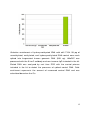
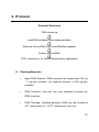
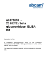
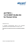
ab117134
EpiSeeker
Hydroxymethylated DNA
Immunoprecipitation
(hMeDIP) Kit
Instructions for Use
For selective enrichment of DNA fragments
containing 5-hydroxymethylcytosine in a high
throughput format using DNA isolated from
various species
This product is for research use only and is not
intended for diagnostic use.
1
Table of Contents
1.
Overview
3
2.
Background
7
3.
Components and Storage
10
4.
Protocol
12
5.
Troubleshooting
18
6.
Related Products
21
2
1. Overview
EpiSeeker hydroxymethylated DNA Immunoprecipitation (hMeDIP)
Kit contains all reagents required for carrying out a successful
hMeDIP procedure using DNA isolated from mammalian cells or
tissues. This kit includes a positive control DNA fragment, a negative
control non-immune IgG, and control primers that can be used with
the positive control to demonstrate the enrichment efficacy for
hydroxymethylated DNA with the kit reagents and protocol. The
positive control DNA containing 5-hmC can be immunoprecipitated
by a 5-hmC antibody but not by a non-immune IgG. In this hMeDIP,
immunoprecipitation of 5-hmC-enriched DNA fragments is processed
in a microplate under optimized reaction conditions, which enables
hMeDIP to be completed within 3 hours with high efficiency.
Immunoprecipitated hydroxymethylated DNA is then cleaned,
released, and eluted. Eluted DNA can be used for various
downstream
applications
including
PCR
(hMeDIP-PCR)
and
microarray (hMeDIP-chip).
3
Selective enrichment of hydroxymethyated DNA with ab117134. 50 pg of
unmethylated, methylated, and hydroxymethylated DNA control were each
spiked into fragmented human genomic DNA (500 ng). hMeDIP was
processed with the 5-hmC antibody and non-immune IgG included in the kit.
Eluted DNA was analyzed by real time PCR with the control primers
included in the kit to detect the presence of spiked control DNA. Foldenrichment represents the amount of recovered control DNA and was
calculated based on the Cts.
4
Sensitive detection of gene-specific hydroxymethylation by hMeDIP-QPCR.
Human brain DNA (500 ng) was fragmented to 200-600 bps with a
sonicator. The fragmented DNA was used for hydroxymethylated DNA
enrichment with the hMeDIP Kit. Eluted DNA was analyzed by real time
PCR with primers specifically for OCT4 or GAPDH sequences in the
promoter
regions.
Results
show
that
the
promoter
region
is
hydroxymethylated in OCT4 but not in GAPDH. Fold-enrichment represents
the amount of recovered DNA and was calculated based on the Cts.
ab117134 is suitable for selective enrichment of DNA fragments
containing 5-hydroxymethylcytosine in a high throughput format
using DNA isolated from various species. The hydroxymethylated
DNA that is enriched with this kit can be used for various
downstream
applications
including
PCR
(hMeDIP-PCR)
and
microarray (hMeDIP-chip).
5
The starting material should be good quality purified DNA. The
amount of DNA for each reaction can be 0.1 µg (approximately 1 x
4
10 cells) to 1 µg. For an optimal reaction, the input DNA amount
should be 0.5 µg per well.
Genomic DNA should be sheared by sonication before starting
hydroxymethylated DNA immunoprecipitation. The sheared DNA
fragments should range in size from 200-600 base pairs.
Negative (Non-Immune IgG) and positive controls (Control DNA) are
provided in this kit. The Control DNA is a 200 base pair DNA
fragment
containing
44
cytosine
residues
which
are
hydroxymethylated. The kit also includes control PCR primers that
can
be
used
for
verifying
the
enrichment
efficiency
of
hydroxymethylated control DNA.
The 5-hydroxymethylcytosine rabbit polyclonal antibody used in this
kit is highly specific against hydroxymethylated DNA fragments and
is not cross-reactive to methylated and unmethylated DNA
fragments.
To avoid cross-contamination, carefully pipette the sample or
solution into the strip wells. Use aerosol-barrier pipette tips and
always change pipette tips between liquid transfers. Wear gloves
throughout the entire procedure. In case of contact between gloves
and sample, change gloves immediately.
6
2. Background
DNA methylation occurs by the covalent addition of a methyl group
at the 5-carbon of the cytosine ring, resulting in 5-methylcytosine (5mC). In somatic cells, 5-mC is found almost exclusively in the
context of paired symmetrical methylation of the dinucleotide CpG,
whereas in embryonic stem (ES) cells, a substantial amount of 5-mC
is also observed in non-CpG contexts. The biological importance of
5-mC as a major epigenetic modification in phenotype and gene
expression has been widely recognized.
Quite recently, a novel modified nucleotide called 5-hydroxymethylcytosine (5-hmC) has been detected to be abundant in mouse brains
and embryonic stem cells. In mammals, it can be generated by the
oxidation of 5-methylcytosine, a reaction mediated by the Tet family
of enzymes and DNMT proteins. It is a hydroxylated and methylated
form of cytosine.
7
A line of evidence showed that 5-hmC also plays an important and
different role from 5-mC in regulation of DNA methylation, chromatin
remodeling, and gene expression, particularly in brain-specific gene
regulation. For example, it was shown that 5-hmC inhibits the
binding of the methyl-CpG binding domain proteins to DNA,
suggesting a potential gene regulatory function of 5-hmC. 5-hmC
was observed to be linked with epigenetic reprogramming in
mammalian zygotes. However, the exact functions of 5-hmC have
not yet been fully identified since gene-specific distribution of 5-hmC
is unknown due to the inability of currently used DNA methylation
analysis methods in distinguishing 5-hmC from 5-mC
Because of the presence of 5-hmC in DNA with unclear functions in
gene regulation and because of the discovery of enzymes that
produce 5-hmC, it is crucial to identify hydroxymethylation status in
specific gene loci, which would help to better understand
methylation-based epigenetic regulation of gene functions. To
achieve this, an innovative method has been developed to capture
DNA fragments containing 5-hmC, and this method has been
incorporated
into
the
EpiSeeker
hydroxymethylated
DNA
Immunoprecipitation (hMeDIP) Kit. This kit uses a high affinity 5hmC antibody to selectively capture double-stranded or single
stranded DNA fragments containing 5-hmC.
8
The kit has the following features:
•
Extremely fast and convenient protocol with a total
procedure
time
(from
input
sample
to
ready-to-use
hydroxymethylated DNA) of less than 3 hours, which
includes a minimal handling time of less than 20 minutes.
•
Flexible 96 strip well microplate format makes the assay very
easy to handle: manual method with one reaction at a time
or high throughput method with 96 reactions at a time.
•
Highly efficient enrichment ratio of positive/negative control
> 1000.
•
Low DNA input requirement of as low as 0.1 µg per reaction.
•
High
reproducibility
through
pre-optimized
hMeDIP
conditions.
•
Compatible with various downstream analysis workflows
including hMeDIP-PCR and hMeDIP-chip.
9
3. Components and Storage
A. Kit Components
Item
Quantity
(24 tests)
Quantity
(48 tests)
Quantity
(96 tests)
EMH1 (10X Wash Buffer)
5 mL
10 mL
20 mL
EMH2 (Antibody Buffer)
4 mL
8 mL
16 mL
EMH3 (hMeDIP Solution)
3 mL
6 mL
12 mL
EMH4 (DNA Release Buffer)
7 mL
14 mL
28 mL
EMH5 (Non-Immune IgG, 0.6 mg/ml)*
10 µL
20 µL
40 µL
EMH6 (5-hmC Antibody, 0.6 mg/ml)*
25 µL
50 µL
100 µL
EMH7 (Control DNA, 500 ng/ml)*
5 µL
10 µL
20 µL
EMH8 (Proteinase K, 10 mg/ml)*
28 µL
56 µL
112 µL
EMH9 (Control Primer-Forward, 20 µM)*
5 µL
10 µL
20 µL
EMH10 (Control Primer-Reverse, 20 µM)*
5 µL
10 µL
20 µL
8-Well Assay Strips (With Frame)
3
6
12
Adhesive 8-Well Strip Film
3
6
12
*Spin the solution down to the bottom prior to use.
10
B. Additional Materials Required
•
Variable temperature waterbath or incubator oven
•
Thermalcycler with 48- or 96-well block
•
Sonication device
•
Orbital shaker
•
Adjustable pipette and multiple-channel pipette
•
Aerosol resistant pipette tips
•
Parafilm M
•
0.2 ml or 0.5 ml PCR vials
C. Storage
•
Store EMH7 at –20°C away from light.
•
Store EMH1, EMH5, EMH6, EMH8, EMH9, EMH10, and
8-Well Assay Strips at 4°C away from light.
•
Store remaining components at room temperature away
from light.
•
All components of the kit are stable for 6 months from
the date of shipment, when stored properly.
Note: Check if EMH1 contains salt precipitates before
use. If so, briefly warm at room temperature or 37°C and
shake the buffer until salts are re-dissolved.
11
4. Protocol
Protocol Summary
DNA shearing
hmeDNA/antibody immunoprecipitation
Release of hmeDNA from hmeDNA/Ab complex
Elution of hmeDNA
PCR, microarray, or similar downstream application
A. Starting Materials
•
Input DNA Amount: DNA amount can range from 100 ng
- 1 µg per reaction. An optimal amount is 500 ng per
reaction.
•
DNA Isolation: You can use your method of choice for
DNA isolation.
•
DNA Storage: Isolated genomic DNA can be stored at
4°C (short term) or –20°C (long term) until use.
12
B. Preparation of 1x Wash Buffer
1. Prepare Diluted EMH1 1X Wash Buffer: Add 5 ml of
EMH1 10X Wash Buffer to 45 ml of distilled water
(pH 7.2-7.5). This Diluted EMH1 1X Wash Buffer can
now be stored at 4°C for up to six months.
C. Preparation of Antibody Coated Wells
1. Predetermine the number of strip wells required for your
experiment. Carefully remove un-needed strip wells from
the plate frame and place them back in the bag (seal the
bag tightly and store at 4°C)..
2. Add 100 µl of EMH2 to each well and then add the
following antibodies: 1 µl of EMH5 to the negative
control well, 1 µl of EMH6 to the sample wells, and 1 µl
of EMH6 to the positive control wells.
3. Cover the wells with parafilm M and incubate at room
temperature for 60 min. Meanwhile, prepare fragmented
DNA as described in the next step.
D. Shearing of Genomic DNA
1. For the best results, DNA should be fragmented by a
suitable sonication method:
13
a. Probe-based Sonication: You will need to
optimize the sonication settings. For example,
DNA of 200-1000 bp size can be obtained by
sonicating 3-4 pulses of 10-12 sec each at level
2 using a microtip probe, followed by a
30-40 sec rest period on ice between each
pulse.
b. Waterbath-based
Sonication:
Follow
the
manufactuerer’s user manual for DNA shearing
at a size range of 200-600 bp.
Note: If desired, remove 10 µl of sheared DNA for
purification and agarose gel analysis along with a DNA
marker on a 1-2% agarose gel, stained with ethidium
bromide. Visualize it under ultraviolet light.
E. Preparation of hMeDIP Reaction
1. Remove EMH2 from the wells and wash the wells two
times with 200 µl of Diluted EMH1 each time.
2. Dilute the EMH7 (Control DNA) to 50 ng/ml (50 pg/µl) by
adding 1 µl of EMH7 to 9 µl of EMH3 hMeDIP Solution
and dilute your sample DNA with EMH3 hMeDIP
Solution to 10 µg/ml (10 ng/µl). Setup the hMeDIP
reactions by adding the appropriate reagents to each
corresponding well according to the following chart.
14
Reagents
Sample
Positive
Negative
Negative
well
Control
Control
Control Well
Well
Well for
for Control
Sample
DNA
EMH3
50 µL
99 µL
50 µL
99 µL
Sample DNA
50 µL
N/A
50 µL
N/A
EMH7
0 µL
1 µL
0 µL
1 µL
Note:
(1) The final amount of each component should be 500 ng/well
for sample DNA and 50 pg/well for control DNA;
(2) An input DNA control is only used for estimating enrichment
efficiency of hMeDIP and is generally not needed as the included
positive and negative controls can be used for estimating the
same objective more accurately;
(3) If an input DNA control is to also be included, remove 5 µl of
the sonicated DNA solution prepared at Step D to a 0.5 ml vial,
label as “input DNA”, and place on ice.
3. Cover the wells with parafilm M and incubate at room
temperature for 90 min on an orbital shaker at 50100 rpm.
15
F. Wash of the Reaction Wells
1. Carefully remove and discard the solution containing the
reagents by pipetting out each well.
2. Thoroughly wash each well five times with 200 µl of the
Diluted EMH1 each time. This can be done by simply
pipetting Diluted EMH1 in and out of the wells.
3. Wash each well with 200 µl of EMH4 one time by
pipetting EMH4 in and out.
G. Release and Elution of DNA
1. Prepare EMH4-EMH8 Solution by adding 1 µl of EMH8
to every 39 µl of EMH4. Mix.
2. Add 40 µl of the EMH4-EMH8 Solution to each well.
3. Separate and insert the wells into a thermalcycler with a
48 well block.
4. Tightly seal the wells with Adhesive 8-Well Strip Film
and incubate at 60°C for 15 min, followed by incubation
at 95°C for 3 min.
16
Note: If only a thermalcycler with a 96 well block is available,
then:
(1) incubate the wells at 65°C for 20 min and quickly transfer
the DNA solution from each well to 0.2 ml strip PCR tubes.
Cap the PCR tubes and then incubate the PCR tubes
containing the DNA solution at 95°C for 3 min in the
thermalcycler;
(2) place the PCR tubes in room temperature. If liquid is
collected on the inside of the caps, briefly centrifuge the
liquid down to the bottom.
DNA is now ready for use or storage at –20°C.
For real time PCR analysis, we recommend using 1-2 µl of
eluted DNA in a 20 µl PCR reaction. Input DNA can be
added directly to a PCR reaction after appropriate dilution.
For end point PCR, the number of PCR cycles may need to
be optimized for better PCR results. In general, the
amplification difference between “EMH5” and “EMH6” may
vary from 3 to 8 cycles, depending on experimental
conditions.
For hMeDIP-chip, additional DNA cleanup/concentration and
whole genome amplification (WGA) steps may be needed.
17
5. Troubleshooting
Problem
Cause
Solution
Little or no
PCR products
generated from
samples.
Poor DNA quality due
to insufficient cell
amounts, extraction, or
degradation.
To obtain the best
results, the amount of
DNA per hMeDIP should
be 0.1-1 µg with 260/280
ratio >1.6.
Inappropriate DNA
fragmenting
conditions.
DNA fragment size
should be between 2001000 bp with an optimal
size range of 200-600
bp. Oversized DNA
fragments may reduce
targeted DNA capturing
via antibody and
undersized DNA
fragments may decrease
PCR efficiency.
Incorrect temperature
and/or insufficient time
during DNA release.
Ensure the incubation
time and temperature
described in the protocol
are followed correctly.
Improper PCR
program settings.
Ensure PCR program
settings are properly
programmed.
Inappropriate PCR
reaction solution.
If using a homemade
PCR reaction solution,
check if each
component is correctly
mixed. If using a PCR
Fast Kit, check if it is
suitable for your PCR.
Little or no
PCR products
generated from
samples.
18
Problem
Cause
Solution
Little or no
PCR products
generated from
samples.
Inappropriate primers.
Confirm the species
specificity of your
primers. Primers should
be designed to cover a
short sequence region
(70-150 bp) for more
efficient and exact
amplification of target
DNA regions.
Improper sample
storage.
DNA samples should be
stored at –20°C (3-6
months).
Insufficient washing of
wells.
Check if washing
recommendations at
each step is performed
according to the
protocol. If the signal
intensity in the negative
control is still high,
washing stringency can
be increased in the
following ways:
1. Increase wash time at
each wash step: after
adding Diluted EMH1,
leave it in the
tubes/wells for 2-3 min
before removing it.
2. Add an additional one
or two wash steps: The
volume of Diluted EMH1
is sufficient for at least
two extra washes for
each sample.
No difference
in signal
intensity
between
negative
control and
positive control
19
Problem
Cause
Solution
No difference
in signal
intensity
between
negative
control and
positive control
Too many PCR cycles.
Plateau phase of
amplification caused by
over-increased number
of PCR cycles in
endpoint PCR may
mask the difference in
signal intensity between
negative control and
positive control.
Decreasing the number
of PCR cycles (ex: 3235 cycles) to keep
amplification at
exponential phase will
reduce high background
in endpoint PCR and
allow differences in
amplification to be seen.
Real time PCR is
another alternative in
such cases.
Little or No
PCR products
generated from
positive control
only
PCR conditions are
not optimized.
Make sure the PCR
conditions include
correct and appropriate
temperature, cycles, and
solutions.
For further technical questions please do not hesitate to
contact us by email ([email protected]) or phone (select
“contact us” on www.abcam.com for the phone number for
your region).
20
6. Related Products
EpiSeeker methylated DNA Immunoprecipitation (MeDIP) Kit DNA (ab117133)
EpiSeeker methylated DNA Immunoprecipitation (MeDIP) Kit
(ab117135)
EpiSeeker methylated DNA Immunoprecipitation (MeDIP) Kit Tissue (ab117136)
EpiSeeker
hydroxymethylated
DNA
Quantification
Kit
DNA
Quantification
Kit
(Colorimetric) (ab117130)
EpiSeeker
hydroxymethylated
(Fluorometric) (ab117131)
21
22
UK, EU and ROW
Email: [email protected]
Tel: +44 (0)1223 696000
www.abcam.com
US, Canada and Latin America
Email: [email protected]
Tel: 888-77-ABCAM (22226)
www.abcam.com
China and Asia Pacific
Email: [email protected]
Tel: 108008523689 (中國聯通)
www.abcam.cn
Japan
Email: [email protected]
Tel: +81-(0)3-6231-0940
www.abcam.co.jp
Copyright © 2012 Abcam, All Rights Reserved. The Abcam logo is a registered trademark.
23
All information / detail is correct at time of going to print.