1
3430 Schmon Parkway
Thorold, ON, Canada L2V 4Y6
Phone: 866-667-4362 • (905) 227-8848
Fax: (905) 227-1061
Email: [email protected]
Novel Influenza A Virus (H1N1) RT-PCR Detection Kit
Product Insert
Product # 27900
INTENDED USE
Norgen’s Novel Influenza A Virus (H1N1) RT-PCR Detection Kit is used for the purification and
detection of the novel Influenza A virus (H1N1) of swine origin that caused a worldwide pandemic
(Phase 6, declared by WHO) in 2009. The kit contains two parts: (1) a novel Influenza A virus (H1N1)
RNA purification kit using Norgen’s proprietary spin column technology, and (2) a ready-to-use
detection kit for rapid and accurate detection of the novel Influenza A virus (H1N1) by end-point RTPCR.
The kit is designed for the rapid extraction and qualitative detection of the H1 Hemagglutinin viral RNA
transcript of the novel Influenza A virus (H1N1) in nasopharyngeal swabs, nasal swabs, throat swabs
and nasal aspirates from human patients with signs and symptoms of respiratory infection in
conjunction with clinical and epidemiological risk factors. This kit is intended to be used in clinical labs
with the ability to perform RT-PCR.
Negative results do not preclude influenza virus infection and should not be used as the sole basis for
treatment or other patient management decisions.
SUMMARY AND EXPLANATION
Influenza is caused by three immunologic types of RNA viruses (A, B and C) within the
Orthomyxoviridae family. Seasonal influenza is typically caused by three major subtypes of
hemaglutinin (H1, H2 and H3) and two subtypes of neuraminidase (N1 and N2). A novel sub-type of
influenza A virus called pandemic H1N1 2009 virus was identified in Mexico and reported by the CDC
and WHO in April, 2009 (Novel swine-origin influenza A (H1N1) virus investigation team, 2009; CDC,
2009-a; and Fraser et al., 2009). H1N1 2009 is a novel sub-type virus that transmits easily between
humans with 21 countries reporting cases within a month of initial identification (CDC, 2009-b). It is
essential that public health laboratories around the world undertake detailed surveillance to monitor the
spread and impact of pandemic H1N1 2009 virus as well as try to predict future changes in virulence
(Fraser et al., 2009). Methods for the rapid diagnosis, case identification and tracking of this novel
pathogen in the human population are therefore required to develop appropriate management
strategies to mitigate morbidity and mortality.
H1N1 2009 typically presents with a combination of upper and lower respiratory signs and symptoms,
including fever, headache, myalgia and general malaise. Illness severity can range from symptoms
which resemble the common cold to severe pneumonia requiring hospitalization. Persons at risk for
hospitalization include children under the age of two, adults aged 65 and older and persons with
significant co-morbidities. Duration of illness is 2-5 days, but symptoms may last for a week or longer.
Novel influenza viruses are first identified by amplification and sequencing of specific genes and these
sequences can be compared to those already deposited in databases. Such a methodological
approach is important for identification of new viruses but generally requires high viral loads (or
cultured virus) for good quality sequence data to be obtained. The turn-around-time required for this
type of analysis negates its use as a frontline diagnostic test, especially for large numbers of clinical
samples. Since sequences are now available, it is important that public health laboratories develop
and validate high throughput sensitive, specific and rapid assays for use in diagnostics and
surveillance.
As a rapid response to the outbreak of pandemic H1N1 2009, the Centers for Disease Control (CDC,
Atlanta, USA) provided detailed methodology for real-time PCR amplification and detection of this virus
(Novel swine-origin influenza A (H1N1) virus investigation team, 2009). Other conventional and realtime RT-PCR protocols have also been published recently for detection of this virus (Poon et al.,
2009). Norgen Biotek Corp. has developed a viral RNA extraction and RT-PCR assay for the
detection of pandemic H1N1 2009 virus using primers targeting the hemaglutinin (HA) gene from swab
samples. This detection kit will aid in the identification of new cases of pandemic H1N1 2009 virus and
ensure optimal management to minimize the transmission to vulnerable individuals as well as aid in
surveillance activities to understand the full impact of the virus in the community.
PRINCIPLES OF THE PROCEDURES
Norgen’s Novel Influenza A Virus (H1N1) RT-PCR Detection Kit contains two parts: (1) a novel
Influenza A virus (H1N1) RNA purification kit using Norgen’s proprietary spin column technology, and
(2) a ready-to-use detection kit for rapid and accurate detection of the novel Influenza A virus (H1N1)
by end-point RT-PCR. In the first part, the provided RNA purification kit allows the extraction of viral
RNA from the various acceptable respiratory specimens. Purification is based on spin column
chromatography using Norgen’s proprietary resin. The RNA isolated is of the highest quality and free
of other cellular components. An Isolation control is provided for spike-in isolation for the subsequent
determination of extraction efficiency. In the second part, the purified RNA is subjected to RT-PCR
using the provided ready-to-use RT-PCR detection kit. The RNA is directly added to the provided onestep RT-PCR master mix. The reaction involves first the reverse transcription of the viral RNA into
cDNA followed by PCR amplification of the H1-specific fragment using a thermo-stable polymerase. An
RT-PCR control is provided in all reactions for the determination of RT-PCR efficiency. In addition, a
detection control is provided as a positive control for detection. The RT-PCR detection kit is provided
with a molecular weight marker for the DNA agarose gel electrophoresis. The detection of the novel
Influenza H1N1 virus is determined by the presence of the H1 specific PCR product, and confirmed by
the presence of the Isolation Control product as well as the RT-PCR control product.
Kit Components:
Component
Contents
Lysis Solution
30 mL
Wash Solution (concentrate)
11 mL
Elution Buffer
2 mL
Mini Filter Spin Columns
24
Collection Tubes
24
Elution Tubes (1.7 mL)
24
H1N1 2X RT- PCR Master Mix
0.35 mL
a
1.4 mL
b
H1N1 PosC*
0.05 mL
Nuclease Free-Water
1.25 mL
Norgen’s DNA Marker
0.1 mL
Product Insert
1
H1N1 IsoC*
* IsoC = Isolation Control ; PosC= Positive Control
a
b
The positive control is an in vitro transcribed H1 RNA fragments.
The isolation control is an in vitro transcribed cloned RNA product.
Customer-Supplied Reagents and Equipment
• Benchtop microcentrifuge
• 95-100% ethanol
• Thermocycler and or Real-Time PCR System
• Micropipettes with an accuracy range between 1-10 µL, 10-100 µL and 100-1000 µL
• Laminar flow hood for extractions
• Vortex
• Sterile, nuclease-free aerosol-barrier micropipettor tips
• Microcentrifuge tube rack
• Disposable latex gloves
• β-mercaptoethanol
Storage Conditions and Product Stability
• The Positive Control (H1N1 PosC, blue cap) and Isolation Control (H1N1 IsoC, orange cap)
should be stored at -70°C. If needed, make aliquots of the controls according to the volume
used in the protocol (10 µL of H1N1 PosC or 20 µL of H1N1 IsoC) prior to freezing.
• The 2X RT-PCR Mastermix should be stored at -20°C. Make appropriate aliquots if needed
• All other kit components may be stored at room temperature
• The 2X RT-PCR Mastermix, Postive Control and Isolation Control should not undergo
repeated freeze-thaw (a maximum freeze-thaw of three times).
• Do not use kit or reagents past their expiration dates
• Allow reagents to thaw at room temperature prior to use
• After addition of RT-PCR master mix use within one hour
• Kit reagents are stable through the end of the expiration month indicated on the packaging
label when stored at the recommended temperatures.
General Precautions
• Follow universal precautions. All patient specimens should be considered as potentially
infectious and handled accordingly.
• Diagnostic laboratory work on clinical samples from patients who are suspected of having
H1N1 influenza virus infection should be conducted in a BSL2 laboratory. All sample
manipulations should be carried out in a biosafety cabinet. Viral isolation on clinical
specimens from patients who are suspected of having H1N1 influenza virus infection should
be performed in a BSL2 laboratory with BSL3 practices (H1N1 Influenza Virus Biosafety
Guidelines for Laboratory Workers: www.cdc.gov/h1n1flu/guidelines_labworkers.htm).
• Wear personal protective equipment, including gloves and lab coats when handling kit
reagents. Wash hands thoroughly when finished performing the test.
• Do not smoke, drink or eat in areas where kit reagents and/or human specimens are being
used.
• Dispose of unused kit reagents and human specimens according to local, provincial or federal
regulations.
• Workflow in the laboratory should proceed in a uni-directional manner, beginning in the preamplification area(s) (i.e. specimen collection and RNA extraction) and moving to the
amplification / detection area(s) (RT-PCR and gel electrophoresis).
• Do not use supplies and equipment across the dedicated areas of specimen extraction and
sample preparation. No cross-movement should be allowed between the different areas.
• Supplies and equipment used for specimen preparation should not be used for pipetting or
processing amplified DNA or other sources of target nucleic acids.
• All amplification supplies and equipment should be kept in the amplification / detection area at
all times.
• Personal protective equipment, such as laboratory coats and disposable gloves, should be
area specific.
• As contamination of patient specimens or reagents can produce erroneous results, it is
essential to use aseptic techniques.
• Pipette and handle reagents carefully to avoid mixing of the samples.
• Use proper pipetting techniques and maintain the same pipetting pattern throughout the
procedure to ensure optimal and reproducible values.
• Do not substitute or mix reagents from different kit lots or from other manufacturers.
• Do not interchange reagent tube / bottle caps as this may lead to contamination and
compromise test results.
• Only use the protocol provided in this insert. Alterations to the protocol and deviations from
the times and temperatures specified may lead to erroneous results.
Quality Control
In accordance with Norgen’s ISO 9001 and ISO 13485-certified Quality Management System, each lot
of Norgen’s Novel Influenza A Virus (H1N1) RT-PCR Detection Kit, including the H1N1 2x RT-PCR
Master Mix, H1N1 Isolation Control and H1N1 Positive Control are tested against predetermined
specifications to ensure consistent product quality.
Product Use Limitations
Norgen’s Novel Influenza A Virus (H1N1) RT-PCR Detection Kit is designed for research purposes
only. It is not intended for human or diagnostic use.
Product Warranty and Satisfaction Guarantee
NORGEN BIOTEK CORPORATION guarantees the performance of all products in the manner
described in our product manual. The customer must determine the suitability of the product for its
particular use.
Safety Information
Biosafety level 2 practices are recommended for works involving clinical samples from patients who
are suspected of having H1N1 influenza virus infection. Ensure the appropriate containment
equipment and facilities are used for activities involving cultures or potentially infectious clinical
materials. Ensure that a suitable lab coat, disposable gloves and protective goggles are worn when
working with chemicals. For more information, please consult the appropriate Material Safety Data
Sheets (MSDSs). These are available as convenient PDF files online at www.norgenbiotek.com.
CAUTION: DO NOT add bleach or acidic solutions directly to the sample-preparation waste.
The Lysis Solution contains guanidine salts, and should be handled with care. Guanidine salts form
highly reactive compounds when combined with bleach, thus care must be taken to properly dispose of
any of these solutions.
If liquid containing these buffers is spilt, clean with suitable laboratory detergent and water. If the spilt
liquid contains potentially infectious agents, clean the affected area first with laboratory detergent and
water, and then with 1% (v/v) sodium hypochlorite.
INSTRUCTIONS FOR USE
Notes Prior to Use
• All centrifugation steps are carried out in a benchtop microcentrifuge at 14,000 x g
(~ 14,000 RPM) except where noted. All centrifugation steps are performed at room
temperature.
• A variable speed centrifuge should be used for maximum kit performance. If a variable
speed centrifuge is not available a fixed speed centrifuge can be used, however reduced
yields may be observed.
• Ensure that all solutions are at room temperature prior to use.
• Prepare a working concentration of the Wash Solution by adding 25 mL of 95% ethanol
(provided by the user) to the supplied bottle containing the concentrated Wash Solution.
This will give a final volume of 36 mL. The label on the bottle has a box that may be
checked to indicate that the ethanol has been added.
• Add 10 µL of β -mercaptoethanol (provided by the user) to each 1 mL of Lysis Solution
required. β-mercaptoethanol is toxic and should be dispensed in a fume hood.
• It is important to work quickly during this procedure.
A. SPECIMEN COLLECTION AND LYSATE PREPARATION
Acceptable specimen types include nasal swabs, nasopharyngeal swabs, throat swabs or nasal
aspirates in viral transport medium tube or equivalent. If using swabs, use only sterile Dacron, nylon
or rayon swabs with plastic shafts. Note: Do not use calcium alginate swabs as they may contain
substances that are inhibitory to PCR.
This kit is also compatible with samples collected using Norgen’s Sample Collection Kit For Upper
Respiratory Tract Infectious Agents (Cat #29100). Please follow the instructions provided with that kit
for specimen collection and preservation.
i. Specimens previously collected using Norgen’s Sample Collection Kit For Upper Respiratory
Tract Infectious Agents (Cat #29100)
1) Transfer 300 µL of preserved specimen to an RNase-free microcentrifuge tube.
2) Add 300 µL of the Lysis Solution and vortex for 10 seconds to mix.
3) Add 20 µL of the Isolation Control (H1N1 IsoC) to the lysate. Vortex for 10 seconds to mix.
4) Add 300 µL of 95% ethanol to the lysate. Vortex for 10 seconds to mix.
5) Proceed to RNA Isolation (Step B).
ii. Specimen collection from swab in media:
1) If swab is transported in media the user should transfer 200 µL to an RNase-free
microcentrifuge tube.
2) Add 500 µL of the Lysis Solution and vortex for 10 seconds to mix.
3) Add 20 µL of the Isolation Control (H1N1 IsoC) to the lysate. Vortex for 10 seconds to mix.
4) Add 400 µL of 95% ethanol to the lysate. Vortex for 10 seconds to mix.
5) Proceed to RNA Isolation (Step B).
iii. Specimen collection directly from swab:
1) Alternatively, dry swabs can be placed directly into an RNase-free microcentrifuge tube
containing 1 mL of the Lysis Solution.
2) Using sterile techniques, cut the tip where the nasal or throat cells were collected and place
into microcentrifuge tube containing the Lysis Solution.
3) Close the tube and vortex for 1 minute to release the virus particles.
4) Using a sterile pipette transfer, 400 µL of the lysate into another RNase-free microcentrifuge
tube.
5) The extraction control (EC) (20 µL) should then be added to the Lysis tube and the tube
vortexed for 10 seconds to mix.
6) Add 200 µL of 95% ethanol to the lysate. Vortex for 10 seconds to mix.
7) Proceed to RNA Isolation (Step B).
B. SPECIMEN RNA PURIFICATION
Following the lysate preparation viral RNA can be extracted from the patient specimens using the
supplied buffers and solutions according to the following protocol:
1.
2.
Assemble a column with one of the provided collection tubes.
Apply the lysate with ethanol (up to 650 µL) to the column and centrifuge for 1 minute at
14,000 rpm.
Note: Ensure the entire lysate volume has passed through into the collection tube by
inspecting the column. If the entire lysate volume has not passed through, spin for an
additional minute.
3.
4.
5.
Discard the flowthrough and reassemble the spin column with its collection tube.
Depending on lysate volume, repeat steps B2 and B3.
Apply 400 µL of Wash solution and centrifuge for one minute at 14,000 rpm.
Note: Ensure the entire wash solution has passed through into the collection tube by
inspecting the column. If the entire wash volume has not passed through, spin for an
additional minute.
6.
7.
8.
Discard the flowthrough and reassemble the spin column with its collection tube.
Repeat steps B5 and B6 two more times.
Spin the column for 2 minutes to thoroughly dry the resin at 14,000 rpm. Discard the
collection tube.
9. Place the column into a new 1.7 mL Elution tube.
10. Add 50 µL of Elution Solution to the column.
11. Centrifuge for 2 minutes at 2,000 rpm followed by a 2 minute spin at 14,000 rpm. Note the
volume eluted from the column. If the entire 50 µL has not been eluted, spin the column for
an additional minute at 14,000 rpm.
12. The purified RNA sample could be used immediately for RT-PCR as described below. It is
recommended that samples be placed at -70°C for long term storage.
C. H1N1 RT-PCR Assay Preparation
Notes:
• Before use, suitable amounts of all RT-PCR components should be completely thawed at room
temperature, vortexed and centrifuged briefly.
• The amount of H1N1 2X RT-PCR Master Mix provided is enough for up to 32 RT-PCR reactions (24
sample PCR, 4 positive control PCR and 4 no template control PCR).
• For every PCR run, one reaction containing H1N1 Positive Control (H1N1 PosC) and one reaction
as no template control must be included for proper interpretation of results.
• The recommended minimum number of RNA samples tested per PCR run is 6.
1. Prepare the RT-PCR for sample detection as shown in Table 1 below. The recommended amount of
sample RNA to be used is 5 µL. However, a volume between 1 and 10 µL of sample RNA may be
used as template. Adjust the final volume of the RT-PCR reaction to 20 µL using the Nuclease-Free
Water provided.
Table 1. RT-PCR Assay Preparation
RT-PCR Components
Volume Per RT-PCR Reaction
H1N1 2X RT-PCR Master Mix
10 µL
Sample RNA
1 to 10 µL
Nuclease-Free Water
Up to 10 µL
Total Volume
20 µL
2. For every RT-PCR run, prepare one positive control RT-PCR as shown in Table 2 below:
Table 2. RT-PCR Positive Control Preparation
RT-PCR Components
Volume Per RT-PCR Reaction
H1N1 2X RT-PCR Master Mix
10 µL
H1N1 PosC
10 µL
Total Volume
20 µL
3. For every RT-PCR run, prepare one no template control RT-PCR as shown in Table 3 below:
Table 3. RT-PCR Negative Control Preparation
PCR Components
Volume Per PCR Reaction
H1N1 2X RT-PCR Master Mix
10 µL
Nuclease-Free Water
10 µL
Total Volume
20 µL
D. H1N1 RT-PCR Assay Programming
1. Program the thermocycler according to the program shown in Table 4 below.
2. Run RT-PCR.
Table 4. H1N1 Assay Program
PCR Cycle
Step
Temperature
Duration
Cycle 1
Step 1
50oC
25 min
Cycle 2
Cycle 3 (40x)
o
Step 1
95 C
5 min
Step 1
94oC
15 sec
o
Step 2
64 C
30 sec
Step 3
72oC
45 sec
o
Cycle 4
Step 1
72 C
5 min
Cycle 5
Step 1
4o C
∞
E. H1N1 RT-PCR Assay Results Interpretation
1. For the analysis of the PCR data, the entire 20 µL PCR reaction should be loaded on a 1X TAE
2 % Agarose DNA gel along with 10 µL of Norgen’s DNA Marker (provided).
2. The PCR products should be resolved on the 1X TAE 2 % Agarose gel at 150V for 30 minutes.
3. Sample results are provided below:
M
A
B
C
D
2000
1500
1000
750
500
H1N1 IsoC
300
H1N1 Target
150
H1N1 PCRC
50
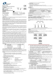
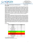
Figure 1: A representative 1X TAE 2% agarose gel showing the amplification of H1N1 under different
conditions (H1N1 Target). The size of the H1N1 target amplicon corresponds to 222bp as represented by
the provided DNA Marker (M). The size of the H1N1 Isolation Control (H1N1 IsoC) corresponds to 499bp as
represented by the provided DNA Marker (M). The H1N1 2X RT-PCR Master Mix contains an H1N1 RTPCR Control (H1N1 PCRC). The H1N1 PCRC Controls for PCR inhibition. The size of the H1N1 PCRC
corresponds to 142bp as represented by the provided DNA Marker (M). The amplification from each lane is
interpreted as follows:
Lane A: Positive Control or H1N1 Detected – All three RT-PCR amplicons were detected
Lane B: No Template Control – Only H1N1 PCRC was detected
Lane C: H1N1 Detected – Lane C showed detection of both the H1N1 Target and H1N1 PCRC. The
result is considered positive detection of H1N1.
Lane D: H1N1 Not Detected – Detection of H1N1 IsoC and H1N1 PCRC, suggesting that the RNA
isolation was successful but no H1N1 RNA was present in the sample
Table 5. Interpretation of RT-PCR Assay Results
Input Type
H1N1 IsoC
Band (499 bp)
H1N1 Target
Band (222 bp)
H1N1 PCRC
Band (142 bp)
Interpretation
Positive Control
X
X
X
Valid
X
Valid
X
Positive
X
Negative
X
Positive
Negative Control
Sample
X
Sample
X
Sample
X
X
Sample
X
Sample
X
Positive
X
Positive
** For results obtained that are not covered in Table 5 above, please refer to the Troubleshooting
Section.
F. H1N1 RT-PCR Assay Specificity
•
The specificity of Norgen’s Novel Influenza A Virus (H1N1) RT-PCR Detection Kit is first and
foremost ensured by the selection of the H1-specific primers, as well as the selection of
stringent reaction conditions. The primers were checked for possible homologies to all in
GenBank published sequences by sequence comparison analysis. The specific detectability of
all relevant strains has thus been ensured by a database alignment and by PCR amplification
with the following commonly found seasonal flu viruses:
-
H1N1
H3N2
Avian H5N1
Influenza B
G. H1N1 RT-PCR Assay Specificity
•
•
•
The linear range (analytical measurement) of Norgen’s Novel Influenza A Virus (H1N1) RTPCR Detection Kit was determined by analysing a dilution series of a H1N1 quantification
standard ranging from 1 x 10⁷ copies/µl to 1 x 10⁻¹ copies/µl.
Each dilution has been tested in replicates (n = 4) using Norgen’s Novel Influenza A Virus
(H1N1) RT-PCR Detection Kit on 1X TAE 2% Agarose gel.
The linear range of Norgen’s Novel Influenza A Virus (H1N1) RT-PCR Detection Kit has been
determined to cover concentrations from 5 x 10⁴ copies/mL to at least 1 x 10⁹ copies/mL
Frequently Asked Questions
1. How many samples should be included per RT-PCR run?
• Norgen’s Novel Influenza A Virus (H1N1) RT-PCR Detection Kit is designed to test 24 samples.
For every 6 samples, a non-template control and a Positive Control must be included. It is
preferable to pool and test 6 samples at a time.
2.
How can I interpret my results if neither the H1N1 PCR control nor the H1N1 Isolation
Control amplifies?
• If neither the H1N1 PCR control nor the H1N1 Isolation Control amplifies, the sample must be retested. If the positive control showed amplification, then the problem occurred during the isolation,
where as if the Positive control did not amplify, therefore the Problem has occurred during the
setup of the PCR assay reaction.
3. How should it be interpreted if only the H1N1 PCR control showed amplification but neither
the H1N1 target nor the H1N1 Isolation control amplified for a sample?
• This indicates a poor isolation. The isolation procedure must be repeated.
4. How should it be interpreted if only the H1N1 Isolation Control was amplified in a sample?
• The sample tested can be considered as H1N1 negative.
5. How should it be interpreted if only the H1N1 target and the H1N1 PCR control were amplified
in a sample?
• The sample tested can be considered as H1N1 positive.
6. How should it be interpreted if only the H1N1 target was amplified in a sample?
• The sample tested should be considered as H1N1 positive. At high H1N1 copies input, the H1N1
amplicon will be predominant and thus the H1N1 PCR control as well as the H1N1 Isolation
control may not amplify as they compete for PCR resources.
7. How should it be interpreted if only the H1N1 PCR control and the H1N1 Isolation control
showed amplification in a sample?
• The sample tested can be considered negative
8. What If I forgot to do a dry spin after my second wash?
• Your RNA elution will be contaminated with the Wash Solution. This may dilute the RNA yield in
your elution and it may interfere with the RT-PCR detection, as ethanol is known to be a PCR
inhibitor.
9. What If I forgot to add the H1N1 Isolation control during the Isolation?
• It is recommended that the isolation is repeated.
Reference
Centers for Disease Control. 2009-a. Outbreak of swine-origin influenza A (H1N1) virus infection
– Mexico, March-April 2009. Morb. Mortal. Wkly. Rep. 58: 467-70.
Centers for Disease Control. 2009-b. Update: novel influenza A (H1N1) virus infections –
worldwide, May 6, 2009. Morb. Mortal. Wkly. Rep. 58: 453-8.
Fraser, C., C. A. Donnelly, S. Cauchemez, W. P. Hanage, K. Van, T. D. Hollingsworth, J. Griffin,
R. F. Baggaley, H. E. Jenkins, E. J. Lyons, T. Ferguson, A. Rambaut, O. G. Pybus, H. LopezGatell, C. M. pluche-Aranda, I. B. Chapela, E. P. Zavala, D. M. Guevara, F. Checchi, E. Garcia,
S. Hugonnet and C. Roth. 2009. Pandemic potential of a strain of Influenza A (H1N1): Early
Findings. Science. 324: 1557-61.
Novel swine-origin influenza A (H1N1) virus investigation team. 2009. Emergence of a novel
swine-origin influenza A (H1N1) virus in humans. N. Engl. J. Med. 360: 2605-15.
Poon, L. L., K. H. Chan, G. J. Smith, C. S. Leung, Y. Guan, K. Y. Yuen and J. S. Peiris. 2009.
Molecular detection of a novel human influenza (H1N1) of pandemic potential by conventional
and real-time quantitative RT-PCR assays. Clin. Chem. 55: in press.
Technical Assistance
NORGEN’s Technical Service Department is staffed by experienced scientists with extensive practical
and theoretical expertise in sample and assay technologies and the use of NORGEN products. If you
have any questions or experience any difficulties regarding Norgen’s Urine DNA Isolation Mini Kit
(Slurry Format) or NORGEN products in general, please do not hesitate to contact us.
NORGEN customers are a valuable source of information regarding advanced or specialized uses of
our products. This information is helpful to other scientists as well as to the researchers at NORGEN.
We therefore encourage you to contact us if you have any suggestions about product performance or
new applications and techniques.
For technical assistance and more information, please contact our Technical Support Team between
the hours of 8:30 and 5:30 (Eastern Standard Time) at (905) 227-8848 or Toll Free at 1-866-667-4362.
or call one of the NORGEN local distributors (www.norgenbiotek.com) or through email at
[email protected].
3430 Schmon Parkway, Thorold, ON Canada L2V 4Y6
Phone: (905) 227-8848
Fax: (905) 227-1061
Toll Free in North America: 1-866-667-4362
©2010 Norgen Biotek Corp.
PI27900-3