1
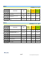
USER MANUAL
Genequality
AZF MX
ref. 04-23C
ref. 04-23A
ref. 04-23R
Kit for the identification of
deletions in AZF locus
Compliant to the European Guidelines
“EAA/EQMN best practice guidelines for molecular diagnosis
of Y-chromosomal microdeletions. State of the art 2004”
Simoni M., Bakker E., Krausz C. Int. J. Andrology, 2004, 27: 240-249.
04-23A-25(8033622781004)-EN.doc
1 PRODUCT INFORMATION
3
2 KIT CONTENT
4
3 STORAGE AND STABILITY OF THE REAGENTS
6
4 PRECAUTIONS FOR USE
6
5 SAFETY RULES
7
5.1
General safety rules
7
5.2
Safety rules about the kit
8
6 MATERIALS REQUIRED, BUT NOT PROVIDED
10
6.1
Reagents
10
6.2
Instruments
10
6.3
Materials
10
7 PREPARATION OF THE REAGENTS
11
8 INTRODUCTION
12
9 TEST PRINCIPLE
15
10 PRODUCT DESCRIPTION
16
11 COLLECTION, MANIPULATION AND PRE-TREATMENT OF SAMPLE 17
12 PROCEDURE
17
12.1 DNA EXTRACTION
17
12.1.1 DNA extraction from fresh or frozen whole blood
18
12.2 DNA AMPLIFICATION
19
12.3 VISUALIZATION OF THE AMPLIFICATION PRODUCTS
20
12.3.1 High Resolution agarose gel electrophoresis
20
12.3.2 Sample loading
21
13 INTERPRETATION OF THE RESULTS
22
14 TROUBLESHOOTING
24
15 DEVICE LIMITS
27
pag.1
04-23A-25(8033622781004)-EN.doc
16 DEVICE PERFORMANCES
27
16.1 Specificity
27
16.2 Diagnostic sensitivity
27
17 BIBLIOGRAPHIC REFERENCES
28
pag.2
04-23A-25(8033622781004)-EN.doc
1 PRODUCT INFORMATION
This user manual describes the instructions for use of the following products:
AZF-MX COMPLETE (cod 04-23C)
Complete system for the identification of deletions in the AZF locus,
involved in male infertility.
The kit includes all the reagents for DNA extraction, amplification and
visualization by agarose gel electrophoresis, the internal control of sample
amplificability and the reference DNA.
Code
04-23C-12
04-23C-24
Product
AZF-MX COMPLETE
AZF-MX COMPLETE
Pkg
12 test
24 test
AZF-MX (cod 04-23A)
Kit for the identification of deletions in the AZF locus, involved in male
infertility.
The kit includes the reagents for amplification and visualization by agarose
gel electrophoresis, the internal control of sample amplificability and the
reference DNA.
Code
04-23A-12
04-23A-24
Product
AZF-MX
AZF-MX
Pkg
12 test
24 test
AZF-MX – amplification reagents
(cod 04-23R)
Kit for the identification of deletions in the AZF locus, involved in male
infertility.
The kit includes the reagents for amplification, the internal control of sample
amplificability and the reference DNA.
Code
04-23R-12
04-23R-24
Product
AZF-MX – amplification reagents
AZF-MX - amplification reagents
Pkg
12 test
24 test
pag.3
04-23A-25(8033622781004)-EN.doc
2 KIT CONTENT
NOTE:
In the kits with different codes (C,A or R) different components are included.
(legenda: X = component included in the kit; 0 = component not included in
the kit)
BOX P
cod.
04-23C
cod.
04-23A
cod.
04-23R
STORE AT – 20°C
DESCRIPTION
LABEL
TUBE (T)
OR LID
COLOUR
12 test
24 test
X
X
X
Single-dose premix tubes
for Multiplex I.
colourless
(T)
12
24
X
X
X
Single-dose premix tubes
for Multiplex II.
Blue (T)
12
24
X
X
X
Single-dose premix tubes
for Multiplex III.
Yellow (T)
12
24
X
X
X
Thermostabile Hot-start
Taq DNA polimerase
Super AB Taq
5 U/μL
Red
1X 25 μL
1X 50 μL
X
0
0
Protease
Proteasi
Green
3X 100 μL
6X 100 μL
SMALL BAG
cod.
04-23C
cod.
04-23A
cod.
04-23R
STORE AT – 20°C
X
X
X
DESCRIPTION
Reference DNA
(from an undeleted male)
LABEL
TUBE (T)
OR LID
COLOUR
12 test
24 test
Reference DNA
(undeleted
male)
Blue
1X 20 μL
1X 40 μL
pag.4
04-23A-25(8033622781004)-EN.doc
BOX F
cod.
04-23C
cod.
04-23A
cod.
04-23R
STORE AT +2°/ +8°C
X
X
DESCRIPTION
0
Electrophoresis loading buffer
(6X solution)
X
X
0
Ethidium Bromide solution
(2,5 mg/mL)
X
X
0
DNA Molecular Weight Marker
(MW)
LABEL
TUBE (T)
OR LID
COLOUR
12 test
24 test
Bromophenol
blue
Blue
1X 200 μL
1X 350μL
Red
1X 100 μL
1X 180 μL
Yellow
1X 100 μL
1X 180 μL
Ethidium
Bromide
TOXIC
R 23 68
S 36/37 45
MW Marker
BOX A
cod.
04-23C
cod.
04-23A
cod.
04-23R
STORE AT +15°/ +25°C
DESCRIPTION
LABEL
Agarose molecular biology grade
TUBE (T)
OR LID
COLOUR
12 test
24 test
AGAROSE
10 g
20 g
50 X TAE
40 mL
70 mL
X
X
X
X
X
X
Electrophoresis buffer
TRIS-Acetate -EDTA pH 8,0
X
0
0
Cell Lysing Solution
SOLUTION 1
1X 3 mL
1X 6 mL
X
0
0
Washing Solution
SOLUTION 2
1X 11 mL
1X 22 mL
X
0
0
Washing Solution
SOLUTION 3
1X 10,5 mL
1X 21 mL
X
0
0
Eluting Solution
Eluting
Solution
1X 3 mL
1X 6 mL
X
0
0
Filter columns
12
24
X
0
0
2 mL tubes
24
48
pag.5
04-23A-25(8033622781004)-EN.doc
3 STORAGE AND STABILITY OF THE REAGENTS
Each component of the kit should be stored according to the directions
indicated on the label of the single boxes.
In particular:
Box P
Small bag
Box F
Box A
store at -20°C
store at -20°C
store at +2/+8°C
store at +15/+25°C
(room temperature)
When stored at the recommended temperature, all test reagents are stable
until their expiration date, indicated on the labels.
4 PRECAUTIONS FOR USE
• The kit should be handled by investigator qualified through education and
training in molecular biology techniques applied to diagnostics.
• Before starting the kit procedure, read carefully and completely the
instruction manual.
• Keep the product out of heating sources;
• Do not use any part of the kit if over the expiration date;
• In case of any doubt about the storage conditions, box integrity or method
application,
contact
AB
ANALITICA
technical
support
at:
[email protected] before using the kit.
In the amplification of nucleic acids, the investigator has to take the following
special precautions:
• Use filter-tips;
pag.6
04-23A-25(8033622781004)-EN.doc
• Store the biologic samples, the purified DNA, the reference DNA included
in the kit and all the amplification products in different places from where
amplification reagents are stored.
• Organise the space in different pre- and post-PCR units; do not share
consumables (pipets, tips, tubes,…) between them.
• Change frequently the gloves;
• Wash the bench surfaces with 5% sodium hypochloride;
• Store the extracted DNA at +2° / +8°C for short term, or freeze them at
-20°C for long term storage.
• Thaw the PCR premixes at room temperature before use. Add Taq DNA
polymerase and purified DNA very quickly at room temperature, better if in
an ice-bath.
5 SAFETY RULES
5.1 General safety rules
• Wear disposable gloves to handle reagents and clinical samples, wash
your hands at the end of work.
• Do not pipet with mouth.
• Since no known diagnostic method can assure the absence of infective
agents, it is a good rule to consider every clinical sample as potentially
infectious and handle it as such.
• All the devices that get directly in touch with clinical samples should be
considered as contaminated and disposed as such. In case of accidental
spilling of the samples, clean up with 10% Sodium Hypochloride. The
materials used to clean up should be disposed in special containers for
contaminated products
• Clinical samples, materials and contaminated products should be disposed
after decontamination by:
pag.7
04-23A-25(8033622781004)-EN.doc
immersion in a solution of 5% Sodium Hypochloride (1 volume of
Sodium Hypochloride solution every 10 volumes of contaminated fluid)
for 30 minutes
OR
autoclaving at 121°C at least for 2 hours (NOTE: do not autoclave
solutions containing Sodium Hypochloride!!)
5.2 Safety rules about the kit
The risks for the use of this kit are related to the single components:
Dangerous components:
ETHIDIUM BROMIDE (included in 04-23C and 04-23A)
3,8-diamino-1-ethyl-6-phenylphenantridiumbromide (Ethidium Bromide) <2%
Description of risk:
T (Toxic)
RISK SENTENCES AND S SENTENCES
R 23 and R 68
S 36/37 45
Toxic for inhalation.
Risk of irreversible effects.
Wear laboratory coat and disposable gloves.
In case of accident or discomfort, seek for medical
assistance and show the container or label.
R and S sentences refer to the concentrated product, as provided in the kit.
In particular for Ethidium Bromide, until the dilution in the agarose gel.
In manipulating concentrated Ethidium Bromide, use a chemical dispensing
fume cabinet. Always wear disposable gloves and laboratory coat in
manipulating the diluted Ethidium solution as well.
The product can not be disposed with the common waste. It must not reach
the drainer system. For the disposal, follow the local law.
In case of accidental spilling of Ethidium Bromide, clean with Sodium
hypochloride and water.
pag.8
04-23A-25(8033622781004)-EN.doc
SOLUTION 1 and SOLUTION 2 (included in 04-23C)
Solution 1 and 2 contain guanidinium hydrochloride
Description of risk:
Harmful and irritating
RISK SENTENCES AND S SENTENCES
R 22-36
and R 38
S 13-26-36-46
Toxic if swallowed. Irritating for skin and eyes.
Store away of food and beverage. In case of
contact with eyes, rinse immediately with plenty of
water and seek medical advise. Use protective
cloth and suitable gloves.
In case of accident or discomfort, seek for medical
advice immediately and show the container or
label.
PROTEASE (included in 04-23C)
Description of risk:
May cause sensitization, irritating.
RISK SENTENCES AND S SENTENCES
R 37
and R 38-41-42
S 24-26-36
S 37; S 46
Toxic if swallowed. Irritating for skin and eyes.
Avoid contact with skin and eyes. In case of
contact with eyes, rinse immediately with plenty of
water and seek medical advise. Use protective
cloth and suitable gloves.
In case of accident or discomfort, seek for medical
advice immediately and show the container or
label.
Safety data sheet (MSDS) of the kit is available upon request.
pag.9
04-23A-25(8033622781004)-EN.doc
6 MATERIALS REQUIRED, BUT NOT PROVIDED
6.1 Reagents
•
•
•
•
•
Reagents for DNA extraction (necessary for cod. 04-23A and 04-23R)
Sterile DNase and RNase free water;
Distilled water;
96% – 100% Ethanol (necessary for the kit cod. 04-23C)
Reagents for agarose gel electrophoresis (necessary for cod. 04-23R)
6.2 Instruments
• Laminar flow cabinet (use is recommended while adding TAQ polymerase
to the amplification premix to avoid contamination; it would be
recommended to use another laminar flow cabinet to add the extracted
DNA);
• Micropipettes (range: 0,2-2 µL; 0,5-10 µL; 2-20 µL; 20-200 µL; 100-1000
µL);
• Thermal cycler;
• Thermoblock or thermal bath;
• Microcentrifuge (max 12-14.000 rpm);
• Balance;
• Vortex;
• Magnetic heating stirrer or microwave.
• Chemical cabinet (its use is recommended in handling Ethidium Bromide);
• Horizontal electrophoresis chamber for agarose minigel;
• Power supply (50-150 V);
• UV Transilluminator;
• Photo camera or image analyzer.
6.3 Materials
• Disposable gloves;
• Disposable sterile filter-tips (range: 0,2-2 µL; 0,5-10 µL; 2-20 µL; 20-200
µL; 100-1000 µL)
• Graduate cylinders (1 L) for of TAE dilution;
• Pyrex bottle or Becker for agarose gel preparation;
• Parafilm.
• Microtubes (1,5 – 2,0 mL)
pag.10
04-23A-25(8033622781004)-EN.doc
7 PREPARATION OF THE REAGENTS
Preparation of 1 L of 1X TAE buffer:
Mix 20 mL of 50X TAE with 980 mL of distilled water.
pag.11
04-23A-25(8033622781004)-EN.doc
8 INTRODUCTION
Nowadays, the analysis of microdeletions of Y chromosome is considered an
essential diagnostic approach to study male infertility.
Recent studies have divided the Y chromosome long arm into three regions,
called AZF (Azoospermia Factors), frequently found to be deleted in some
azoospermatic and serious oligozoospermatic subjects.
These loci (AZFa, AZFb e AZFc) contain genes that control the correct
course of spermatogenesis (Fig.1) Alterations in one or more AZF loci cause
a drastic reduction of germinal cells up to their complete absence.
Several studies marked the relation between the deletions and the presence
of repeated DNA sequences showing that a recombinational event between
the two proviral sequences is the basis of AZFa deletion (Kamp et al., 2000;
Sun et al., 2000).
AZFc deletions are generally more uniform and follow the recombination
between blocks of repeated DNA sequences flanking the region. (KurodaKawaguchi et al., 2001).
A diagnosis of Y-related infertility is suspected in males with serious
Azoospermia or Oligozoospermia, associated with anomalous morphology
and/or spermatic motility, in absence of other known causes. In 5 to 10% of
these subjects microdeletions of Y chromosome are identified by molecular
analysis.
To evaluate the integrity of Y euchromatic region, at least four genes should
be investigated: USP9Y (or DFFRY), DBY, RBMY and DAZ. These genes
are currently considered to be directly or indirectly involved in male fertility.
USP9Y is a single copy gene located in AZFa region, it has its homologous
on the X chromosome and it is expressed ubiquitously. (Brown et al., 1998).
DBY, located in AZFa region, is a single copy gene with an homologous on
the X chromosome as well, but it gives a specific transcript at the testicular
level (Lahn & Page, 1997).
RBMY is a multicopy gene mainly located in AZFb region and expressed
exclusively at testicular level (Ma et al, 1993).
DAZ is a gene located in AZFc region in four copies arranged in two clusters
(Saxena et al., 2000).
Recently, PCR amplification of short DNA sequences (STS) within AZF loci
has been recognised as the best method to detect microdeletions in these
regions.
pag.12
04-23A-25(8033622781004)-EN.doc
Fig.1:
SCHEMATIC VISUALIZATION OF Y
CHROMOSOME AND AZF LOCI
Markers
for
the
detection
of
microdeletions in AZF locus.
SRY
ZFY
AZFa
sY86
sY84
DFFRY, in USP9Y gene
DBY, in DEAD BOX Y gene
sY95
AZFb
AZFc
sY117
sY125
sY127
sY134
sY254, in DAZ gene
sY255, in DAZ gene
Published data demonstrated that the routinely performed diagnostic
protocols were very different and often leading to inaccurate or incomplete
diagnosis. The need of standardization was highlighted in a preliminary
publication of the European Guidelines that described the main
characteristics of the amplification reaction and the markers required to have
a complete and reliable diagnosis (Simoni et al., 1999).
Four years after this preliminary publication, the complete sequencing of Y
chromosome (Skaletsky et al., 2003) and the knowledge about the molecular
mechanisms of deletions (Kampo et al., 2000; Sun et al., 2000; Repping et
al., 2002) the new European Guidelines were published (Simoni et. Al.,
2004).
The European Guidelines give precise indications for the selection of the
patients to be screened. The authors suggest.
•
•
•
A multiplex PCR amplification of genomic DNA must be performed.
The amplification of the ZFX/ZFY gene is an appropriate internal PCR
control because the primers amplify a unique fragment both in male and
female DNA, respectively.
The basic characteristics of the selected markers are: Y-specificity,
absence of other homologous sequences, non-polymorphic.
pag.13
04-23A-25(8033622781004)-EN.doc
•
At least two loci in each AZF region should be analysed:
for AZFa: sY84, sY86
for AZFb: sY127, sY134
for AZFc: sY254, sY255
•
The SRY gene should be included in the analysis as a control for the
testis-determining factor on the short arm of the Y chromosome and for
the presence of Y-specific sequences when the ZFY gene is absent (e.g.
in XX males).
pag.14
04-23A-25(8033622781004)-EN.doc
9 TEST PRINCIPLE
PCR method (Polymerase Chain Reaction) has been the first method of DNA
amplification described in literature. (Saiki RK et al., 1985). It can be defined
as an in vitro amplification reaction of a specific part of DNA (target
sequence) by a thermo-stable DNA polymerase.
Three nucleic acid segments are involved in the reaction: double stranded
DNA template to be amplified (target DNA) and two single-stranded
oligonucleotides “primers” that are designed in order to anneal specifically to
the template DNA.
The DNA polymerase begins the synthesis process at the region marked by
the primers and synthesizes new double stranded DNA molecules, identical
to the original double stranded target DNA region, by facilitating the binding
and joining of the complementary nucleotides that are free in solution
(dNTPs). After several cycles, one can get millions of DNA molecules which
correspond to the target sequence.
The sensitivity of this test makes it particularly suitable for the application in
laboratory diagnostics.
The multiplex amplification allows the simultaneous amplification of different
DNA sequences in the same reaction, by mean of a selected primers mix.
pag.15
04-23A-25(8033622781004)-EN.doc
10 PRODUCT DESCRIPTION
The amplification of short DNA sequences (STS) in AZF loci is the best
method to verify the presence of microdeletions in Y chromosome.
In particular, the strategy of the proposed method consists of the amplification
of eleven markers by mean of 3 multiplex PCR.
AZF MX method is compliant to the indications of the recently proposed
European Guidelines for diagnostic testing of Y-chromosomal microdeletions
(Simoni M et al., 2004) that define the basic characteristics of the primer set
and of the amplification protocol to enable the detection of almost all the
clinically relevant deletions.
The amplification of the internal control (in the ZFX/ZFY gene) allows to verify
the good quality of the extracted DNA and, at the same time, the absence of
amplification inhibitors, avoiding false positive results.
The kit includes reference DNA from a non-deleted male. The amplification of
the reference DNA (with the presence of all the expected bands) is a
guarantee of a correct course of the reaction.
The reference DNA is of human origin but it is not dangerous for the operator.
The kit is in premix format: all the reagents for the amplification are pre-mixed
and aliquoted in monodose test tubes to which Taq polymerase and the
extracted DNA will be added.
This premix format allows the reduction of the manipulation in preamplification steps, with a considerable time saving for the operator, the
repeated freezing/thawing of reagents (that could alter the product
performances) is avoided and, above all, this form reduces at minimum the
risk of sample contamination and the risk to get false positive results.
Nevertheless, it is always recommended to use all the proper amplification
controls.
pag.16
04-23A-25(8033622781004)-EN.doc
11 COLLECTION, MANIPULATION AND PRETREATMENT OF SAMPLE
The procedure for the analysis of microdeletions of Y chromosome starts
from the collection of whole blood samples.
The sample collection should follow all the usual sterility precautions.
Blood should be treated with EDTA. Other coagulating agents, as heparin,
are strong inhibitors of TAQ polymerase and so they could alter the efficiency
of the amplification reaction.
Fresh blood can be stored at +2 / +8°C for short time; if DNA is not extracted
shortly, it is necessary to freeze the sample.
12 PROCEDURE
12.1 DNA EXTRACTION
AB ANALITICA suggests the use of AZF MX COMPLETE kit (cod. 04-23C)
that includes the extraction system, with which the method has been
standardized.
In any case, any DNA extraction method can be used, provided that allows
the extraction of pure and integral DNA.
Extracted DNA should be quantified by spectrophotometric measurement.
The amount of DNA to be used in the amplification is 180 ng, therefore, the
purified DNA solution should be diluted taking into account that the available
volume in each premix tube is 6 μL.
For any problem in method application you can contact AB ANALITICA
technical support at: [email protected] .
The extraction method below is referred to the kit AZF MX COMPLETE (cod
04-23C).
pag.17
04-23A-25(8033622781004)-EN.doc
12.1.1 DNA extraction from fresh or frozen whole blood
1.
Add 20 μL of Protease into a 1.5 mL tube.
2.
Add 200 μL of fresh or frozen whole blood (treated with EDTA).
3.
Vortex and mix the Solution 1; then add to the sample 200 μL of this
solution. Mix by vortexing for 15 sec.
4.
Incubate at 56°C for 10 min.
5.
Centrifuge briefly to remove droplets from the inside of the lid; then add
200 μL of Ethanol (96%-100%) and mix by vortexing for 15 sec.
6.
Centrifuge briefly; then transfer the mix into a filter column; close the
cap and centrifuge at 8000 rpm for 1 min.
7.
Transfer the filter column into a new 2 mL tube and discard the column
containing the filtrate.
8.
Open the cap of the filter column and add 500 μL of Solution 2; close
the cap and centrifuge at 8000 rpm for 1 min.
9.
Transfer the filter column into a new 2 mL tube and discard the column
containing the filtrate.
10. Open the cap of the filter column and add 500 μL of Solution 3; close
the cap and centrifuge at 14000 rpm for 3 min.
11. Transfer the filter column into a 1.5 mL tube, paying attention not to wet
the lower part of the filter with the filtrate. If this happens centrifuge briefly.
12. Open the cap of the filter column and add 200 μL of the Elution Solution
or sterile distilled water. Incubate for 1-5 min at room temperature and then
centrifuge at 8000 rpm for 1 min.
Extracted and purified DNA contained in the filtrate is stable for at least 1 year
if stored at – 20° C.
pag.18
04-23A-25(8033622781004)-EN.doc
12.2 DNA AMPLIFICATION
For each sample, add to each premix tube:
0,2 µL
6 µL
Super AB Taq
extracted DNA
It is important to include in each experiment a negative control to monitor the
contamination (add distilled water to the mix instead of extracted DNA) and a
reference DNA (DNA of a undeleted male) that will show all the bands of the
different markers.
Put the microtubes into the thermalcycler programmed as below:
1 cycle
94°C
5 min
94°C
1 min
60°C
1 min
72°C
1 min
1 cycle
72°C
7 min
storage
4°C
40 cycles
Tab. 1: Lenghts of the amplification products
MX 1
bp
MX 2
bp
MX 3
bp
ZFX/ZFY
SRY
sY254
sY86
sY127
sY255
495
472
380
320
274
120
ZFX/ZFY
SRY
sY95
sY117
sY125
495
472
303
262
200
DBY
ZFX/ZFY
SRY
sY84
sY134
DFFRY
689
495
472
326
301
155
NOTE: The amplification of ZFX/ZFY gene is the internal PCR control
because the primers amplify an unique fragment both in male and female
DNA respectively.
pag.19
04-23A-25(8033622781004)-EN.doc
12.3 VISUALIZATION OF THE AMPLIFICATION PRODUCTS
12.3.1 Agarose gel electrophoresis
Preparation of a 3% agarose gel:
Weight 1.5 g of supplied agarose powder and pour it into 50 mL of 1X TAE.
Leave the solution on a magnetic stirring heater or in a microwave until the
solution becomes clear. Allow the gel to cool for a few minutes (3-5 min),
then add 10 µL of the Ethidium Bromide solution.
CAUTION: Ethidium Bromide is a strong mutagenic agent: Always wear
gloves and preferably work under a chemical safety cabinet during the
handling of this reagent or gels containing it.
Pour the gel into the appropriate gel casting tray, with the comb placed in and
allow the gel to cool at room temperature or in a fridge until the gel becomes
solid.
Remove the comb carefully (pay attention to not damage the gel’s wells),
transfer the tray into the electrophoresis chamber and pour the appropriate
amount of 1X TAE buffer so that it covers the gel completely (about 1-2 mm
over the gel surface).
pag.20
04-23A-25(8033622781004)-EN.doc
12.3.2 Sample loading
For visualization of the amplification products, mix into a tube or directly on a
parafilm layer:
2 µL
10 µL
6X Blue*
Amplification product of each multiplex PCR
2 µL
10 µL
6X Blue*
DNA Molecular Weight Marker*
and
NOTE:
6X Blue* and DNA Molecular Weight Marker * are included in cod. 04-23A
and 04-23C only; if other loading buffers or molecular weight markers are
used, refer to the manufacturer’s instructions.
Load the mixture in the gel wells; switch on the power supply and set the
voltage between 80-90 V.
Run the gel for about 2 hours, then place the gel on an UV transilluminator
and analyze the results by comparing the size of the amplification products
with the reference Molecular Weight Marker.
* DNA Molecular Weight Marker (Marker MW):
501- 489, 404, 331, 242, 190, 147, 111, 110, 67, 34x2, 26 bp.
(NOTE: In a 2.5% agarose gel the 501-489 bp bands usually are not clearly
resolved and appear as an unique band; the 26 and 34 bp bands are
sometimes too small to be visible in a 2.5% agarose gel (because of their low
molecular weight).
NOTICE:
UV rays are dangerous for skin and, above all, eyes: always wear
gloves and safety glass or make use of the protection screen of UV
transilluminator.
pag.21
04-23A-25(8033622781004)-EN.doc
13 INTERPRETATION OF THE RESULTS
The included controls should show the following results:
CONTROL
RESULT
INTERPRETATION
reference DNA
presence of all
expected bands
negative control
absence of bands
the The multiplex PCR amplification
works correctly.
Absence of contaminations
Then the interpretation of the bands on agarose gel follows the table below:
AZF MARKERS
RESULT
ZFX/ZFY band
AZF markers
absent
absent (some or all of sample not amplificable*
them)
present
amplificable sample, without any
present
deletion at the AZF locus, and
present
presence of the testis determining
factor.
present
amplificable sample, without AZF
absent
locus and presence of the testis
present
determining factor (e.g XX-male).
ZFX/ZFY band
all AZF markers
SRY band
ZFX/ZFY band
all AZF markers
SRY band
ZFX/ZFY band
AZF markers
INTERPRETATION
present
amplificabile
sample,
absent (one or more of deletions at the AZF locus.
them)
with
*if the results show that the sample is not amplificable (absence of the band
of the internal control ZFX/ZFY) the complete analysis should be repeated.
The parallel amplification of the SRY marker and the internal control ZFX/ZFY
allows the monitoring of any female contamination.
pag.22
04-23A-25(8033622781004)-EN.doc
MARKERS /
INTERNAL
CONTROL
AZF MARKERS
NOT-DELETED
XY MALE
XX MALE
XX FEMALE
DELETED
XY MALE
+
+
+
-+
+
-+
--
+ / -+
+
ZFX / ZFY
SRY
In case of absence of the band of one or more markers, it is suggested to
repeat the amplification.
1
2
3
Fig 2.
2.5
%
high
resolution
agarose
gel
electrophoresis of the three multiplex PCR
amplifications.
1.
2.
3.
4.
DNA Molecular Weight Marker
Multiplex I
Multiplex II
Multiplex III
The analysed patient is not deleted for any of the markers.
For any problem you can contact AB ANALITICA technical support:
e-mail: [email protected]
fax N +39 049-8709510.
pag.23
04-23A-25(8033622781004)-EN.doc
14 TROUBLESHOOTING
Neither amplification products, nor reference DNA band
•
TAQ polymerase was not correctly added to the premix
- Use pipets and tips of suitable volumes (pipet range 0,2 - 2 μL) and suitable
tips.
- Check visually that TAQ polymerase diffuses in the premix: this is easy
because the enzyme is in dissolved glycerol that has a higher density.
- Alternatively, put the drop of TAQ polymerase on the tube wall, then
centrifuge briefly.
•
The thermalcycler was not programmed correctly.
Check the conformity of the thermalcycler program and the temperature
profile in the instruction manual.
•
The kit doesn’t work properly
- Store the premixes, TAQ polymerase and reference DNA at -20°C;
- Avoid repeated freezing and thawing of the reagents.
No amplification bands (or only for some markers), absence of the
ZFX/ZFY band, but a good band for reference DNA
Possible problems during the extraction step:
If you used the kit AZF MX COMPLETE (cod. 04-23C) please consult the
table in the following page.
pag.24
04-23A-25(8033622781004)-EN.doc
Possible causes
Comments and suggestions
Inefficient cell lysis, due to an insufficient Repeat the extraction with a new sample;
mixing of Solution 1 with the sample.
pay attention to mix the sample with
Solution 1 by vortexing. (Point 11, par.
11.1)
Inefficient cell lysis, due to a decreased Repeat the extraction with a new sample
protease activity.
by using a new aliquot of Protease; avoid
repeated freezing/thawing of the solution.
Ethanol (96%-100%) was not added to Repeat correctly the extraction by starting
the lisate before its transfer into the filter from a new sample.
column.
Leukopenic patient.
Do not use whole blood, but enrich the
leukocitary fraction of the sample (buffy
coat, Ficoll-Hypaque).
Ethanol (96%-100%) was not added to Repeat correctly the extraction by starting
the lisate before its transfer into the filter from a new sample
column (point 6, par.11.1) or a solution
with a low percentage of Ethanol has
been used.
The filter column has not been incubated The filter column has to be incubated for
at room temperature (+15°C/+25°C) for 1 at least 1 min at room temperature after
min (point 12, par.11.1)
adding the Elution Solution.
DNA has not been efficiently eluted.
In order to increase the efficiency of DNA
elution, incubate the filter column at room
temperature for 5 min.
Solution 1 and 2 have not been correctly Verify that both the solutions have been
used.
employed in the correct order. Repeat the
extraction by starting from a new sample.
Elution with an excessive volume of Repeat correctly the extraction, starting
Elution Solution.
from a new sample.
Presence of a little volume of DNA in the For a good result of the amplification
eluate.
reaction the DNA amount has to be
180 ng.
If the concentration of eluted DNA
(revealed
with
spectrophotometric
measures) is too low, it is suggested to
repeat the extraction, and elute in less
than 200 μL volume. Note that decreasing
the elution volume to <200 μL, DNA
concentration increases in the eluate, but
the total yield may be reduced.
pag.25
04-23A-25(8033622781004)-EN.doc
In case you used the kit AZF MX cod. 04-23A and
following points and proceed as suggested:
•
•
•
04-23R, verify the
Verify that you followed very careful the manufactures’s instructions of
the extraction kit.
Consult the “troubleshooting” section of the user manual of the extraction
kit.
Repeat the DNA extraction from a new sample.
Some inhibitors or other factors which interfere with amplification
reaction are present.
•
The obtained DNA solution is not pure (the ratio A260/A 280 is
low).Verify to have correctly followed the extraction protocol and repeat
the extraction by starting from a new sample;
•
In the obtained DNA solution there is residual RNA (the ratio A260/A 280
is too high) which can be eliminated by introducing a digestion step with
RNAase.
In case you used the kit AZF MX COMPLETE cod. 04-23C:
•
If the lower part of the filter has been wetted with the filtrate of Solution 3
(Point 14, par 11.1), transfer the filter column in a new 2 mL tube and
centrifuge at 14000 rpm for 1 min.
•
Verify that Solutions 2 and 3 have been employed in the correct order, as
indicated in the protocol.
For any problem you can contact AB ANALITICA technical support:
e-mail: [email protected]
pag.26
04-23A-25(8033622781004)-EN.doc
15 DEVICE LIMITS
The kit can have reduced performances if the clinical sample is not suitable
for this analysis (blood sample non properly stored or treated with heparin as
anti-coagulant)
16 DEVICE PERFORMANCES
16.1 Specificity
The primers used in this method were studied in a way that they do not
amplify repeated sequences in the chromosome or DNA sequences that have
big nucleotidic differences among the population.
Primer sequence alignment in the most important databanks shows the
absence of unspecific alignment. Moreover, experimental data (total absence
of feminine DNA amplification) revealed that the primers used for each
marker are Y-specific.
16.2 Diagnostic sensitivity
The use of a basic set of STS primers, as suggested in the European
Guidelines (Simoni et al., 2004) enables the detection of almost all the
clinically relevant deletions and over 95% of the deletions reported in
literature in the three AZF loci.
pag.27
04-23A-25(8033622781004)-EN.doc
17 BIBLIOGRAPHIC REFERENCES
Brown GM, Furlong RA, Sargent CA, Erickson RP, Longepied G, Mitchell M,
Jones MH, Hargreave TB, Cooke HJ, Affara NA.
Hum Mol Genet. Jan;7(1):97-107, 1998
Kamp C, Hirschmann P, Voss H, Huellen K, Vogt PH.,
Hum Mol Genet. Oct 12;9(17):2563-72, 2000.
Kuroda-Kawaguchi T, Skaletsky H, Brown LG, Minx PJ, Cordum HS,
Waterston RH, Wilson RK, Silber S, Oates R, Rozen S, Page DC,
Nat Genet. Nov;29(3):279-86, 2001.
Lahn BT, Page DC,
Science. Oct 24;278(5338):675-80, 1997
Ma K, Inglis JD, Sharkey A, Bickmore WA, Hill RE, Prosser EJ, Speed RM,
Thomson EJ, Jobling M, Taylor K,
Cell. Dec 31;75(7):1287-95, 1993
Saiki RK, S Scharf, F Faloona, KB Mullis, GT Horn, HA Erlich and N Arnheim,
Science 230, 1350-1354, 1985.
Saxena R, de Vries JW, Repping S, Alagappan RK, Skaletsky H, Brown LG,
Ma P, Chen E, Hoovers JM, Page DC,
Genomics. Aug 1;67(3):256-67, 2000
Simoni M., Bakker E., Eurlings M. C. M., Matthijs G., Moro E., Muller C. R. et
Vogt P. H.,
Int. J. Andrology, 1999; 22:292-299.
Simoni M., Bakker E., Krausz C.
Int. J. Andrology, 2004, 27: 240-249.
Sun C, Skaletsky H, Rozen S, Gromoll J, Nieschlag E, Oates R, Page DC.,
Hum Mol Genet., Sep 22;9(15):2291-6. 2000
Vogt P.H., Edelmann A., Kirsch S.;
Hum. Mol. Genet., 5:933-943, 1996
pag.28
04-23A-25(8033622781004)-EN.doc
t
AB ANALITICA srl
Via Svizzera 16 - 35127 PADOVA, (ITALY)
Tel +39 049 761698 - Fax +39 049 8709510
e-mail: [email protected]