1
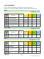
USER MANUAL
AMPLIQUALITY
CMV
ref. 03-22A
ref. 03-22R
Kit for the detection of
Human Cytomegalovirus
0123
03-22A-50(8033622781578)-EN.doc
1. PRODUCT INFORMATION...................................................................... 3
2. KIT CONTENT.......................................................................................... 4
3. STORAGE AND STABILITY OF THE REAGENTS................................. 5
4. PRECAUTIONS FOR USE....................................................................... 6
5. SAFETY RULES ...................................................................................... 7
5.1. General safety rules ............................................................................ 7
5.2. Safety rules about the kit .................................................................... 7
6. MATERIALS REQUIRED, BUT NOT PROVIDED ................................... 9
6.1. Reagents............................................................................................... 9
6.2. Instruments .......................................................................................... 9
6.3. Materials ............................................................................................... 9
7. PREPARATION OF THE REAGENTS................................................... 10
8. INTRODUCTION .................................................................................... 11
9. TEST PRINCIPLE................................................................................... 13
10.
PRODUCT DESCRIPTION ................................................................. 14
11. COLLECTION, MANIPULATION AND PRE-TREATMENT OF THE
SAMPLES..................................................................................................... 15
11.1.
Biological fluids .............................................................................. 15
11.2.
Blood ............................................................................................... 16
12.
12.1.
EXTRACTION PROTOCOL ................................................................ 17
DNA extraction................................................................................ 17
12.2. DNA amplification........................................................................... 17
12.2.1. BG DNA amplification
17
pag. 1
03-22A-50(8033622781578)-EN.doc
12.2.2. Direct amplification (1st amplification) of Cytomegalovirus DNA:
17
12.3. Visualization of the amplification products ................................. 18
12.3.1. Agarose gel electrophoresis
18
12.3.2. Sample Loading
19
12.4. INTERPRETATION OF THE RESULTS: BG DNA amplification and
CMV first amplification ............................................................................... 20
12.5.
2nd Cytomegalovirus DNA amplification (nested PCR) ............... 21
12.6. RESULTS INTERPRETATION: second amplification of
Cytomegalovirus DNA ................................................................................ 22
13.
TROUBLESHOOTING ........................................................................ 23
14.
DEVICE LIMITS................................................................................... 25
15.
DEVICE PERFORMANCES ................................................................ 25
15.1.
Specificity........................................................................................ 25
15.2.
Sensitivity........................................................................................ 25
16.
BIBLIOGRAPHIC REFERENCES....................................................... 26
pag. 2
03-22A-50(8033622781578)-EN.doc
1. PRODUCT INFORMATION
This manual is related to the following products:
CMV:
Kit for the detection of Cytomegalovirus by DNA amplification in the UL122
region (IE2 exon 4).
The kit contains the reagents for DNA amplification and visualization by
agarose gel electrophoresis, the internal control of sample amplificability and
positive control.
Cod.
03-22A-25
03-22A-50
Prod
CMV
CMV
Pkg
25 test
50 test
CMV - amplification reagents
Reagents for the detection of Cytomegalovirus by DNA amplification in the
UL122 region (IE2 exon 4).
The kit contains the reagents for DNA amplification, the internal control of
sample amplificability and the positive control.
Cod.
03-22R-25
03-22R-50
Prod
CMV- amplification reagents
CMV- amplification reagents
pag. 3
03-22A-50(8033622781578)-EN.doc
Pkg
25 test
50 test
2. KIT CONTENT
NOTE: In the kits with different codes different components are included.
(legenda: X = component included in the kit; 0 = component not included in the kit)
BOX P
cod.
03-22A
cod.
03-22R
STORE AT – 20°C
DESCRIPTION
LABEL
COLOUR
OF TUBE
(T)
OR LID
25 test
50 test
8 test
X
X
monodose premixes for
direct CMV amplification
Red (T)
25
50
8
X
X
monodose premixes for
nested CMV amplification
Green (T)
25
50
8
X
X
monodose premixes for
β-globin gene amplification
Blue (T)
25
50
8
X
X
Thermostable Hot-start TAQ
DNA polymerase
Red
1 x 40μL
1 x 80μL
1 x 20μL
AB
SuperTaq
5 U/μL
SMALL BAG
cod.
03-22A
cod.
03-22R
STORE AT – 20°C
X
X
DESCRIPTION
Plasmid DNA containing part
of Cytomegalovirus genome
LABEL
COLOUR
OF TUBE
(T)
OR LID
25 test
50 test
8 test
CMV
positive
control
Blue
1 x 30 μL
1 x 50 μL
1 x 10 μL
BOX F
cod.
03-22A
cod.
03-22R
STORE AT +2°/ +8°C
X
0
LABEL
COLOUR
OF TUBE
(T)
OR LID
25 test
50 test
8 test
Loading Buffer for
electrophoresis
6X Blue
Blue
1X250μL
1X450μL
1X100μL
Ethidium
Bromide
TOXIC
R 23 68
S 36/37 45
Red
1X170μL
1X250μL
1X100μL
Marker PM
Yellow
1X200μL
1X400μL
1X70μL
DESCRIPTION
X
0
Ethidium Bromide solution
(2,5 mg/mL)
X
0
DNA Molecular Weight
Marker
pag. 4
03-22A-50(8033622781578)-EN.doc
BOX A
cod.
03-22A
cod.
03-22R
STORE AT +15°/ +25°C
DESCRIPTION
LABEL
COLOUR
OF TUBE
(T)
OR LID
25 test
50 test
8 test
X
0
Agarose Molecular Biology
grade
AGAROSE
1X20g
1X40g
1X6g
X
0
Electrophoresis buffer
TRIS-Acetate-EDTA, pH 8,00
50X TAE
1X60mL
1X125mL
1X30mL
3. STORAGE AND STABILITY OF THE REAGENTS
Each component of the kit should be stored according to the directions
indicated on the label of the single boxes.
In particular:
Box P
Small bag
Box F
Box A
store at -20°C
store at -20°C
store at a +2 / +8°C
store at +15 / +25°C
When stored at the recommended temperature, all test reagents are stable
until their expiration date.
pag. 5
03-22A-50(8033622781578)-EN.doc
4. PRECAUTIONS FOR USE
• The kit should be handled by investigator qualified through education and
training in molecular biology techniques applied to diagnostics;
• Before starting the kit procedure, read carefully and completely the
instruction manual;
• Keep the product away from sources of heat product out of heating sources;
• Do not use any part of the kit if over the expiration date;
• In case of any doubt about the storage conditions, box integrity or method
application,
contact
AB
ANALITICA
technical
support
at:
[email protected] .
In the amplification of nucleic acids, the investigator has to take the following
special precautions:
• Use filter-tips;
• Store the biological samples, the extracted DNA, positive control included in
the kit and all the amplification products in different places from where
amplification reagents are stored.
• Organise the space in different pre- and post-PCR units; do not share
consumables (pipets, tips, tubes, etc) between them.
• Change the gloves frequently;
• Wash the bench surfaces with 5% sodium hypochloride;
• Thaw the PCR premixes at room temperature before use.
Add the Taq DNA polymerase and purified DNA very quickly at room
temperature or in an ice-bath.
pag. 6
03-22A-50(8033622781578)-EN.doc
5. SAFETY RULES
5.1. General safety rules
• Wear disposable gloves to handle the reagents and the clinical samples and
wash the hands at the end of work.
• Do not pipet with mouth.
• Since no known diagnostic method can assure the absence of infective
agents, it is a good rule to consider every clinical sample as potentially
infectious and handle it as such.
• All the devices that get directly in touch with clinical samples should be
considered as contaminated and disposed as such. In case of accidental
spilling of the samples, clean up with 10% Sodium Hypochloride. The
materials used to clean up should be disposed in special containers for
contaminated products.
• Clinical samples, materials and contaminated products should be disposed
after decontamination by:
immersion in a solution of 5% Sodium Hypochloride (1 volume of 5% Sodium
Hypochloride solution every 10 volumes of contaminated fluid) for 30 minutes
OR
autoclaving at 121°C at least for 2 hours (NOTE: do not autoclave solutions
containing Sodium Hypochloride!!).
5.2.
Safety rules about the kit
The risks for the use of this kit are related to the single components:
Dangerous components:
ETHIDIUM BROMIDE (included in the 03-22A kit)
3,8-diamino-1-ethyl-6-phenylphenantridiumbromide <2%
Description of risk:
T (Toxic)
pag. 7
03-22A-50(8033622781578)-EN.doc
RISK SENTENCES AND S SENTENCES
ETHIDIUM BROMIDE
R 23 and R 68
S 36/37 45
Toxic for inhalation.
Risk of irreversible effects.
Wear laboratory coat and disposable gloves.
In case of accident or discomfort, seek for medical
assistance and show the package label.
R and S sentences refer to the concentrated product, as provided in the kit.
In particular, for Ethidium Bromide, until the dilution in the agarose gel.
In manipulating concentrated Ethidium Bromide, use a chemical dispensing
fume cabinet. Always wear disposable gloves and laboratory coat in
manipulating the diluted Ethidium solution as well.
The product can not be disposed with the common waste. It must not reach
the drainer system. For the disposal, follow the local law.
In case of accidental spilling of Ethidium Bromide, clean with Sodium
hypochloride and water.
Safety data sheet (MSDS) of the product is available upon request.
.
pag. 8
03-22A-50(8033622781578)-EN.doc
6. MATERIALS REQUIRED, BUT NOT PROVIDED
6.1.
Reagents
• Reagents for DNA extraction;
• Sterile DNase and RNase free water;
• Distilled water;
• Reagents for DNA visualization by agarose gel electrophoresis (necessary
for 03-22R kit).
6.2.
Instruments
• Laminar flow cabinet (use is recommended while adding TAQ polymerase to
the amplification premix to avoid contamination; it would be recommended
to use another laminar flow cabinet to add the extracted DNA);
• Micropipettes (range: 0,2-2 µL; 0,5-10 µL; 2-20 µL);
• Thermalcycler;
• Microcentrifuge (max 12.000-14.000 rpm);
• Balance;
• Magnetic heating stirrer or microwave;
• Chemical cabinet (use is recommended in handling Ethidium Bromide);
• Horizontal electrophoresis chamber for agarose minigel;
• Power supply (50-150 V);
• UV Transilluminator;
• Photo camera or image analyzer.
6.3.
Materials
• Disposable gloves;
• Disposable sterile filter-tips (range: 0,2-2 µL; 0,5-10 µL; 2-20 µL;);
• Graduate cilinders (1 L) for TAE dilution;
• Pyrex bottle or Becker for agarose gel preparation;
• Parafilm.
pag. 9
03-22A-50(8033622781578)-EN.doc
7. PREPARATION OF THE REAGENTS
Preparation of 1 L of 1X TAE buffer:
Mix 20 mL of 50X TAE with 980 mL of distilled water.
pag. 10
03-22A-50(8033622781578)-EN.doc
8. INTRODUCTION
The understanding about biology and pathogenesis of Cytomegalovirus
(CMV) infection have been tremendously improved first by the development
of cellular culture techniques and recently by molecular biology.
It has been registered that the spread of the infection among population has
been constantly high (40%-100%), but recently the number and the variety of
the symptomatically evident infections (cytomegalic desease) have shown a
great increase, as consequence of the increased number of
immunocompromised or immuno-immature subjects (immature newborn,
transplants, malignant tumours, splenectomy, transfusions, HIV infections, old
age) (Vancikova Z. and Dvorak P., 2001).
In healthy subjects the CMV infection is usually asymptomatic.
Cytomegalovirus is a virus belonging to the Herpesviridae family: it is an
ubiquitous agent that infects several animal species, human included, and it
is highly species-specific.
CMV virus has a very slow replicative cycle and produces nuclear and
cytoplasmic inclusion bodies. It has a double-stranded linear genome, that
measures about 240 Kb; during the infection process, in the host a specific
DNA polymerase of alfa-specific type is synthesized, associated to a highly
immunogenic protein with 3’-5’ exonucleasic activity is associated to it.
CMV genome can survive in the host cell in a latent stage (with the possibility
of successive reactivations), otherwise it can induce a persistent infection or a
lytic cycle; during the productive inflammation the sequential expression of
the viral genome sequence is time-regulated by genes classified in three
categories: immediate early (IE or alfa), early (E or beta) and late (L or
gamma) (Mocarski ES, 1996).
In CMV infected patients, the virus replicates in different organs as lungs,
liver, kidney, gastrointestinal tract and it is excreted through saliva, breast
milk, spermatic fluid, urine, cervical and vaginal secretions, tears, blood and
faeces.
The affected districts are different, depending on the cause of the
immunodeficiency (tab.1). CMV has cytopathic effect: it causes tissue
damages with the formation of cytomegalic cells containing intranuclear
inclusions.
pag. 11
03-22A-50(8033622781578)-EN.doc
Tab 1. CMV disease in immunosuppressed patients.
(Razonable and Emery, 2004)
Symptom
Fever or Hepatitis
Gastrointestinal symptoms
Retinitis
Pneumonia
Encephalopathy
Deafness
Polyradiculopathy
Addison disease’s symptoms
Transplant rejection
Atherosclerosis
Death
Newborn
+
+
SOT
++
+
+
+
HSCT
+
++
+
++
++
++
AIDS
+
+
++
+
+
+
+
+
+
+
+
SOT: Solid Organ Transplant; HSCT: Haematopoietic stem Cell Transplant;
AIDS: Acquired Immunodeficiency Syndrome
In the past, the standard methods to diagnose CMV included several
techniques for virus isolation from cell cultures and for the search of pp65
antigen from peripheral blood leukocytes (Razonable et al., 2002a).
Since the therapy is fundamental in the prevention of cytomegalic disease in
particular subjects, the application of more sensitive and reproducible
methods, such as molecular techniques, was required.
pag. 12
03-22A-50(8033622781578)-EN.doc
9. TEST PRINCIPLE
PCR method (Polymerase Chain Reaction) has been the first method of DNA
amplification described in literature (Saiki RK et al., 1985). It can be defined
as an in vitro amplification reaction of a specific part of DNA (target
sequence) by a thermostable DNA polymerase.
Three nucleic acid segments are involved in the reaction: double stranded
DNA template to be amplified (target DNA) and two single-stranded
oligonucleotides “primers” that are designed in order to anneal specifically to
the template DNA.
The DNA polymerase begins the synthesis process at the region marked by
the primers and synthesizes new double stranded DNA molecules, identical
to the original double stranded target DNA region, by facilitating the binding
and joining of the complementary nucleotides that are free in solution
(dNTPs). After several cycles, one can get millions of DNA molecules which
correspond to the target sequence.
The sensitivity of this test makes it particularly suitable for the application in
laboratory diagnostics.
Moreover, the amplification reaction can be executed from a wide range of
biological samples and since it allows to amplify very small DNA fragments,
the starting DNA can be also partially degraded.
pag. 13
03-22A-50(8033622781578)-EN.doc
10. PRODUCT DESCRIPTION
The method proposed in this kit consists of the amplification of the viral DNA
in the UL122 region.
The method allows also to evaluate the suitability of extracted DNA, by the
amplification of β-globin (BG) gene (amplification control). A negative result in
BG gene amplification indicates either the presence of inhibitors of the
amplification reaction in the extracted DNA or that the DNA is highly
degraded. This method helps the operator to recognize possible false
negative results for Cytomegalovirus.
The kit also provides with a positive control of amplification. When the
amplification of the positive control is successful, it is guaranteed that the
reaction is correct. This control is not dangerous for the operator because it is
plasmid DNA containing only a part of Cytomegalovirus genome.
The kit is in premix format: all the reagents for the amplification are pre-mixed
and aliquoted in monodose test tubes (0,2 or 0,5 mL) to which Taq
polymerase and the extracted DNA will be added.
This premix format allows the reduction of the manipulation steps in preamplification, with considerable time saving for the operator; the repeated
freezing/thawing of reagents (that could alter the product performances) is
avoided and, above all, this format minimizes the risk of contamination, so the
risk to get false positive results.
Nevertheless, it’s always recommended to use all the proper amplification
controls.
pag. 14
03-22A-50(8033622781578)-EN.doc
11. COLLECTION, MANIPULATION AND PRETREATMENT OF THE SAMPLES
CMV can be in free, episomal or integrated form and this is determinant in the
choice of the protocol to use for isolation and subsequent virus identification.
CMV DNA can be directly extracted from several biologic fluids like spermatic
fluid, maternal milk, vaginal and cervical secretions, saliva, serum, etc.;
If starting from urine or liquor (CSF) a preliminary centrifugation of the sample
is required, to obtain a pellet enriched of cells. Sometimes also the
supernatant is tested.
If starting from peripheral blood to search CMV in leukocytes (PBL) or in
mononuclear cells (PBMC), a further step for obtaining a lymphocyte pellet
(by FICOLL Method or red blood cells lysis) is necessary.
CMV DNA can be extracted also from whole blood.
11.1.
Biological fluids
In the case of cervix and vaginal swabs, sample collection is performed by
the use of a little swab or a little sterile brush (cyto-brush). The collected cells
are diluted in a suitable transport media (1X PBS, physiological solution or
other medium).
The sample can be stored at +2 / +8°C for max 48 h; after then, it is better to
proceed with nucleic acid extraction.
If it is not possible to extract the sample in a short time, the specimen must be
stored at -20°C.
When sample cellularity is too low, it could be necessary, before starting DNA
extraction, to concentrate the sample by repeated centrifugation steps. For
sample resuspension, sterile PBS can be used. It can be useful also to
remove mucus and red cells, if present.
Collect the seminal liquid in a sterile container and store it at +4°C for some
hours (maximum overnight); alternatively, it can be frozen with addition of a
suitable media for DNA extraction.
Urine should be collected in a sterile container, stored at +2/+8°C for max 24
h and then processed.
pag. 15
03-22A-50(8033622781578)-EN.doc
The application of molecular technique allowed a better diagnosis and
characterization of clinical Central Nervous System syndromes associated to
CMV infection. In this case, the sample of choice is liquor (CSF).
The liquor sample can be stored at room temperature or at +2/+8°C, if
processed in a short time; alternatively it is suggested to freeze the sample.
11.2.
Blood
Since long time it was discussed about which was the blood compartment
that was best indicated for CMV analysis. A recent prospective study
(Razonable et al., 2002b) compared 4 blood compartments (whole blood,
plasma, PBL, PBMC) of 17 SOT (Solid Organ Transplant) and HSCT
(Haematopoietic Stem Cell Transplant) patients and demonstrated that even
if all this sample types are suitable for CMV search both for prognosis and
therapy monitoring aims, whole blood seems to be more sensitive when CMV
is present in low number of copies. Later, other publications supported these
data (Weinbeerg et al., 2002, Cortez et al., 2003, Mengelle et al., 2003).
The IHMF (International Herpes Management Forum) guidelines
suggest, where possibile, the use of whole blood for CMV search in
transplanted patients (Razonable and Emery, 2004).
Sample collection should follow all the usual sterility precautions, as routine; it
should be transported in sterile boxes, without transport medium.
Blood should be treated with EDTA. Other anticoagulating agents, as heparin,
are strong inhibitors of TAQ polymerase and so they could alter the efficiency
of the amplification reaction.
Fresh blood can be stored at +2 / +8°C (processed within 4 hours from the
collection); if DNA is not shortly extracted, the sample must be frozen at 20°C.
If the investigator prefers to start the analysis from a leukocyte pellet, the
Ficoll-Hypaque method or the erythrocyte lysis protocol for lymphocytes
isolation can be used.
Alternatively, a buffy coat can be prepared by whole blood centrifugation at
3300 x g for 10 min at room temperature. After centrifugation, three different
fractions can be observed: the upper clear phase is the plasma, the
intermediate phase is the buffy coat , that contains concentrated leukocytes,
and the lowest contains erythrocytes.
pag. 16
03-22A-50(8033622781578)-EN.doc
12. EXTRACTION PROTOCOL
12.1.
DNA extraction
Any extraction method can be used, provided that it extracts pure and integral
DNA.
It is necessary to remember that the volumes of the extracted DNA, indicated
in paragraph 12.2 for BG and CMV amplification, are referred to the extracts
obtained with AB ANALTICA methods. Using an alternative extraction
method, if the volume of the DNA solution to amplify is less than 10 μL, it will
be necessary to adjust the volume of the amplification mix by adding water.
12.2.
DNA amplification
12.2.1.
BG DNA amplification
For each sample add to each premixed tube (blue tube):
0,2 µL AB Super Taq
10 µL extracted DNA
12.2.2.
Direct amplification (1st amplification) of CMV DNA
For each sample add to each premixed red tube:
0,2 µL AB Super Taq
10 µL extracted DNA
It is important to include in each experiment a negative control (add to the
mix distilled water instead of DNA) and a positive control (10 μL of the
positive control included in the kit).
pag. 17
03-22A-50(8033622781578)-EN.doc
Centrifuge briefly, put the tubes (BG and CMV) into the thermalcycler
programmed as below:
1 cycle
40 cycles
1 cycle
95°C
5 min
95°C
60 sec
56°C
60 sec
72°C
60 sec
72°C
5 min
Amplification products lenght:
CMV
BG
12.3.
472 bp.
268 bp.
Visualization of the amplification products
12.3.1.
Agarose gel electrophoresis
Prepare a 3% agarose gel:
Weight 1.5 g of Agarose and pour it into 50 mL of 1x TAE.
Leave the solution on a magnetic stirring heater or in a microwave, until the
solution becomes clear.
Allow the gel to cool to “hand warm” and then add 10 µL of Ethidium Bromide
solution (2.5 mg/ml).
NOTICE: Ethidium Bromide is a strong mutagenic agent; always wear
gloves and preferably work under a chemical safety cabinet during the
handling of this reagent or gels containing it.
Place the gel into the appropriate gel casting tray, with the comb placed in
and allow the gel to cool at room temperature or in a fridge until the gel
becomes solid.
When the gel is solidified, remove carefully the comb (pay attention not to
damage the gel wells) transfer the tray into the electrophoresis chamber and
pour the appropriate amount of 1x TAE buffer so that it covers completely the
gel (about 1-2 mm over the gel surface).
pag. 18
03-22A-50(8033622781578)-EN.doc
12.3.2.
Sample Loading
Mix into a tube or directly on a parafilm layer:
2 μL
10 μL
of 6X Blue*
of amplification product or Molecular weight Marker (PM*marker)
Load the mixture in the gel wells; switch on the power supply and set the
voltage between 80-100 V.
Run the gel for about 40 min, then place the gel on an UV transilluminator
and analyze the results by comparing the size of the amplification products
with the reference Molecular Weight Marker.
*: 6X Blue and PM Marker are included in 03-22A kit; for the use of another
loading buffer or of another Molecular Weight Marker, follow the indications
specified by the supplier.
DNA Molecular Weight Marker (PM Marker, included in 03-22A kit)
fragment sizes: 501-489, 404, 353, 242, 190, 147, 110, 89, 67, 34, 26 bp.
NOTE: In a 3% agarose gel the 501-489 bp bands are usually not clearly
resolved and appear as an unique band; the 26 and 34 bp bands are
sometimes too small to be visible in a 3% agarose gel (because of their low
molecular weight).
NOTICE: UV rays are dangerous for skin and, above all, eyes: always
wear gloves and safety glass or make use of the protection screen of
the UV transilluminator.
pag. 19
03-22A-50(8033622781578)-EN.doc
12.4.
INTERPRETATION OF THE RESULTS: BG DNA
amplification and CMV first amplification
The controls should show the following results:
CONTROL
RESULT
Positive control POSITIVE
Negative
NEGATIVE
control
INTERPRETATION
The PCR amplification is correct
Absence of contaminations
Then, for the interpretation of the band pattern, follow the table below:
DNA BAND
RESULT
INTERPRETATION
BG gene band
absent
BG gene band
CMV band
present
absent
BG gene band
CMV band
present
present
Sample not suitable for amplification
(repeat the DNA extraction)
Amplificable sample
Proceed with the second
amplification to value the true
sample negativity
Amplificable sample
positive sample for CMV.
pag. 20
03-22A-50(8033622781578)-EN.doc
12.5.
CMV nested amplification (2nd DNA amplification)
Add to each premixed green tube:
0,2 µL AB Super Taq
1 µL
First amplification product
It is important to include in this step the negative and the positive controls
used in the CMV direct amplification. When considered useful, it is possible
to include also a negative control for the nested amplification, by adding to
the nested mix 1 µL of sterile water, instead of the direct amplification
product.
Centrifuge briefly, then put the tubes into the thermalcycler programmed as
below:
1 cycle
35 cycles
1 cycle
95°C
5 min
95°C
30 sec
62°C
30 sec
72°C
30 sec
72°C
5 min
Nested amplification:
CMV fragment lenght:
183 bp.
At the end of the amplification run, visualize the nested amplification
products by 3% agarose gel electrophoresis.
Weight 1.5 g of Agarose and pour it into 50 mL of 1x TAE.
Follow the instructions indicated at paragraph 12.3.
pag. 21
03-22A-50(8033622781578)-EN.doc
12.6.
INTERPRETATION OF THE RESULTS: second
amplification of Cytomegalovirus DNA
The controls should show the following results:
CONTROL
RESULT
Positive control
POSITIVE
Negative Control
NEGATIVE
Negative control
Nested amplification
NEGATIVE
INTERPRETATION
The first and the second
amplification are correct
Absence of contaminations in the
first amplification
Absence of contaminations in the
second amplification
Then, for the interpretation of the band pattern, follow the table below:
DNA BAND
RESULT
CMV band in the first and
absent
second amplification
CMV band in the second
present
amplification
1
2
3
INTERPRETATION
Negative sample for
Cytomegalovirus
Positive sample for
Cytomegalovirus
4
Fig. 1:
3% agarose gel electrophoresis of the amplification
products.
1
DNA Molecular Weight Marker (MW)
2
Positive control for CMV DNA
3
CMV positive sample
4
Negative control
pag. 22
03-22A-50(8033622781578)-EN.doc
13. TROUBLESHOOTING
1. Neither amplification products, nor positive control DNA band
• TAQ polymerase was not correctly added to the premix
– Use pipets and tips of suitable volume range (pipet range 0,2 - 2 μL);
– Check visually that TAQ polymerase diffuses in the premix: this is
easy because the enzyme is dissolved in glycerol that has a higher
density;
– Alternatively, check visually the drop of TAQ polymerase put on the
tube wall, then centrifuge briefly.
________________________________________________________________________________
• The thermalcycler was not correctly programmed
– Check the conformity of the thermalcycler program and the
temperature profile in the instruction manual; then repeat the
amplification with the correct program.
________________________________________________________________________________
• The kit doesn’t work properly
– Store the premix, TAQ polymerase and positive control at -20°C;
– Avoid repeated freezing/thawing of the premix and the reagents.
________________________________________________________________________________
2. No amplification bands nor for BG neither Cytomegalovirus in the
tested sample, but a good band for positive controls
• Problems during the extraction step
– Be sure that the extraction kit is adequate and that you followed
correctly all the instructions;
– Consult the troubleshooting section of the extraction kit user manual;
– Repeat the DNA extraction starting from a new sample.
________________________________________________________________________________
• The amplification was inhibited
– Dilute the starting sample with distilled water and TE;
– Repeat the DNA extraction from a smaller amount of clinical sample;
pag. 23
03-22A-50(8033622781578)-EN.doc
– Use an adequate extraction system.
________________________________________________________________________________
3. Presence of aspecific products or extrabands after the visualization
of the amplified products in agarose gel
• The thermalcycler makes temperature changes too slowly
– Do a thermalcycler revision.
________________________________________________________________________________
• The preparation of the amplification reaction has been executed in a long
time at room temperature
– Accelerate the work time at room temperature;
– Work on ice.
_______________________________________________________________________________
• The starting sample contained degraded DNA
– Repeat the extraction step using another clinical specimen;
– Be sure that the sample had been collected and stored in appropriate
way.
For any further problem contact AB ANALITICA technical support (e-mail:
[email protected].
pag. 24
03-22A-50(8033622781578)-EN.doc
14. DEVICE LIMITS
The kit can have reduced performances if:
• the clinical sample is not suitable for this analysis (not correct transport
media or not correct sample storage);
• the DNA is not amplifiable because of the presence of inhibitors of the
amplification reaction or due to an inadequate extraction system;
• the kit has not been stored at the suggested temperature.
15. DEVICE PERFORMANCES
15.1.
Specificity
Primer sequence alignment in the most important databanks shows the
absence of unspecific alignment, and it has guaranteed the amplification of
Cytomegalovirus.
Cross reactions with genomic DNA or other pathogenic microorganism
nucleic acid have not been revealed.
15.2.
Sensitivity
With reference to other nested PCR diagnostic methods, it was assumed that
the sensitivity of the kit is less than 50 DNA copies.
pag. 25
03-22A-50(8033622781578)-EN.doc
16. BIBLIOGRAPHIC REFERENCES
Cortez KJ, Fisher SH, Fahle GA et al. J Infect Dis 188, 967-972, 2003.
Mengelle C, Sandres-Saune K, Pasquier C et al. J Clin Microbiol 41, 38403845, 2003.
Mocarski ES. Virology (Fields BN, Knipe DM, Howley PM, eds). Philadelphia:
Lippincott-Raven, 1996, 2447-2492.
Razonable RR, Brown RA, Wilson J et al. Transplantation 73, 968-973,
2002b.
Razonable RR and Emery VC. Herpes 11(3), 77-86, 2004.
Razonable RR, Paya CV, Smith TF. J Clin Microbiol 40, 746-752, 2002a.
Saiki RK, Scharf S, Faloona F et al. Science 230, 1350-1354, 1985.
Vancikova Z and Dvorak P. Curr Drug Targets Immune Endocr Metabol
Disord 1, 179-187, 2001.
Weinberg A, Schissel D, Giller R. J Clin Microbiol 40, 4203-4206, 2002.
pag. 26
03-22A-50(8033622781578)-EN.doc
pag. 27
03-22A-50(8033622781578)-EN.doc
pag. 28
03-22A-50(8033622781578)-EN.doc
pag. 29
03-22A-50(8033622781578)-EN.doc
AB ANALITICA srl - Via Svizzera 16 - 35127 PADOVA, (ITALY)
Tel +39 049 761698 - Fax +39 049 8709510
e-mail: [email protected]