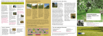
1
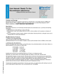
CD90.2 MicroBeads
mouse
Order no. 130-049-101
Contents
1.3Applications
1.Description
●
Isolation of T cells for the analysis of their cytokine expression
in a mouse model of lung fibrosis,1 or after adoptive
transfer of T cells from wild-type and knock-out mice into
immunodeficient mice.2
●
Isolation of tumor-infiltrating T cells to evaluate their
immunotherapeutic potential, 3 or functional properties.4
1.1 Principle of the MACS® Separation
1.2 Background information
1.3Applications
1.4 Reagent and instrument requirements
2.Protocol
2.1 Sample preparation
2.2 Magnetic labeling
2.3 Magnetic separation
1.4 Reagent and instrument requirements
●
Buffer: Prepare a solution containing phosphate-buffered saline
(PBS), pH 7.2, 0.5% bovine serum albumin (BSA), and 2 mM
EDTA by diluting MACS BSA Stock Solution (# 130-091-376)
1:20 with autoMACS™ Rinsing Solution (# 130-091-222). Keep
buffer cold (2−8 °C). Degas buffer before use, as air bubbles
could block the column.
▲ Note: EDTA can be replaced by other supplements such as anticoagulant citrate
dextrose formula-A (ACD-A) or citrate phosphate dextrose (CPD). BSA can be
replaced by other proteins such as mouse serum albumin, mouse serum, or fetal
bovine serum. Buffers or media containing Ca 2+ or Mg2+ are not recommended for
use.
●
MACS Columns and MACS Separators: CD90.2+ cells can be
enriched by using MS, LS, or XS Columns or depleted with the
use of LD, CS, or D Columns. Cells which strongly express
the CD90.2 antigen can also be depleted using MS, LS, or XS
Columns. Positive selection or depletion can also be performed
by using the autoMACS or the autoMACS Pro Separator.
3. Example of a separation using the CD90.2 MicroBeads
4.References
1.Description
Components
2 mL CD90.2 MicroBeads, mouse:
MicroBeads conjugated to monoclonal rat antimouse CD90.2 antibodies (isotype: rat IgG2b).
Capacity
For 2×10⁹ total cells, up to 200 separations.
Product format CD90.2 MicroBeads are supplied in buffer
containing stabilizer and 0.05% sodium azide.
Storage
Store protected from light at 2 − 8 °C. Do not
freeze. The expiration date is indicated on the
vial label.
1.1 Principle of the MACS® Separation
First, the CD90.2 (Thy1.2)+ cells are magnetically labeled with
CD90.2 MicroBeads. Then, the cell suspension is loaded onto a
MACS® Column, which is placed in the magnetic field of a MACS
Separator. The magnetically labeled CD90.2+ cells are retained
within the column. The unlabeled cells run through; this cell
fraction is thus depleted of CD90.2+ cells. After removing the
column from the magnetic field, the magnetically retained CD90.2+
cells can be eluted as the positively selected cell fraction.
Column Max. number
of labeled cells
Max. number Separator
of total cells
Positive selection
2 ×10⁸
MS10⁷
MiniMACS, OctoMACS, VarioMACS, SuperMACS
LS10⁸
2 ×10⁹
MidiMACS, QuadroMACS,
VarioMACS, SuperMACS
XS10⁹
2 ×10¹⁰
SuperMACS
Depletion
5 ×10⁸
LD10⁸
CS
2×10⁸
MidiMACS, QuadroMACS,
VarioMACS, SuperMACS
VarioMACS, SuperMACS
1.2 Background information
SuperMACS
D10⁹
The mouse CD90.2 alloantigen, also known as Thy1.2, is a panT cell marker for the most common inbred mouse strains. CD90.2 is
expressed on thymcoytes, peripheral T cells, on some intraepithelial
T cells, and at lower levels on early hematopoietic stem cells in bone
marrow.
Positive selection or depletion
140-000-054.07
Mouse CD90.2 MicroBeads are suitable for positive selection or
depletion of mouse T lymphocytes from single-cell suspensions of
lymphoid and non-lymphoid tissues or peripheral blood, in CD90.2expressing mouse strains. The mouse CD90.2 antibody does not
cross-react with CD90.1 (Thy1.1).
Miltenyi Biotec GmbH
Friedrich-Ebert-Straße 68, 51429 Bergisch Gladbach, Germany
Phone +49 2204 8306-0, Fax +49 2204 85197
[email protected]
www.miltenyibiotec.com
autoMACS2×10⁸
4 ×10⁹
autoMACS, autoMACS Pro
▲ Note: Column adapters are required to insert certain columns into the
VarioMACS™ or SuperMACS™ Separators. For details see the respective MACS
Separator data sheet.
●
(Optional) Fluorochrome-conjugated CD90.2 antibody for
flow cytometric analysis, e.g., CD90.2-FITC (# 130-091-602),
CD90.2-PE (# 130-091-601), or CD90.2-APC (# 130-091-790)
For more information about other fluorochrome conjugates
see www.miltenyibiotec.com.
Miltenyi Biotec Inc.
2303 Lindbergh Street, Auburn, CA 95602, USA
Phone 800 FOR MACS, +1 530 888 8871, Fax +1 530 888 8925
[email protected]
page 1/3
Order no. 130-049-101
●
(Optional) Propidium iodide (PI) or 7-AAD for flow cytometric
exclusion of dead cells.
●
(Optional) Dead Cell Removal Kit (# 130-090-101) for the
depletion of dead cells.
9. Proceed to magnetic separation (2.3).
●
(Optional) Pre-Separation Filters (# 130-041-407) to remove
cell clumps.
2.Protocol
2.1 Sample preparation
Prepare a single-cell suspension from lymphoid organs, nonlymphoid tissues, or peripheral blood using standard methods.
For details see the General Protocols section of the respective
separator user manual. The General Protocols are also available at
www.miltenyibiotec.com/protocols.
▲ Dead cells may bind non-specifically to MACS MicroBeads. To
remove dead cells, we recommend using density gradient centrifugation
or the Dead Cell Removal Kit (# 130-090-101).
2.2 Magnetic labeling
▲ Work fast, keep cells cold, and use pre-cooled solutions. This will
prevent capping of antibodies on the cell surface and non-specific
cell labeling.
▲ Volumes for magnetic labeling given below are for up to
10⁷ total cells. When working with fewer than 10⁷ cells, use the same
volumes as indicated. When working with higher cell numbers,
scale up all reagent volumes and total volumes accordingly (e.g.
for 2×10⁷ total cells, use twice the volume of all indicated reagent
volumes and total volumes).
▲ For optimal performance it is important to obtain a single‑cell
suspension before magnetic separation. Pass cells through 30 µm
nylon mesh (Pre-Separation Filters, # 130-041-407) to remove cell
clumps which may clog the column. Wet filter with buffer before
use.
▲ Note: For depletion with LD Columns, resuspend up to 1.25×10⁸ cells in
500 µL of buffer.
2.3 Magnetic separation
▲ Choose an appropriate MACS Column and MACS Separator
according to the number of total cells and the number of CD90.2+ cells.
For details see table in section 1.4.
Magnetic separation with MS or LS Columns
1. Place column in the magnetic field of a suitable MACS
Separator. For details see the respective MACS Column data
sheet.
2. Prepare column by rinsing with the appropriate amount of
buffer:
MS: 500 µL
LS: 3 mL
3. Apply cell suspension onto the column.
4. Collect unlabeled cells that pass through and wash
column with the appropriate amount of buffer. Collect total
effluent; this is the unlabeled cell fraction. Perform washing
steps by adding buffer three times. Only add new buffer when
the column reservoir is empty.
MS: 3×500 µL
LS: 3×3 mL
5. Remove column from the separator and place it on a suitable
collection tube.
6. Pipette the appropriate amount of buffer onto the column.
Immediately flush out the magnetically labeled cells by firmly
pushing the plunger into the column.
MS: 1 mL
LS: 5 mL
7. (Optional) To increase the purity of CD90.2+ cells, the eluted
fraction can be enriched over a second MS or LS Column.
Repeat the magnetic separation procedure as described in
steps 1 to 6 by using a new column.
▲ The recommended incubation temperature is 2–8 °C. Working
on ice may require increased incubation times. Higher temperatures
and/or longer incubation times may lead to non-specific cell
labeling.
For instructions on the column assembly and the separation refer to
the XS Column data sheet.
1.
Depletion with LD Columns
Determine cell number.
Magnetic separation with XS Columns
2. Centrifuge cell suspension at 300×g for 10 minutes. Aspirate
supernatant completely.
1. Place LD Column in the magnetic field of a suitable MACS
Separator. For details see LD Column data sheet.
3. Resuspend cell pellet in 90 µL of buffer per 10⁷ total cells.
2. Prepare column by rinsing with 2 mL of buffer.
4. Add 10 µL of CD90.2 MicroBeads per 10⁷ total cells.
3. Apply cell suspension onto the column.
5. Mix well and incubate for 15 minutes in the refrigerator
(2−8 °C).
4. Collect unlabeled cells that pass through and wash
column with 2×1
mL of buffer. Collect total effluent;
this is the unlabeled cell fraction. Perform washing
steps by adding buffer two times. Only add new buffer when
the column reservoir is empty.
6. (Optional) Add staining antibodies, e.g., 10 µL of CD90.2-FITC
(# 130-091-602), and incubate for 5 minutes in the dark in the
refrigerator (2−8 °C).
140-000-054.07
7. Wash cells by adding 1−2 mL of buffer per 10⁷ cells and
centrifuge at 300×g for 10 minutes. Aspirate supernatant
completely.
8. Resuspend up to 10⁸ cells in 500 µL of buffer.
Depletion with CS Columns
1. Assemble CS Column and place it in the magnetic field of a
suitable MACS Separator. For details see CS Column data
sheet.
▲ Note: For higher cell numbers, scale up buffer volume accordingly.
Unless otherwise specifically indicated, Miltenyi Biotec
products and services are for research use only and not for
diagnostic or therapeutic use.
page 2/3
Order no. 130-049-101
2. Prepare column by filling and rinsing with 60 mL of buffer.
Attach a 22G flow resistor to the 3-way stopcock of the
assembled column. For details see CS Column data sheet.
CD90.2– cells
Spleen cells before separation
3. Apply cell suspension onto the column.
Depletion with D Columns
For instructions on column assembly and separation refer to the
D Column data sheet.
CD90.2-FITC
Magnetic separation with the autoMACS™ Separator or the
autoMACS™ Pro Separator
Relative cell number
▲ Buffers used for operating the autoMACS Separator or the
autoMACS Pro Separator should have a temperature of ≥ 10 °C.
▲ Program choice depends on the isolation strategy, the strength
of magnetic labeling, and the frequency of magnetically labeled
cells. For details refer to the section describing the cell separation
programs in the respective user manual.
Magnetic separation with the autoMACS™ Separator
1. Prepare and prime the instrument.
2. Apply tube containing the sample and provide tubes for
collecting the labeled and unlabeled cell fractions. Place
sample tube at the uptake port and the fraction collection
tubes at port neg1 and port pos1.
3. For a standard separation choose one of the following
programs:
Positive selection: “Possel”
Collect positive fraction from outlet port pos1.
Depletion: “Depletes”
Collect negative fraction from outlet port neg1.
CD90.2-FITC
CD90.2+ cells
▲ Refer to the respective user manual for instructions on how to use
the autoMACS™ Separator or the autoMACS Pro Separator.
Relative cell number
Relative cell number
4. Collect unlabeled cells that pass through and wash column
with 30 mL buffer from the top. Collect total effluent; this is
the unlabeled cell fraction.
CD90.2-FITC
4.References
1.
Huaux, F. et al. (2003) Dual role of Il-4 in lung injury and fibrosis. J. Immunol.
170: 2083–2092. [4357]
2.
Roggia, C. et al. (2001) Up-regulation of TNF-producing T cells in the bone
marrow: A key mechanism by which estrogen deficiency induces bone loss in
vivo. Proc. Nat. Acad. Sci. 98: 13960–13965. [2000]
3.
Prévost-Blondel, A. et al. (1998) Tumor-Infiltrating Lymphocytes Exhibiting
High ex vivo Cytolytic Activity Fail to Prevent Murine Melanoma Tumor
Growth in vivo. J. Immunol. 161: 2187–2194. [548]
4.
Radoja, S. et al. (2000) Mice Bearing Late-Stage Tumors Have Normal
Functional Systemic T Cell Response in vitro and in vivo. J. Immunol. 164:
2619–2623. [1054]
Magnetic separation with the autoMACS™ Pro Separator
1. Prepare and prime the instrument.
All protocols and data sheets are available at www.miltenyibiotec.com.
2. Apply tube containing the sample and provide tubes for
collecting the labeled and unlabeled cell fractions. Place
sample tube in row A of the tube rack and the fraction
collection tubes in rows B and C.
Warnings
3. For a standard separation choose one of the following
programs:
Positive selection: “Possel”
Collect positive fraction in row C of the tube rack.
Depletion: “Depletes”
Collect negative fraction in row B of the tube rack.
3. Example of a separation using the CD90.2
MicroBeads
140-000-054.07
CD90.2+ cells were isolated from a mouse spleen suspension using
the CD90.2 MicroBeads, an MS Column, and a MiniMACS™
Separator. Cells are fluorescently stained with CD90.2-FITC
(# 130-091-602). Cell debris and dead cells are excluded from the
analysis based on scatter signals and PI fluorescence.
Reagents contain sodium azide. Under acidic conditions sodium azide yields
hydrazoic acid, which is extremely toxic. Azide compounds should be diluted with
running water before discarding. These precautions are recommended to avoid
deposits in plumbing where explosive conditions may develop.
Warranty
The products sold hereunder are warranted only to be free from defects in workmanship
and material at the time of delivery to the customer. Miltenyi Biotec GmbH
makes no warranty or representation, either expressed or implied, with respect to
the fitness of a product for a particular purpose. There are no warranties, expressed
or implied, which extend beyond the technical specifications of the products.
Miltenyi Biotec GmbH’s liability is limited to either replacement of the products or
refund of the purchase price. Miltenyi Biotec GmbH is not liable for any property
damage, personal injury or economic loss caused by the product.
MACS is a registered trademark and autoMACS, MidiMACS, MiniMACS, OctoMACS,
QuadroMACS, SuperMACS, and VarioMACS are trademarks of Miltenyi Biotec
GmbH.
Copyright © 2008 Miltenyi Biotec GmbH. All rights reserved.
Unless otherwise specifically indicated, Miltenyi Biotec
products and services are for research use only and not for
diagnostic or therapeutic use.
page 3/3