1
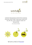
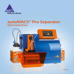
CD34 MicroBead Kit human 2 mL 10 mL 130-046-702 130-046-703 Contents 1.1 Principle of the MACS® Separation 1.Description First, the CD34+ cells are magnetically labeled with CD34 MicroBeads. Then, the cell suspension is loaded onto a MACS® Column which is placed in the magnetic field of a MACS Separator. The magnetically labeled CD34+ cells are retained within the column. The unlabeled cells run through; this cell fraction is thus depleted of CD34+ cells. After removing the column from the magnetic field, the magnetically retained CD34+ cells can be eluted as the positively selected cell fraction. 1.1 Principle of the MACS® Separation 1.2 Background information 1.3Applications 1.4 Reagent and instrument requirements 2.Protocol 2.1 Sample preparation 2.2 Magnetic labeling 2.3 Magnetic separation 2.4 (Optional) Evaluation of hematopoietic progenitor cell purity 3. Example of a separation using the CD34 MicroBead Kit 4.References 1.Description Components 2 mL CD34 MicroBeads, human: MicroBeads conjugated to monoclonal mouse anti-human CD34 antibodies (isotype: mouse IgG1). 2 mL FcR Blocking Reagent, human: Human IgG. or 10 mL CD34 MicroBeads, human: MicroBeads conjugated to monoclonal mouse anti-human CD34 antibodies (isotype: mouse IgG1). 1.2 Background information The CD34 antigen is a single chain transmembrane glycoprotein expressed on human hematopoietic progenitor cells, endothelial progenitor cells, vascular endothelial cells, embryonic fibroblasts, and some cells in fetal and adult nervous tissue. The CD34 MicroBead Kit contains MicroBeads directly conjugated to CD34 antibodies for magnetic labeling of CD34-expressing cells from peripheral blood, cord blood, bone marrow, apheresis harvest, or differentiated ES and iPS cells. Hematopoietic progenitor cells, present at a frequency of about 0.05−0.2% in peripheral blood, 0.1−0.5% in cord blood, and 0.5−3% in bone marrow, can be rapidly and efficiently enriched. 1.3Applications ● Positive selection or depletion of cells expressing human CD34 antigen.¹,⁵ ● Isolation of hematopoietic progenitor cells. ● Isolation of endothelial progenitor cells (EPCs).²,³ ● Isolation of CD34+ progenitor cells from differentiated ES and iPS cell cultures.⁴ ● In vitro differentiation studies.¹,⁵ Studies on hematologic malignancies. 10 mL FcR Blocking Reagent, human: Human IgG. ● Capacity 2 mL: For 2×10⁹ total cells, up to 20 separations. 1.4 Reagent and instrument requirements or 10 mL: For 10¹⁰ total cells, up to 100 separations. Product format CD34 MicroBeads are supplied in buffer containing stabilizer and 0.05% sodium azide. Storage Store protected from light at 2−8 °C. Do not freeze. The expiration date is indicated on the vial label. ● Buffer: Prepare a solution containing phosphate-buffered saline (PBS), pH 7.2, 0.5% bovine serum albumin (BSA), and 2 mM EDTA by diluting MACS BSA Stock Solution (# 130‑091‑376) 1:20 with autoMACS® Rinsing Solution (# 130‑091‑222). Keep buffer cold (2−8 °C). Degas buffer before use, as air bubbles could block the column. ▲▲ Note: EDTA can be replaced by other supplements such as anticoagulant citrate dextrose formula-A (ACD-A) or citrate phosphate dextrose (CPD). BSA can be replaced by other proteins such as human serum albumin, human serum, or fetal bovine serum (FBS). Buffers or media containing Ca 2+ or Mg 2+ are not recommended for use. 140-001-950.05 Miltenyi Biotec GmbH Friedrich-Ebert-Straße 68, 51429 Bergisch Gladbach, Germany Phone +49 2204 8306-0, Fax +49 2204 85197 [email protected] www.miltenyibiotec.com Miltenyi Biotec Inc. 2303 Lindbergh Street, Auburn, CA 95602, USA Phone 800 FOR MACS, +1 530 888 8871, Fax +1 530 888 8925 [email protected] page 1/4 Order no. 130-046-702 Order no. 130-046-703 ● MACS Columns and MACS Separators: CD34+ cells can be enriched by using MS, LS, or XS Columns (positive selection). Cells that strongly express the CD34 antigen can also be depleted using MS, LS, or XS Columns. Positive selection or depletion can also be performed by using the autoMACS Pro or the autoMACS Separator. Column Max. number of labeled cells Max. number Separator of total cells Preparation of cells from leukapheresis material 1. Filter apheresis harvest through 30 µm nylon mesh (PreSeparation Filters, 30 µm # 130-041-407), in order to remove cell clumps. 2. Wash cells once with buffer and resuspend in a final volume of 300 µL of buffer for up to 10⁸ cells. Proceed to magnetic labeling. Positive selection MS 10⁷ 2 ×10⁸ MiniMACS, OctoMACS, VarioMACS, SuperMACS II LS 10⁸ 2 ×10⁹ MidiMACS, QuadroMACS, VarioMACS, SuperMACS II XS 10⁹ 2 ×10¹⁰ SuperMACS II Positive selection autoMACS 2×10⁸ 4 ×10⁹ autoMACS Pro, autoMACS ▲▲ Note: Column adapters are required to insert certain columns into the VarioMACS™ or SuperMACS™ II Separators. For details refer to the respective MACS Separator data sheet. ● (Optional) MC CD34 Stem Cell Cocktail (# 130-093-427) for flow cytometric analysis of separated cells. ● (Optional) Fluorochrome-conjugated antibodies for flow cytometric analysis, e.g., CD34-FITC (# 130-081-001), CD34-PE (# 130-081-002), CD34-APC (# 130-090-954), CD133 (293C3)-PE (# 130-090-853), CD133 (293C3)-PE (# 130-090-854), CD45-FITC (# 130-080-202), CD45-PE (# 130-080-201), or CD45-APC (# 130-091-230). For more information about antibodies refer to www.miltenyibiotec.com/ antibodies. ● (Optional) Propidium Iodide Solution (# 130-093-233) or 7-AAD for flow cytometric exclusion of dead cells. ● (Optional) Dead Cell Removal Kit (# 130-090-101) for the depletion of dead cells. ● (Optional) Pre-Separation Filters, 30 µm (# 130-041-407) to remove cell clumps. 2.Protocol 2.1 Sample preparation When working with anticoagulated peripheral blood or buffy coat, peripheral blood mononuclear cells (PBMCs) should be isolated by density gradient centrifugation, for example, using Ficoll-Paque™. ▲▲ Note: To remove platelets after density gradient separation, resuspend cell pellet in buffer and centrifuge at 200×g for 10−15 minutes at 20 °C. Carefully aspirate supernatant. Repeat washing step. When working with tissues or lysed blood, prepare a single-cell suspension using standard methods. For details refer to the protocols section at www.miltenyibiotec.com/ protocols. ▲ Dead cells may bind non-specifically to MACS MicroBeads. To remove dead cells, we recommend using density gradient centrifugation or the Dead Cell Removal Kit (# 130-090-101). 2.2 Magnetic labeling ▲ Work fast, keep cells cold, and use pre-cooled solutions. This will prevent capping of antibodies on the cell surface and non-specific cell labeling. ▲ Volumes for magnetic labeling given below are for up to 10⁸ total cells. When working with fewer than 10⁸ cells, use the same volumes as indicated. When working with higher cell numbers, scale up all reagent volumes and total volumes accordingly (e.g. for 2×10⁸ total cells, use twice the volume of all indicated reagent volumes and total volumes). ▲ For optimal performance it is important to obtain a single‑cell suspension before magnetic labeling. Pass cells through 30 µm nylon mesh (Pre-Separation Filters, 30 µm # 130-041-407) to remove cell clumps which may clog the column. Moisten filter with buffer before use. ▲ The recommended incubation temperature is 2–8 °C. Higher temperatures and/or longer incubation times may lead to nonspecific cell labeling. Working on ice may require increased incubation times. 1. Determine cell number. 2. Centrifuge cell suspension at 300×g for 10 minutes. Aspirate supernatant completely. 3. Resuspend cell pellet in 300 µL of buffer for up to 10⁸ total cells. 4. Add 100 µL of FcR Blocking Reagent for up to 10⁸ total cells. 5. Add 100 µL of CD34 MicroBeads for up to 10⁸ total cells. 6. Mix well and incubate for 30 minutes in the refrigerator (2−8 °C). 7. (Optional) Add fluorochrome-conjugated CD34 antibody recognizing another epitope than QBEND/10 (e.g. clone AC136: CD34-PE, # 130-081-002) or fluorochrome-conjugated CD45 antibody (e.g. CD45-FITC, # 130-080-202), and incubate for 5 minutes in the dark in the refrigerator (2−8 °C). 8. Wash cells by adding 5−10 mL of buffer for up to 10⁸ cells and centrifuge at 300×g for 10 minutes. Aspirate supernatant completely. 9. Resuspend up to 10⁸ cells in 500 µL of buffer. ▲ Note: For higher cell numbers, scale up buffer volume accordingly. ▲ Note: For depletion with LD Columns, resuspend up to 1.25×10⁸ cells in 500 µL of buffer. 10. Proceed to magnetic separation (2.3). 140-001-950.05 Unless otherwise specifically indicated, Miltenyi Biotec products and services are for research use only and not for diagnostic or therapeutic use. page 2/4 Order no. 130-046-702 Order no. 130-046-703 2.3 Magnetic separation Magnetic separation with the autoMACS® Pro Separator 1. Prepare and prime the instrument. ▲ Choose an appropriate MACS Column and MACS Separator according to the number of total cells and the number of CD34+ cells. For details refer to the table in section 1.4. ▲ Always wait until the column reservoir is empty before proceeding to the next step. 2. Apply tube containing the sample and provide tubes for collecting the labeled and unlabeled cell fractions. Place sample tube in row A of the tube rack and the fraction collection tubes in rows B and C. 3. For a standard separation choose one of the following programs: Magnetic separation with MS or LS Columns 1. Place column in the magnetic field of a suitable MACS Separator. For details refer to the respective MACS Column data sheet. 2. Prepare column by rinsing with the appropriate amount of buffer: MS: 500 µL LS: 3 mL 3. Apply cell suspension onto the column. Collect flow-through containing unlabeled cells. 4. Wash column with the appropriate amount of buffer. Collect unlabeled cells that pass through and combine with the flowthrough from step 3. MS: 3×500 µL 5. Remove column from the separator and place it on a suitable collection tube. 6. Pipette the appropriate amount of buffer onto the column. Immediately flush out the magnetically labeled cells by firmly pushing the plunger into the column. MS: 1 mL Positive selection of CD34+ cells from cord blood: Posseld2. Collect positive fraction in row C of the tube rack. Magnetic separation with the autoMACS® Separator 1. Prepare and prime the instrument. 2. Apply tube containing the sample and provide tubes for collecting the labeled and unlabeled cell fractions. Place sample tube at the uptake port and the fraction collection tubes at port neg1 and port pos2. 3. For a standard separation choose one of the following programs: LS: 3×3 mL ▲ Note: Perform washing steps by adding buffer aliquots only when the column reservoir is empty. LS: 5 mL 7. (Optional) To increase the purity of CD34+ cells, the eluted fraction can be enriched over a second MS or LS Column. Repeat the magnetic separation procedure as described in steps 1 to 6 by using a new column. Magnetic separation with XS Columns For instructions on the column assembly and the separation refer to the XS Column data sheet. Magnetic separation with the autoMACS® Pro Separator or the autoMACS® Separator ▲ Refer to the respective user manual for instructions on how to use the autoMACS® Pro Separator or the autoMACS Separator. Positive selection of CD34+ cells from peripheral blood, bone marrow or leukapheresis: Posseld. Positive selection of CD34+ cells from peripheral blood, bone marrow or leukapheresis: Posseld. Positive selection of CD34+ cells from cord blood: Posseld2. Collect positive fraction from outlet port pos2. 2.4(Optional) Evaluation of hematopoietic progenitor cell purity The purity of the isolated hematopoietic progenitor cells can be evaluated by flow cytometry or fluorescence microscopy. Analysis of CD34+ cells can be accomplished by direct immunofluorescent staining using an antibody recognizing an epitope different from that recognized by the CD34 monoclonal antibody QBEND/10 (e.g. CD34-PE, clone: AC136, # 130-081-002). For optimal discrimination of CD34+ cells from other leukocytes, counterstain cells with an antibody against CD45 (e.g. CD45‑FITC, # 130-080-202). CD34+ cells express CD45 at a lower level as compared to lymphocytes. Use the antibodies in appropriate concentrations as recommended by the manufacturers. Typically, staining for 5 minutes at 2−8 °C should be sufficient. After fluorescent staining, cells should be washed and resuspended in buffer. ▲ Buffers used for operating the autoMACS Pro Separator or the autoMACS Separator should have a temperature of ≥10 °C. ▲ Program choice depends on the isolation strategy, the strength of magnetic labeling, and the frequency of magnetically labeled cells. For details refer to the section describing the cell separation programs in the respective user manual. 140-001-950.05 Unless otherwise specifically indicated, Miltenyi Biotec products and services are for research use only and not for diagnostic or therapeutic use. page 3/4 Order no. 130-046-702 Order no. 130-046-703 3. Example of a separation using the CD34 MicroBead Kit Isolation of CD34+ cells from PBMCs using the CD34 MicroBead Kit, two MS Columns, and a MiniMACS™ Separator. Cells were stained with CD34-PE (# 130-081-002) and CD45-FITC (# 130‑080-202). Cell debris and dead cells are excluded from the analysis based on scatter signals and propidium iodide fluorescence. CD45-FITC Before separation Warnings Reagents contain sodium azide. Under acidic conditions sodium azide yields hydrazoic acid, which is extremely toxic. Azide compounds should be diluted with running water before discarding. These precautions are recommended to avoid deposits in plumbing where explosive conditions may develop. Warranty The products sold hereunder are warranted only to be free from defects in workmanship and material at the time of delivery to the customer. Miltenyi Biotec GmbH makes no warranty or representation, either expressed or implied, with respect to the fitness of a product for a particular purpose. There are no warranties, expressed or implied, which extend beyond the technical specifications of the products. Miltenyi Biotec GmbH’s liability is limited to either replacement of the products or refund of the purchase price. Miltenyi Biotec GmbH is not liable for any property damage, personal injury or economic loss caused by the product. autoMACS and MACS are registered trademarks and MidiMACS, MiniMACS, OctoMACS, QuadroMACS, SuperMACS, and VarioMACS are trademarks of Miltenyi Biotec GmbH. Ficoll-Paque is a trademark of GE Healthcare companies. CD34-PE Copyright © 2013 Miltenyi Biotec GmbH. All rights reserved. + CD45-FITC CD34 cells CD34-PE 4.References 1. Giarratana, M. C. et al. (2005) Ex vivo generation of fully mature human red blood cells from hematopoietic stem cells. Nat. Biotechnol. 23: 69–74. 2. Timmermans, F. et al. (2007) Endothelial outgrowth cells are not derived from CD133+ cells or CD45+ hematopoietic precursors. Arterioscler. Thromb. Basc. Biol. 27: 1572–1579. 3. O, E. et al. (2011) Efficient nonadhesive ex vivo expansion of early endothelial progenitor cells derived from CD34+ human cord blood fraction for effective therapeutic vascularization. FASEB J. 25: 159–169. 4. Wang, Z. Z. et al. (2007) Endothelial cells derived from human embryonic stem cells form durable blood vessels in vivo. Nat. Biotechnol. 25: 317–318. 5. Bonanno, G. et al. (2009) Interleukin-21 induces the differentiation of human umbilical cord blood CD34-lineage– cells into pseudomature lytic NK cells. BMC Immunol. 10: 46. All protocols and data sheets are available at www.miltenyibiotec.com. 140-001-950.05 Unless otherwise specifically indicated, Miltenyi Biotec products and services are for research use only and not for diagnostic or therapeutic use. page 4/4