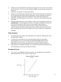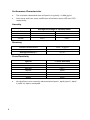
1
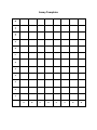
AssayMaxTM Human ApoA-II ELISA Kit Assaypro LLC 3400 Harry S Truman Blvd St. Charles, MO 63301 T (636) 447-9175 F (636) 395-7419 www.assaypro.com For any questions regarding troubleshooting or performing the assay, please contact our support team at [email protected]. Thank you for choosing Assaypro. Assay Summary Step 1. Add 50 µl of Standard or Sample per well. Incubate 2 hours. Step 2. Wash, then add 50 µl of Biotinylated Antibody per well. Incubate 1 hour. Step 3. Wash, then add 50 µl of SP Conjugate per well. Incubate 30 minutes. Step 4. Wash, then add 50 µl of Chromogen Substrate per well. Incubate 8 minutes. Step 5. Add 50 µl of Stop Solution per well. Read at 450 nm immediately. Symbol Key Consult instructions for use. H G F E D C B A 1 2 3 4 5 6 7 8 9 10 11 12 Assay Template Human Apolipoprotein A-II ELISA Kit Catalog No. EA5222-1 Sample insert for reference use only Introduction Apolipoprotein A-II (ApoA-II) is the second most abundant apolipoproteins in human plasma HDL, comprising about 25% of the protein mass. After being synthesized by the liver and intestine as a preprotein containing 100 amino acids, ApoA-II is processed to 77 amino acids in the mature plasma protein (1-3). ApoA-II is found in plasma as a monomer, homodimer of 17.4 kDa, or heterodimer with ApoE and ApoD (4-7). It has been reported that ApoA-II plays roles in HDL remodeling, cholesterol efflux, modulating HDL interaction with enzymes and receptors, and triglyceride metabolism (7-12). Principle of the Assay The AssayMax Human Apolipoprotein A-II ELISA (Enzyme-Linked Immunosorbent Assay) kit is designed for detection of human apoA-II in plasma, serum, urine, milk, and cell culture samples. This assay employs a quantitative sandwich enzyme immunoassay technique that measures human apoA-II in less than 4 hours. A monoclonal antibody specific for human apoAII has been pre-coated onto a 96-well microplate with removable strips. ApoA-II in standards and samples is sandwiched by the immobilized antibody and biotinylated polyclonal antibody specific for human apoA-II, which is recognized by a streptavidin-peroxidase conjugate. All unbound material is then washed away and a peroxidase enzyme substrate is added. The color development is stopped and the intensity of the color is measured. Caution and Warning This product is for Research Use Only and is Not For Use In Diagnostic Procedures. Prepare all reagents (working diluent buffer, wash buffer, standard, biotinylated antibody, and SP conjugate) as instructed, prior to running the assay. Prepare all samples prior to running the assay. The dilution factors for the samples are suggested in this insert. However, the user should determine the optimal dilution factor. Spin down the SP conjugate vial and the biotinylated antibody vial before opening and using contents. The kit should not be used beyond the expiration date. 1 The Stop Solution is an acidic solution. Reagents Human ApoA-II Microplate: A 96-well polystyrene microplate (12 strips of 8 wells) coated with a monoclonal antibody against human ApoA-II. Sealing Tapes: Each kit contains 3 precut, pressure sensitive sealing tapes that can be cut to fit the format of the individual assay. Human ApoA-II Standard: Human ApoA-II in a buffered protein base (4 g, lyophilized). Biotinylated Human ApoA-II Antibody (50x): A 50-fold concentrated biotinylated polyclonal antibody against ApoA-II (140 l). EIA Diluent Concentrate (10x): A 10-fold concentrated buffered protein base (30 ml). Wash Buffer Concentrate (20x): A 20-fold concentrated buffered surfactant (30 ml, 2 bottles). Streptavidin-Peroxidase Conjugate (SP Conjugate): A 100-fold concentrate (80 l). Chromogen Substrate: A ready-to-use stabilized peroxidase chromogen substrate tetramethylbenzidine (8 ml). Stop Solution: A 0.5 N hydrochloric acid to stop the chromogen substrate reaction (12 ml). Storage Condition Upon arrival, immediately store components of the kit at recommended temperatures up to the expiration date. Store SP Conjugate, Standard, and Biotinylated Antibody at -20°C. Store Microplate, Diluent Concentrate (10x), Wash Buffer, Stop Solution, and Chromogen Substrate at 2-8°C. Unused microplate wells may be returned to the foil pouch with the desiccant packs and resealed. May be stored for up to 30 days in a vacuum desiccator. Diluent (1x) may be stored for up to 30 days at 2-8°C. Store Standard at -20°C for up to 15 days after reconstituting with Diluent. Other Supplies Required 2 Microplate reader capable of measuring absorbance at 450 nm. Pipettes (1-20 l, 20-200 l, 200-1000 l, and multiple channel). Deionized or distilled reagent grade water. Sample Collection, Preparation and Storage Plasma: Collect plasma using one-tenth volume of 0.1 M sodium citrate as an anticoagulant. Centrifuge samples at 3000 x g for 10 minutes and use supernatants. Dilute samples 1:1000 with EIA Diluent and assay. The undiluted samples can be stored at -20°C or below for up to 3 months. Avoid repeated freeze-thaw cycles (EDTA or Heparin can also be used as an anticoagulant). Serum: Samples should be collected into a serum separator tube. After clot formation, centrifuge samples at 3000 x g for 10 minutes, and remove serum. Dilute samples 1:1000 into EIA Diluent and assay. The undiluted samples can be stored at -20°C or below for up to 3 months. Avoid repeated freeze-thaw cycles. Urine: Collect urine using sample pot. Centrifuge samples at 800 x g for 10 minutes and assay. Store samples at -20°C or below for up to 3 months. Avoid repeated freeze-thaw cycles. Cell Culture Media: Centrifuge cell culture media at 3000 x g for 10 minutes to remove debris. Collect supernatants and assay. Store samples at -20°C or below. Avoid repeated freeze-thaw cycles. Cell Lysate: Rinse cell with cold PBS and then scrape the cell into a tube with 5 ml cold PBS and 0.5 M EDTA. Centrifuge suspension at 1500 rpm for 10 minutes at 4°C and aspirate supernatant. Re-suspend pellet in icecold Lysis Buffer (10 mM Tris, pH 8.0, 130 mM NaCl, 1% Triton X-100, 6 protease inhibitor cocktail). For every 1 x 10 cell, add approximately 100 μL of ice-cold Lysis Buffer. Incubate on ice for 60 minutes. Centrifuge at 13000 rpm for 30 minutes at 4°C and collect supernatant for assay. Milk: Collect milk using sample tube. Centrifuge samples at 800 x g for 10 minutes. Milk dilution is suggested at 1:2 in EIA Diluent; however, the user should determine the optimal dilution factor. The undiluted samples can be stored at -20°C or below for up to 3 months. Avoid repeated freeze-thaw cycles. Reagent Preparation Freshly dilute all reagents and bring all reagents to room temperature before use. EIA Diluent Concentrate (10x): If crystals have formed in the concentrate, mix gently until the crystals have completely dissolved. Dilute the EIA Diluent Concentrate 1:10 with reagent grade water. Store for up to 30 days at 2-8°C. Standard Curve: Reconstitute the 4 g of Human ApoA-II Standard with 1 ml of EIA Diluent to generate a 4 g/ml standard solution. Allow the standard to sit for 10 minutes with gentle agitation prior to making dilutions. Prepare duplicate or triplicate standard points by serially diluting the standard solution (4 g/ml) 1:4 with EIA Diluent to produce 1, 3 0.25, 0.063, 0.016, and 0.004 g/ml solutions. EIA Diluent serves as the zero standard (0 g/ml). Any remaining solution should be frozen at -20°C and used within 15 days. Standard Point P1 P2 P3 P4 P5 P6 P7 Dilution Standard (4 g/ml) 1 part P1 + 3 parts EIA Diluent 1 part P2 + 3 parts EIA Diluent 1 part P3 + 3 parts EIA Diluent 1 part P4 + 3 parts EIA Diluent 1 part P5 + 3 parts EIA Diluent EIA Diluent [ApoA-II] (µg/ml) 4.000 1.000 0.250 0.063 0.016 0.004 0.000 Biotinylated Human ApoA-II Antibody (50x): Spin down the antibody briefly and dilute the desired amount of the antibody 1:50 with EIA Diluent. Any remaining solution should be frozen at -20°C. Wash Buffer Concentrate (20x): If crystals have formed in the concentrate, mix gently until the crystals have completely dissolved. Dilute the Wash Buffer Concentrate 1:20 with reagent grade water. SP Conjugate (100x): Spin down the SP Conjugate briefly and dilute the desired amount of the conjugate 1:100 with EIA Diluent. Any remaining solution should be frozen at -20°C. Assay Procedure 4 Prepare all reagents, standard solutions, and samples as instructed. Bring all reagents to room temperature before use. The assay is performed at room temperature (20-25°C). Remove excess microplate strips from the plate frame and return them immediately to the foil pouch with desiccants inside. Reseal the pouch securely to minimize exposure to water vapor and store in a vacuum desiccator. Add 50 l of Human ApoA-II Standard or sample per well. Cover wells with a sealing tape and incubate for 2 hours. Start the timer after the last addition. Wash five times with 200 l of Wash Buffer manually. Invert the plate each time and decant the contents; hit 4-5 times on absorbent material to completely remove the liquid. If using a machine, wash six times with 300 l of Wash Buffer and then invert the plate, decanting the contents; hit 4-5 times on absorbent material to completely remove the liquid. Add 50 l of Biotinylated Human ApoA-II Antibody to each well and incubate for 1 hour. Wash the microplate as described above. Add 50 l of Streptavidin-Peroxidase Conjugate to each well and incubate for 30 minutes. Turn on the microplate reader and set up the program in advance. Wash the microplate as described above. Add 50 l of Chromogen Substrate per well and incubate for 8 minutes or till the optimal blue color density develops. Gently tap plate to ensure thorough mixing and break the bubbles in the well with pipette tip. Add 50 l of Stop Solution to each well. The color will change from blue to yellow. Read the absorbance on a microplate reader at a wavelength of 450 nm immediately. If wavelength correction is available, subtract readings at 570 nm from those at 450 nm to correct optical imperfections. Otherwise, read the plate at 450 nm only. Please note that some unstable black particles may be generated at high concentration points after stopping the reaction for about 10 minutes, which will reduce the readings. Data Analysis Calculate the mean value of the duplicate or triplicate readings for each standard and sample. To generate a standard curve, plot the graph using the standard concentrations on the x-axis and the corresponding mean 450 nm absorbance on the y-axis. The best-fit line can be determined by regression analysis using log-log or four-parameter logistic curve-fit. Determine the unknown sample concentration from the Standard Curve and multiply the value by the dilution factor. Standard Curve The curve is provided for illustration only. A standard curve should be generated each time the assay is performed. 5 Performance Characteristics The minimum detectable dose of ApoA-II is typically ~ 0.004 µg/ml. Intra-assay and inter-assay coefficients of variation were 4.8% and 7.1% respectively. Linearity Sample Dilution 1:500 1:1000 1:2000 Average Percentage of Expected Value Plasma Serum 81% 84% 99% 98% 111% 112% Recovery Standard Added Value Recovery % Average Recovery % 0.01 – 1 g/ml 85 – 109% 97.5% Cross-Reactivity Species Canine Bovine Monkey Mouse Rat Swine Rabbit 6 % Cross Reactivity None None <10% None None None None No significant cross reactivity observed with ApoA-I, ApoB, ApoC-I, ApoCII, ApoC-III, ApoH, and ApoM. References (1) (2) (3) (4) (5) (6) (7) (8) (9) (10) (11) (12) Tsao YK et al. (1985) J. Biol. Chem. 260:15222-15231 Gordon JI et al. (1983) J. Biol. Chem. 258:14054-14059 Brewer HB et al. (1972) Proc. Natl. Acad. Sci. USA. 69:1304-1308 Weisgraber KH and Mahley RW (1978) J. Biol. Chem. 253:6281-6288 Blanco-Vaca F et al. (1992) J. Lipid Res. 33:1785-1796 Gillard BK et al. (2005) Biochemistry 44:471-479 Durbin DM and Jonas A (1999) J. Lipid Res. 40:2293-2302 Forte TM et al. (1995) J. Lipid Res. 36:148-157 Fidge NH (1999) J. Lipid Res. 40:187-201 Warden CH et al. (1993) Science 261:469-471 Schultz JR et al. (1993) Nature 365:762-764 Weng W and Breslow JL (1996) Proc. Natl. Acad. Sci. USA. 93:14788-14794 Version 3.8R www.assaypro.com • E-mail: [email protected] 7