1
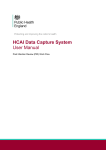
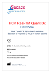
TNF (Human) ELISA Kit
Catalog Number KA0648
96 assays
Version: 02
Intended for research use only
www.abnova.com
Table of Contents
Introduction ................................................................................................... 3
Intended Use ................................................................................................................. 3
Background ................................................................................................................... 3
Principle of the Assay .................................................................................................... 3
General Information ...................................................................................... 4
Materials Supplied ......................................................................................................... 4
Storage Instruction ........................................................................................................ 4
Materials Required but Not Supplied ............................................................................. 5
Precautions for Use ....................................................................................................... 5
Assay Protocol .............................................................................................. 7
Reagent Preparation ..................................................................................................... 7
Sample Preparation ....................................................................................................... 8
Assay Procedure ........................................................................................................... 9
Data Analysis ............................................................................................... 12
Calculation of Results .................................................................................................. 12
Performance Characteristics ....................................................................................... 12
Resources .................................................................................................... 13
Troubleshooting ........................................................................................................... 13
References .................................................................................................................. 14
Plate Layout ................................................................................................................ 15
KA0648 2 / 15
Introduction
Intended Use
For quantitative detection of human TNF-α in cell culture supernatants, human plasma (EDTA, heparin and
citrate), serum, cerebrospinal fluid, urine, synovial fluid or other body fluids.
The assay will recognize both natural and recombinant Hu TNF-α.
Background
Tumor Necrosis Factor- α (TNF- α) is a non-glycosylated 17.5 kDa, 157 amino acid protein. TNF- α is a potent
lymphoid factor and exerts cytotoxic effects on a wide range of tumor cells and other target cells. It is secreted
by macrophages, monocytes, neutrophils, T-cells, and NK-cells following their stimulation by bacterial
lipopolysaccharides. TNF-α has been suggested to play a pro-inflammatory role and has been detected in
synovial fluid of patients with rheumatoid arthritis. Various pathological conditions are associated with the
production of high levels of TNF-α. These include septic shock, cachexia (e.g. HIV, tuberculosis, cancer),
autoimmune diseases, hepatitis, leukemia, myocardialischaemia, organ transplantation rejection, multiple
sclerosis, rheumatoid arthritis, and meningococcal septicemia. Annually, many people die from septic shock
syndrome, triggered by TNF-α following complications from an infectious disease. In many cases elevated
TNF-α serum levels predict a higher mortality.
Principle of the Assay
The TNF-α ELISA kit is an in vitro enzyme-linked immunosorbent assay for the quantitative measurement of
human TNF-α in cell culture supernatants, serum, plasma, cerebrospinal fluid, urine, synovial fluid and other
body fluids. This assay employs an antibody specific for human TNF-α coated onto a 96-well plate. Standards,
samples and biotinylated anti-human TNF-α are pipetted into the wells. TNF-α present in a sample is captured
by the antibody immobilized to the wells and by the biotinylated TNF-α specific detection antibody. After
washing away unbound biotinylated antibody, HRP-conjugated streptavidin is pipetted to the wells. The wells
are again washed. Following the second wash step, TMB substrate solution is added to the wells, resulting in
color development proportional to the amount of TNF-α bound. The Stop Solution changes the color from blue
to yellow, and the intensity of the color is measured at 450 nm.
KA0648 3 / 15
General Information
Materials Supplied
List of component
Component
State
Amount
ready to use
1 frame
Lyophilized
4 vials
Biotinylated TNF-α antibody
ready to use
10 ml
HRP-Conjugated Avidin
ready to use
12 ml
20x Wash solution concentrate (sufficient for 1000 ml): Dilute 1:20.
concentrated
50 ml
Dilution buffer
ready to use
100 ml
Stop solution: 0,9 N H2SO4
ready to use
8 ml
TMB-Substrate
ready to use
8 ml
96 Well Plate with 12 Strips Break apart microtiter test strips each with
TNF-α antibody coated single wells.
TNF-α Standard: Lyophilized & stabilized human TNF-α, reconstitute
with Sample Diluent volume shown on the label.
Storage Instruction
Reagent
Storage
TNF-α antibody coated 96 well
Store at 2-8°C in closed aluminum
plates with 12 strips.
bag with desiccant Strips which are
Break apart microtiter test strips
not used must be stored in the
each with 8 antibody coated
re-sealable aluminum bag in
single wells
humidity free and airtight conditions
TNF- alpha Standard
Store at 2-8°C
Lyophilized
Stability
3 months after opening
Until date of kit expiry in lyophilized
format. Unstable. Use immediately
after dissolving. Keep on ice if not
used within 1 hr after dissolving
Biotinylated antibody.
Store at 2-8°C Avoid contamination
Ready for use.
(Use clean sterile tips)
HRP-Conjugated Avidin. Ready
Store at 2-8°C Avoid contamination
for use.
(Use clean sterile tips)
Sample Diluent
Store at 2-8°C Avoid contamination
3 months after opening
3 months after opening
3 months after opening
(Use clean sterile tips or pipettes)
20x Concentrated Wash Buffer
Store at 2-8°C To avoid crystal
Until expiry date
formation, wash buffer concentrate,
may also be stored at Room
Temperature.
KA0648 4 / 15
Diluted Wash Buffer
1x working dilution
1 week at room temperature or one
Bottles used for the working dilution
month at 2-8 °C
should be cleaned regularly, discard
cloudy solutions
TMB-Substrate Solution
Store at 2-8°C, protected from light!
Until expiry date
Avoid contamination (Use clean
sterile tips)
Stop Solution
Store at 2-8°C. May also be stored
Until expiry date at room
at Room Temperature
temperature
Materials Required but Not Supplied
Micro plate reader capable of measuring absorbance at 450 nm.
Precision pipettes to deliver 2 ul to 1 ml volumes.
Multi-channel pipet (25 ul to 350 ul).
Adjustable 1-25 ml pipettes for reagent preparation.
100 ml and 1 liter graduated cylinders.
Absorbent paper.
Distilled or de-ionized water.
Log-log graph paper or computer and software for ELISA data analysis.
Tubes to prepare standard or sample dilutions.
Timer
Precautions for Use
This kit has been configured for research use only and is not for diagnostic and clinical use.
Caution: TMB substrate (Tetramethylbenzidine) and the Stop solution (H2SO4) are toxic or corrosive and
should be handled with care. Use gloves during handling.
Procedure Note
•
When not in use, kit components should be refrigerated. All reagents should be warmed to room
temperature before use.
Microtiter plates should be allowed to come to room temperature before opening the foil pouches.
Once the desired number of strips has been removed, immediately reseal the pouch and store at 2 8°C to maintain plate integrity. Protect from humidity.
Samples should be collected in pyrogen/endotoxin-free tubes.
Samples should be frozen if not analyzed shortly after collection. Avoid multiple freeze-thaw cycles of
frozen samples. Thaw completely and mix well prior to analysis.
KA0648 5 / 15
When possible, avoid use of badly hemolyzed or lipemic sera. If large amounts of particulate matter
are present, centrifuge or filter prior to analysis.
It is recommended that all standards, controls and samples be run in duplicate.
Samples that are >400 pg/ml should be diluted with Sample Diluent.
When pipetting reagents, maintain a consistent order of addition from well-to-well. This ensures equal
incubation times for all wells.
Cover or cap all reagents when not in use.
Do not use reagents after the kit expiration date.
Read absorbances within 20 minutes of assay completion.
In-house controls should be run with every assay. If control values fall outside pre-established ranges,
the accuracy of the assay is suspect.
All residual wash liquid must be drained from the wells by efficient aspiration or by decantation followed
by tapping the plate forcefully on absorbent paper. Never insert absorbent paper directly into the wells.
Because TMB Chromogen is light sensitive, avoid prolonged exposure to light. Also avoid contact
between Stabilized Chromogen and metal, or color may develop.
KA0648 6 / 15
Assay Protocol
Bring all reagents and samples to room temperature (18-25°C)
(18
before use.
Reagent Preparation
•
Antibody coated plate: Before opening the plastic pouch, determine the number of strips required to test
the desired number of samples,
samples plus 16 wells needed for running standards and blanks in duplicate.
Remove non-used strips
ips from the plate-frame
plate
and return them to the foil pouch containing
cont
the desiccant
for up to 3 month at 2-8°C.
•
TNF standard with Sample Diluent volume shown on
Dilution of test standard: Dissolve the lyophilised TNF-α
the label. TNF-α standard is unstable after dissolving. Use immediately or keep on ice if not used within 1
hr after dissolving.
To obtain a standard curve dilute it as follows:
Add 300 µl of TNF-α standard from
fro kit standard tube containing 1000
000 pg/ml of TNF-α
TNF (Standard
tube 1.)
Add 150 µll of Sample Diluent to all other 6 dilution tubes. Take
Ta 150 µll from the first tube ((1000 pg/ml)
and start 2-fold
fold serial dilutions in dilution tubes
tubes as described in the figure by mixing several times
with the pipet in each tube (Total of 7 dilution tubes).
150 µll of sample Diluent in tube 8 serves as zero standard (0 pg/ml).
300 µl of TNF-α Standard from dissolved stock (1000 pg/ml)
1000 pg/ml
500
250
125
62.5
31.25
15.6
Only Sample
pg/ml
pg/ml
pg/ml
pg/ml
pg/ml
pg/ml
Diluent as a
blank
KA0648 7 / 15
•
Amounts of the reagents needed to perfom the test
Reagents
No of strips used
Biotinylated
Avidin-HRP
TMB substrate
Stop Solution
Wash Buffer
(8 well each)
antibody
50 µl/well
50 µl/well
25 µl/well
300 µl/well
50 µl/well
1 (8 wells)
500 µl
500 µl
500 µl
300 µl
30 ml
2 (16 wells)
1 ml
1 ml
1 ml
600 µl
55 ml
4 (32 wells)
2 ml
2 ml
2 ml
1.2 ml
110 ml
6 (48 wells)
3 ml
3 ml
3 ml
1.8 ml
165 ml
8 (64 wells)
4 ml
4 ml
4 ml
2.4 ml
220 ml
12 (96 wells)
6 ml
6 ml
6 ml
4 ml
350 ml
Sample Preparation
•
Sample preparation and dilution: Dilution of samples is not required for initial screening. Samples that
exceed the measuring range should be diluted in sample diluent serially 1:2, 1:4, or further if necessary,
and measured again. The dilution factor must be taken in account when calculating the results.
Dilute and store all samples in tubes or plates made of material with low binding surface, such as
polypropylene.
•
Sample collection and storage: Serum, EDTA, heparin or citrate anti-coagulated plasmas, cerebrospinal
fluid, urine, synovial fluid, other body fluids and cell culture supernatants are suitable for use in the assay
(caution: separate plasma/serum and blood cells within 4 hours after collection, non-separated samples
must be kept at 2-8°C). Do not use grossly haemolysed or lipemic specimens. If samples are to be run
within 24 hours, they may be stored at 2-8°C; otherwise samples should be stored frozen (at least
between -18 to -32°C, but preferably < -70°C). Up to 3 freeze-thaw cycles have no effect on the TNF-α
levels of samples. Nonetheless, excessive freeze-thaw cycles should be avoided. Prior to the assay,
frozen samples should be thawed as quickly as possible in tap water (18-25°C), do not use 37°C or 56°C
water bath for this purpose.
•
Preparation of reagents:
Wash Buffer: If the 20x concentrated Wash Buffer contains visible crystals, warm it at 37°C and mix
gently until dissolved. Dilute 1:20 with de-ionized or distilled water (e.g. 25 ml of Wash Buffer
Concentrate and 475 ml distilled water to yield 500 ml of 1x Wash Buffer). Check the pH of the
diluted wash buffer and adjust to 7.4 if necessary.
Vortex mix Biotinylated antibody solution gently before use.
Vortex mix peroxidase (HRP) labeled avidin gently before use.
Caution: TMB substrate (Tetramethylbenzidine) and the Stop solution (H2SO4) are toxic or corrosive and
should be handled with care. Use gloves during handling.
KA0648 8 / 15
Assay Procedure
1.
Bring all reagents and samples to room temperature (18 - 25°C) before use. It is recommended that all
standards and samples be run at least in duplicate. Leave some wells as a reagent blank (2 to 4 wells).
FIRST STEP: STANDARD, SAMPLES AND BLANK + BIOTINYLATED ANTIBODY
2.
Pipette 50 µl of sample and 50 µl of each diluted standard starting from 1000 pg/ml into appropriate wells.
Pipette 50 µl of sample diluent to the wells which will be used as a blank. Incubate 1 hr at room
temperature without shaking.
SECOND STEP: BIOTINYLATED ANTIBODY
3.
Wash 5x with 1x Wash Solution (300 µl each)
To wash manually: Empty plate contents. Use a multi-channel pipette to fill each well with 300 µl of
diluted wash buffer, then empty plate contents again. Repeat procedure 4 additional times for a total of
FIVE washes. Gently blot plate onto paper towels or other absorbent material. Never let reaction wells
dry. Continue to the next step without delay or interruption.
For automated washing: Aspirate all wells and wash 5 times with 300 µl diluted wash buffer. Blot plate
onto paper towels or other absorbent material. Never let reaction wells dry. Continue to the next step
without delay or interruption.
4.
Promptly add 50 µl of green colored Biotinylated TNF-alpha detection antibody to all wells. Tap the plate
gently by hand to homogenize your mixture. Avoid touching to the reaction wells with the pipette tip.
Incubate at room temperature for 30 minutes without shaking.
THIRD STEP: HRP-CONJUGATED AVIDIN
5.
Wash 5 times 5x as described in Step 3.
Add 50 µl of prepared HRP-conjugated avidin solution (ready to use) to each well. Incubate for 30
minutes at room temperature.
FOURTH STEP: TMB SUBSTRATE
6.
Wash 5 times as described in Step 3.
7.
Using a multichannel pipette, promptly add 50 µl of TMB ready to use substrate reagent to each well.
Incubate for 20 minutes at room temperature in the dark.
8.
Add 25 µl of Stop Solution to each well. Read at 450 nm within 15 minutes.
Correcting for optical imperfections in the microplates by subtracting A630 nm is recommended, but not an
essential procedure.
FIFTH STEP: READING AND CALCULATION
9.
Calculate the mean of reagent blank absorbance values and subtract it from all test well values (standard
and test samples). Mean reagent blank absorbance value at 450 nm should be less than 0.200.
10.
Calculate your results against standard curve.
KA0648 9 / 15
•
Summary
1.
Prepare all reagents, samples and standards. Dilution of samples not required at initial screening.
2.
Add 50
µl standard (starting from 1000 pg/ml), test samples and sample diluent as a blank into the
appropriate wells of the strips. Incubate 1 hour at room temperature. Wash 5x.
3.
Add 50
µl ready for use biotin antibody promptly to each well. Incubate 30 min. at room temperature.
Wash 5x.
4.
Add 50 µl ready for use HRP-Streptavidin solution. Incubate 30 minutes at room temperature. Wash 5x.
5.
Add 50 µl TMB Substrate Solution to each well. Incubate 20 minutes at room temperature.
6.
Add 25 µl Stop Solution to each well. Read at 450 nm against *630 nm immediately.
Subtract blank values from values for standards and test samples.
*Correcting for optical imperfections in the microplates by subtracting A630 nm is recommended, but not an
essential procedure.
KA0648 10 / 15
KA0648 11 / 15
Data Analysis
Calculation of Results
The standard curve must be determined individually for each experiment. Correct the absorbance values of all
standards by subtracting from them the mean O.D. value of the reagent blank (B1 = only sample diluent).
Calculate the mean absorbance value for each standard from the duplicates.
The standard curve is used to determine the amount of TNF alpha in an unknown sample. The standard curve
is generated by plotting the average O.D. (450 nm) obtained for each of the standard concentrations on the
vertical (Y) axis versus the corresponding TNF alpha concentration (pg/mL) on the horizontal (X) axis.
Construct the standard curve using graph paper or statistical software.
If samples generate values higher than the highest standard, dilute the samples with sample diluent and repeat
the assay. Note that the concentration read from the standard curve must be multiplied by the dilution factor.
Performance Characteristics
TNF-α
Assay range
0-1000 pg/ml
Standard curve points
1000, 500, 250, 125, 62.5, 31.25, 15.6 and 0 pg/ml.
Inter-Lot-Precision
≦6%
≦4%
≦8%
Cross-Reactivity
No cross reactivity was observed with the following recombinant human
Intra-Assay-Precision
Inter-Assay-Precision
proteins: IL-1ß, IL-1α, IL-2, IL-3, IL-4, IL-5, IL-6, IL-7, IL-8, IL-9, IL-10,
IL-12, IL-13, TARC
Interferences
No interferences to bilirubin up to 0.3 mg/mL, haemoglobin up to 8.0
mg/mL and triglycerides up to 5.0 mg/mL
Specificity
Recognizes both natural and recombinant human TNF-α.
Sensitivity
<15 pg/ml
TNF-α levels may vary greatly between different study groups and sample types (such as serum samples, cell
culture supernatant, cell extracts or other biological samples). Each research study should include a proper
control group (age, sex, locality or geographical region matched) to establish more precise TNF-α values.
Disease status or the use of drugs or TNF-α stimulating agents may interfere with the TNF-α levels and should
be taken into careful consideration in all studies.
KA0648 12 / 15
Resources
Troubleshooting
Problem
Poor standard Curve
Low signal
Cause
Solution
1. Inaccurate pipetting or pipetting
Check pipettes and calibrate regularly.
error
Vortex the stock before use and dilute
2. Improper standard dilution
carefully in an eppendorf tube.
1. Shorter
incubation
than
recommended
2. Inadequate reagent volumes or
Ensure
sufficient
incubation
time;
Check pipettes and ensure correct
performance.
improper dilution or pipetting error
Large CV
High background
Inaccurate pipetting and drying of
Check pipettes Fill the wells promptly
wells during test procedure.
with wash buffer and reagents.
1. Plate is insufficiently washed
Review the manual for proper wash. If
2. Contaminated wash Buffer
using a plate washer, check that all
3. Wash buffer volume is less than
ports are unobstructed and clean.
advised
Make a fresh wash buffer Use 300µl
per well
Low sensitivity
1. Improper storage of the ELISA kit
Store test kit components as advised
2. Stop solution
in this user manual. Keep substrate
3. Contamination of reagents
solution protected from light.
Stop solution should be added to each
well before measure.
Use
clean
sterile
tips.
Discard
contaminated reagents.
KA0648 13 / 15
References
1.
Seriolo B, Paolino S, Sulli A, Fasciolo D, Cutolo M.(2006). Effects of anti-TNF-alpha treatment on lipid
profile in patients with active rheumatoid arthritis. Ann N Y Acad Sci. 1069:414-9.
2.
Intiso D, Zarrelli MM, Lagioia G, Di Rienzo F, Checchia De Ambrosio C, Simone P, Tonali P, Cioffi Dagger
RP. (2004). Tumor necrosis factor alpha serum levels and inflammatory response in acute ischemic
stroke patients. Neurol Sci. 24:390-396.
3.
Beutler, B. et al. (1987) Cachectin: more than a tumor necrosis factor. N. Engl. J. Med. 316:379-385.
4.
Tracey, K.J. et al. (1987) Anti cachectin/TNF monoclonal antibodies prevent septic shock during lethal
bacteraemia. Nature 330:662-664.
5.
Piguet, P.F. et al. (1987) Tumor necrosis factor/cachectin is an effector of skin and gut lesions of the acute
phase of graft-versus-host
6.
disease. J. Exp. Med. 166:1280-1289.
7.
Aukrust, P. et al. (1994) Serum levels of tumor necrosis factor-α (TNF-α) and soluble TNF receptors in
human immunodeficiency virus type 1 infection-correlations to clinical immunologic, and virologic
parameters. J. Inf. Dis. 169:420-424.
8.
Waage, A. et al. (1987) Association between tumor necrosis factor in serum and fatal outcome in patients
with meningococcal disease. Lancet 1:355-357.
9.
Noraz, N. et al. (1997) Human cytomegalovirus-associated immunosuppression is mediated through
IFN-α. Blood 89(7):2443-2452.
10.
Lekutis, C. et al. (1998) HIV-1 env DNA vaccine administered to rhesus monkey elicits MHC class
II-restricted CD4+ T helper cells that secrete IFN-γ and TNF-α. J. Immunol. 158:4471-4477.
11.
Neuman, M.G. et al. (1998) Role of cytokines in ethanol-induced cytotoxicity in HepG2 cells.
Gastroenterology 114(7):157-169.
12.
Dong, Z. et al. (1998) Activation of cytokine production, tumoricidal properties, and tyrosine
phosphorylation of MAPKs in human monocytes by a new synthetic lipopeptide, JBT3002. J. Leukocyte
Biol. 63:766-774.
13.
Murato, P.A. et al. (1997) Immunodominance of a low-affinity major histocompatibility complex-binding
myelin basic protein epitope (Residues 111-129) in HLA-DR4 (B1*0401) Subjects is associated with a
restricted T cell receptor repertoire. J. Clin. Invest. 100(2):339-349.
14.
Ludviksson, B.R. et al. (1998) Active Wegener’s granulomatosis is associated with HLA-DR+ CD4+ T
cells exhibiting an unbalanced Th1-type T cell cytokine pattern: Reversal with IL-10. J. Immunol.
160:3602-3609.
KA0648 14 / 15
H
G
F
E
D
C
B
A
1
2
3
4
5
6
7
8
9
10
11
12
Plate Layout
KA0648 15 / 15