1
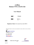
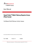
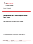
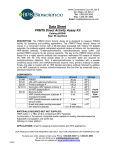
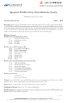
6044 Cornerstone Court West, Suite E
San Diego, CA 92121
Tel: 1.858.829.3082
Fax: 1.858.481.8694
Email: [email protected]
Data Sheet
AP1 Reporter Kit (JNK Signaling Pathway)
Catalog #: 60612
Background
The stress-activated protein kinase/c-jun N-terminal kinase (SAPK/JNK) family of proteins
includes mitogen-activated protein kinases (MAPKs) that are activated by stress, inflammatory
cytokines, mitogens, oncogenes, and inducers of cell differentiation and morphogenesis. Upon
activation of the SAPK/JNK pathway, MAP Kinase Kinases phosphorylate and activate JNKs.
The activated JNKs translocate to the nucleus where they phosphorylate and activate
transcription factors such as c-Jun. The activated c-Jun forms homodimers or heterodimers
with fos family proteins which bind to the activator protein-1(AP1) response element and induce
target gene transcription.
Description
The AP1 Reporter Kit is designed for monitoring the activity of the JNK signaling pathway and
the transcriptional activity of AP1 in cultured cells. The kit contains a transfection-ready AP1
luciferase reporter vector. This reporter contains the firefly luciferase gene under the control of
multimerized AP1 responsive elements located upstream of a minimal promoter. The AP1
reporter is premixed with a constitutively-expressing Renilla luciferase vector that serves as an
internal control for transfection efficiency.
The kit also includes a non-inducible firefly luciferase vector premixed with constitutivelyexpressing Renilla luciferase vector as a negative control. The non-inducible luciferase vector
contains the firefly luciferase gene under the control of a minimal promoter, without any
additional response elements. The negative control is critical for determining pathway-specific
effects and the background luciferase activity.
Applications
•
•
•
Monitor JNK signaling pathway activity and AP1-mediated activity.
Screen for activators or inhibitors of the JNK signaling pathway.
Study effects of RNAi or gene overexpression on the activity of the JNK pathway.
OUR PRODUCTS ARE FOR RESEARCH USE ONLY. NOT FOR DIAGNOSTIC OR THERAPEUTIC USE.
To place your order, please contact us by Phone 1.858.829.3082 Fax 1.858.481.8694
Or you can Email us at: [email protected]
Please visit our website at: www.bpsbioscience.com
140827
6044 Cornerstone Court West, Suite E
San Diego, CA 92121
Tel: 1.858.829.3082
Fax: 1.858.481.8694
Email: [email protected]
Components
Component
Reporter
(Component A)
Specification
AP1 luciferase reporter vector +
constitutively expressing Renilla
luciferase vector
Negative Control Non-inducible luciferase vector +
Reporter
constitutively expressing Renilla
(Component B)
luciferase vector
Amount
500 µl
(60 ng DNA/ µl)
Storage
-20°C
500 µl
(60 ng DNA/ µl)
-20°C
Note: These vectors are designed for transient transfection. They are NOT SUITABLE for
transformation and amplification in bacteria.
Materials Required but Not Supplied
•
•
•
•
•
•
Mammalian cell line and appropriate cell culture medium
96-well tissue culture plate or 96-well tissue culture-treated white clear-bottom assay
plate
Transfection reagent for mammalian cell line [We use Lipofectamine™ 2000 (Life
Technologies #11668027). However, other transfection reagents work equally well.]
Opti-MEM I Reduced Serum Medium (Life Technologies #31985-062)
Dual luciferase assay system:
Dual-Glo® Luciferase Assay System (Promega #E2920): This system assays cells
directly in growth medium. It can be used with any luminometer. Automated injectors
are not required.
OR
Dual-Luciferase® Reporter Assay System (Promega #E1910): This system requires a
cell lysis step. It is ideal for luminometers with automated injectors.
Luminometer
Generalized Transfection and Assay Protocols
The following procedure is designed to transfect the reporter into HEK293 or HeLa cells using
Lipofectamine 2000 in a 96-well format. To transfect cells in different tissue culture formats,
adjust the amounts of reagents and cell number in proportion to the relative surface area. If
using a transfection reagent other than Lipofectamine 2000, follow the manufacturer’s
transfection protocol. Transfection conditions should be optimized according to the cell type and
study requirements.
All amounts and volumes in the following setup are given on a per-well basis.
OUR PRODUCTS ARE FOR RESEARCH USE ONLY. NOT FOR DIAGNOSTIC OR THERAPEUTIC USE.
To place your order, please contact us by Phone 1.858.829.3082 Fax 1.858.481.8694
Or you can Email us at: [email protected]
Please visit our website at: www.bpsbioscience.com
140827
6044 Cornerstone Court West, Suite E
San Diego, CA 92121
Tel: 1.858.829.3082
Fax: 1.858.481.8694
Email: [email protected]
1. One day before transfection, seed cells at a density of ~ 30,000 cells per well in 100 µl of
growth medium so that cells will be 90% confluent at the time of transfection.
2. The next day, for each well, prepare complexes as follows:
a. Dilute DNA mixtures in 15 µl of Opti-MEM I medium (antibiotic-free). Mix gently.
Depending upon the experimental design, the DNA mixtures may be any of following
combinations:
• 1 µl of Reporter (component A); in this experiment, the control transfection is 1 µl of
Negative Control Reporter (component B).
• 1 µl of Reporter (component A) + experimental vector expressing gene of interest; in
this experiment, the control transfections are: 1 µl of Reporter (component A) +
negative control expression vector, 1 µl of Negative Control Reporter (component B)
+ experimental vector expressing gene of interest, and 1 µl of Negative Control
Reporter (component B) + negative control expression vector.
• 1 µl of Reporter (component A) + specific siRNA; in this experiment, the control
transfections are: 1 µl of Reporter (component A) + negative control siRNA, 1 µl of
Negative Control Reporter (component B) + specific siRNA, and 1 µl of Negative
Control Reporter (component B) + negative control siRNA.
Note: we recommend setting up each condition in at least triplicate, and preparing
transfection cocktail for multiple wells to minimize pipetting errors.
b. Mix Lipofectamine 2000 gently before use, then dilute 0.35 µl of Lipofectamine 2000 in
15 µl of Opti-MEM I medium (antibiotic-free). Incubate for 5 minutes at room temperature.
Note: Prepare this dilution cocktail in volumes sufficient for the whole experiment.
c. After the 5 minute incubation, combine the diluted DNA with diluted Lipofectamine 2000.
Mix gently and incubate for 25 minutes at room temperature.
3. Add the 30 µl of the complexes to each well containing cells and medium. Mix gently by
tapping the plate.
4. Incubate cells at 37°C in a CO2 incubator for overnight.
5. The next day, change medium to assay medium (Opti-MEM I, 0.5% FBS, 1% non-essential
amino acids, 1 mM Na-pyruvate, 1% Pen/Strep) containing an activator of AP1 such as
PMA. Incubate cells at 37°C in a CO2 incubator for ~ 6 to 24 hours. After treatment, perform
the dual luciferase assay following the manufacturer’s protocol.
OUR PRODUCTS ARE FOR RESEARCH USE ONLY. NOT FOR DIAGNOSTIC OR THERAPEUTIC USE.
To place your order, please contact us by Phone 1.858.829.3082 Fax 1.858.481.8694
Or you can Email us at: [email protected]
Please visit our website at: www.bpsbioscience.com
140827
6044 Cornerstone Court West, Suite E
San Diego, CA 92121
Tel: 1.858.829.3082
Fax: 1.858.481.8694
Email: [email protected]
Sample protocol to determine the effect of PMA on AP1 reporter activity in HEK293 or
HeLa cells
1. One day before transfection, seed HEK293 or HeLa cells at a density of 30,000 cells per
well into white clear-bottom 96-well plate in 100 µl of growth medium (MEM/EBSS (Hyclone
#SH30024.01), 10% FBS, 1% non-essential amino acids, 1 mM Na-pyruvate, 1%
Pen/Strep). Incubate cells overnight at 37°C in a CO2 incubator.
2. The next day, transfect 1 µl of AP1 reporter (component A) into cells following the
procedure in Generalized Transfection and Assay Protocols.
3. Incubate cells at 37°C in a CO2 incubator overnight.
4. The next day after transfection, prepare 1 mM stock solution of PMA in DMSO. Dilute the
PMA stock in assay medium. Change cell medium to 50 µl of diluted PMA in assay medium
to induce the AP1 reporter. For unstimulated control wells, treat cells with 50 µl of assay
medium (without PMA). Add 50 µl of assay medium (without PMA) to cell-free control wells
to determine the background luminescence.
Set up each treatment in at least triplicate.
5. Incubate cells at 37°C in a CO2 incubator for ~ 6 hours.
6. Perform dual luciferase assay using Dual-Glo® Luciferase Assay System (Promega
#E2920): Add 50 µl of Luciferase reagent per well and rock at room temperature for ~15
minutes, then measure firefly luminescence using a luminometer. Add 50 µl of Stop & Glo
reagent per well. Rock at room temperature for ~15 minutes and measure Renilla
luminescence.
7. To obtain the normalized luciferase activity for the AP1 reporter, subtract the background
luminescence, then calculate the ratio of firefly luminescence from AP1 reporter to Renilla
luminescence from the control Renilla luciferase vector.
OUR PRODUCTS ARE FOR RESEARCH USE ONLY. NOT FOR DIAGNOSTIC OR THERAPEUTIC USE.
To place your order, please contact us by Phone 1.858.829.3082 Fax 1.858.481.8694
Or you can Email us at: [email protected]
Please visit our website at: www.bpsbioscience.com
140827
6044 Cornerstone Court West, Suite E
San Diego, CA 92121
Tel: 1.858.829.3082
Fax: 1.858.481.8694
Email: [email protected]
Figure 1. PMA induced the expression of AP1 reporter. The results are shown as fold
induction of normalized AP1 reporter activity. Fold induction is determined by comparing values
against the mean value for unstimulated control cells.
Figure 2. Dose response of AP1 reporter activity to PMA in HEK293 cells. The results are
shown as fold induction of normalized AP1 reporter activity. Fold induction is determined by
comparing values against the mean value for unstimulated control cells.
The EC50 of PMA is ~5.4 nM
OUR PRODUCTS ARE FOR RESEARCH USE ONLY. NOT FOR DIAGNOSTIC OR THERAPEUTIC USE.
To place your order, please contact us by Phone 1.858.829.3082 Fax 1.858.481.8694
Or you can Email us at: [email protected]
Please visit our website at: www.bpsbioscience.com
140827
6044 Cornerstone Court West, Suite E
San Diego, CA 92121
Tel: 1.858.829.3082
Fax: 1.858.481.8694
Email: [email protected]
Sample protocol to determine the effect of JNK pathway inhibitor on AP1 reporter activity
1. One day before transfection, seed cells at a density of 30,000 cells per well into white clearbottom 96-well plate in 100 µl of growth medium. Incubate cells overnight at 37°C in a CO2
incubator.
2. The next day, transfect 1 µl of AP1 reporter (component A) into cells following the
procedure in Generalized Transfection and Assay Protocols.
3. Incubate cells at 37°C in a CO2 incubator for overnight.
4. The next day after transfection, prepare 10 mM stock solution of JNK inhibitor V in DMSO.
Dilute the inhibitor in assay medium to a final concentration of 10 µM. Carefully remove the
medium from wells and add 45 µl of diluted inhibitor in assay medium to the wells.
Add 45 µl of assay medium (without inhibitor) to inhibitor control wells or unstimulated
control wells.
Add 45 µl of assay medium (without inhibitor) to cell-free control wells (for determining
background luminescence).
Set up each treatment in at least triplicate.
5. Incubate cells at 37°C in a CO2 incubator for 1 hour.
6. Add 5 µl of diluted PMA in assay medium to wells. The final PMA concentration for the cells
is 10 nM.
Add 5 µl of assay medium without PMA to the unstimulated control wells (cells without
inhibitor and without PMA treatment for determining the basal activity).
Add 5 µl of assay medium without PMA to cell-free control wells.
7. Incubate cells at 37°C in a CO2 incubator for 6 hour.
8. Perform dual luciferase assay using Dual-Glo® Luciferase Assay System (Promega
#E2920): Add 50 µl of Luciferase reagent per well and rock at room temperature for ~15
minutes, then measure firefly luminescence using a luminometer. Add 50 µl of Stop & Glo
reagent per well. Rock at room temperature for ~15 minutes and measure Renilla
luminescence.
9. To obtain the normalized luciferase activity for the AP1 reporter, subtract the background
luminescence, then calculate the ratio of firefly luminescence from the AP1 reporter to
Renilla luminescence from the control Renilla luciferase vector.
OUR PRODUCTS ARE FOR RESEARCH USE ONLY. NOT FOR DIAGNOSTIC OR THERAPEUTIC USE.
To place your order, please contact us by Phone 1.858.829.3082 Fax 1.858.481.8694
Or you can Email us at: [email protected]
Please visit our website at: www.bpsbioscience.com
140827
6044 Cornerstone Court West, Suite E
San Diego, CA 92121
Tel: 1.858.829.3082
Fax: 1.858.481.8694
Email: [email protected]
Figure 3. Inhibition of PMA-induced AP1 reporter activity by JNK inhibitor V. The results
are shown as normalized AP1 reporter luciferase activity.
References
Zhou H. et. al. (2005) Frequency and distribution of AP-1 sites in the human genome. DNA
Research. 11: 139-150.
Gaillard P. et.al. (2005) Design and synthesis of the first generation of novel potent, selective,
and in vivo active (benzothiazol-2-yl)acetonitrile inhibitors of the c-Jun N-terminal kinase. J Med
Chem. 48(14):4596-4607.
Related Products
Product Name
AP1 Reporter – HEK 293 cell line
SRE Reporter Kit (MAPK/ERK Signaling Pathway)
MAPK10 (JNK3), human
JNK1-β1(K55M), human
MAP3K14 (NIK), human
MAPKAPK2 (MK2), human
JNK1, mouse
JNK2, human
JNK3, human
ERK1, human
ERK2, human
ERK2, inactive, human
Catalog #
60405
60511
40092
40871
40090
40088
40071
40113
40114
40055
40299
40056
Size
1 vial
500 reactions
10 µg
100 µg
10 µg
100 µg
10 µg
10 µg
10 µg
10 µg
10 µg
10 µg
OUR PRODUCTS ARE FOR RESEARCH USE ONLY. NOT FOR DIAGNOSTIC OR THERAPEUTIC USE.
To place your order, please contact us by Phone 1.858.829.3082 Fax 1.858.481.8694
Or you can Email us at: [email protected]
Please visit our website at: www.bpsbioscience.com
140827