1
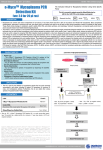
qPCR – MD instructions │ MAY, 2015 (1st Edition)
ISO 9001 / 14001 / 13485 Certified Company
Test for the detection of Mycoplasma by qPCR analysis
e-Myco™ VALiD-Q
-15ºC
Mycoplasma qPCR Detection Kit
RUO
BACKGROUND INFORMATION
PRINCIPLES
• The real-time qPCR(quantitative polymerase chain reaction) DNA amplification technology
shows high sensitivity and specificity for direct detection of pathogen (antigen). iNtRON
developed a novel platform technique about primer design called CLP™ (complementary locking
primer) technology which provides flexibility in Tm (melting temperature) of primer design for
optimization of reaction condition, and maximizes PCR specificity and sensitivity through the
control of non-specific priming.
• The kit contains all reagents required for real-time polymerase chain reaction: nucleotides,
primer and probes, reaction buffer, polymerase and controls. After rehydration of the
components the PCR Master mix is easily prepared by just adding the Inhibition Control DNA
and Taq DNA polymerase to the Primer & Probes.
• The PCR is initiated with a 5 minute high-temperature step to melt all nucleic acids and to
activate the polymerase. In a successful PCR, the presence of mycoplasmas will be indicated by
a signal in the FAM channel.
Contents
50
-25ºC
Storage
Amount
2X qPCR Master Mix Solution
-25 ~ -15ºC
500 μl x 1 vial
(10 μl / test × 50 tests)
Mycoplasma Detection Solution
-25 ~ -15ºC
250 μl x 1 vial
Positive Control (External PC)
-25 ~ -15ºC
25 μl x 1 vial
DNase/RNase Free Water
-
250 μl x 1 vial
Manual
-
1 ea
NOTICE
• To prevent contamination of mycoplasma DNA during experimental procedure, always wear
gloves during sample preparation and PCR reaction setup.
• To avoid false positives, water used in PCR reactions can be UV-irradiated.
• If no internal positive control signal, it shows the problem during PCR process. Please re-test.
• If there is non-specific signal in negative control, it could be due to the contamination or overused template. Please re-test with proper amount of template.
SHELF-LIFE
• 12 months from manufacturing date.
• Within 3 months after opening, within expiry date of the kit.
KIT WORKFLOW
Cultivated Cell
KIT CONTENTS
2X qPCR Master Mix
Solution
Σ
Prototype
PACKAGING INFORMATION AND STORAGE
Mycoplasmas are small, round or filamentous prokaryotic organisms which are a frequent
contaminant of cell cultures. Mycoplasma depend on their hosts for many nutrients due to their
limited biosynthetic capabilities. Up to 30~85% of cell cultures may be contaminated with
mycoplasmas, the main contaminants being the species M.orale, A.laidlawii, M.arginini and
M.hyorhinis. Although these mycoplasmas do not usually kill contaminated cells, they are difficult to
detect and can cause a variety of effects on cultured cells, including changes in metabolism growth,
viability and morphology, there by altering the phenotypic properties of the host cells. Many
methods are available for detection of mycoplasma, including isolation in broth/agar culture, direct
or indirect fluorescence staining, ELISA, immunostaining, direct or indirect PCR. Among those
methods, direct PCR is the highly sensitive, specific and convenient method when the primer
design is optimized.
The e-Myco™ VALiD-Q Mycoplasma qPCR Detection Kit is composed of a set of primers and
probe that are specific for the highly conserved mycoplasma16S-rRNA coding region including
M.pneumoniae, M.argnini, M.hyorhinis, M.fermentans, M.orale and A.laidlawii. The kit is designed
to detect the presence of mycoplasma that might contaminate biological materials such as cultured
cells. Also, the kit can detect mycoplasma within 90minutes sensitively up to 10CFU/ml and
includes internal control for verifying a qPCR run as well as positive control DNA.
Contents
REF
PROTOCOL A
PROTOCOL B
Cell boiling method
Genomic DNA method
Composition
Cell harvest
& washing
• Real-time PCR Reaction solution
< 0.01% dATP, dTTP, dGTP, dCTP
< 0.01% Hot start PCR enzyme
< 0.01% PCR additive materials
• Mycoplasma Detection solution
Detection Solution
DNase/RNase Free Water
< 0.001% Primer /probe for Mycoplasma
< 0.001% Internal control primer pair
< 0.001% Internal control DNA
Boiling
(95ºC, 10min)
• Ultrapure sterilized distilled water
DNA purification using G-spin Total DNA
Extraction Kit(Cat. No. 17046)
positive control
Positive Control (External PC) • Mycoplasma
< 0.001 % Recombinant DNA contained 16S sequence of M. hyorhinis
Instruction Manual
Crude lysate
(Supernatant)
Purified DNA
STORAGE AND STABILTY
• Storage condition: Store the product at -25~-15ºC after receiving.
• Expiration: e-Myco™VALiD-Q Mycoplasma PCR Detection Kit can be stored for up to 12
months without showing any reduction in performance and quality under appropriates or age
condition. The expiration date is labeled on the product box.
APPLICATION
The kit is used for the detection of mycoplasma species that are most commonly encountered in
cell culture, including M.peumoniae, M.arginini, M.fermentans, M.hyorhinis, M.orale, and A. laidlawii.
Furthermore, this kit can detect other various species of mycoplasma.
Perform
Real time PCR
10μl
5μl
2X qPCR
Master Mix
Detection
Solution
5μl
Template
Step
Pre-denaturation
PCR and
Signal Detection
MATERIALS REQUIRED BUT NOT PROVIDED
•
•
•
•
Realtime PCR Instrument
Pipettes
Centrifuge for micro-centrifuge tubes
Disposable gloves
IBT-QMS-Prototype
• G-spin total Extraction Kit (Cat. 17045)
• Sterile pipette tip (with filter)
• Vortex mixer
1/2
Analyze the results
[email protected] www.intronbio.com www.intron-innoplex.com
PCR tube
(optic grade)
Cycle Temp
1
40
Time
Channel setting
95 ºC
5’ 30”
Mycoplasma
FAM
95 ºC
15”
IPC
HEX
58 ºC
1’
qPCR – MD instructions │ MAY, 2015 (1st Edition)
ISO 9001 / 14001 / 13485 Certified Company
PROTOCOL
DATA VALIDATION
You can use this protocol just for detecting the contamination of mycoplasma. However, if you want
to perform genotyping for the detailed determination of species, please purify the genomic DNA of
suspected Mycoplasma-infected cells using our G-spin Total DNA Extraction Kit (Cat.No.17046).
You may use simply this protocol or your other general boiling methods.
[ TECHNICAL TIP ]
1. Use clean, disposable gloves when performing the assay and make sure that the work area is
clean prior to starting the assay setup.
2. Keep your reagents and PCR mixture tubes on a cold block during reaction setup.
3. Use positive displacement pipettes.
4. The amplification and detection areas should be physically separated; i.e., do not use the same
bench area to set up the PCR reactions and run your gels.
1. When the reaction is finished, put a cut-off value according to the below table.
Set Manual baseline
Threshold
Ct Cutoff Value
3 ~15
Auto
Drop after 36 cycle
2. Valid Results : Ct value of control should be as below table
Items
FAM
HEX
Items
FAM
HEX
Positive Control
18 ~ 22
22 ~ 25
Negative Control
<36
<36
DATA INTERPRETATION
PROTOCOL A : Using the Boiling Extract Method
1. Expected Real-time RT-PCR Data
1. Prepare cell suspensions from the test cell culture in a 1.5 ml tube. Then count cell
numbers by general counting methods. You need at least 5x10⁴ cells per test.
Note 1: Harvest adherent cells with trypsin-EDTA solution using standard techniques. Pipette 1
ml of TE-treated adherent cells. Generally, with suspension cells, such as K562, you need not
treat with TE solution. We recommend that you count the cells. You should prepare at least
5x10⁴ cells per test.
Note 2: Strong mycoplasma infections are detected in as little as 10~100 cells, while weak
infections require cells over 5,000~50,000 cells. You can dilute the template according to the
infection rates you suspect
Positive control
Mycoplasma
(FAM)
+
IPC
(HEX)
+
2
Negative control
-
+
Valid
3
Test 1
+
+
Positive
4
Test 2
+
-
Positive (High concentration
of Mycoplasma DNA)
Negative (Mycoplasma Free)
No.
Samples
1
Interpretation
Valid
2. Transfer the counted cells (over 5x10⁴ cells) to a 1.5 ml tube. Spin the tube in a
microcentrifuge for 10~15 seconds. Carefully decant the supernatant.
5
Test 3
-
+
6
Positive Control
-
+
Retest
3. Resuspend the cells in 1 ml of sterile PBS or DPBS solution for washing.
7
Negative Control
+
+
Contamination
4. Spin the tube in a microcentrifuge for 10~15 seconds. Carefully decant the supernatant.
Note : [Option] Repeat this wash step once more.
5. Resuspend the cell pellets in 100 μl of sterile PBS or DPBS solution.
Note : If you want the best result, use of PBS solution is better than Tris (10 mM, pH 8.5), TE
(10 mM Tris, 0.1 mM EDTA), or autoclaved DW.
6. Heat the samples at 95 ºC for 10 min, and vortex for 5-10 sec. Then, centrifuge for 2 min
at 13,000 rpm with a tabletop centrifuge (at RT).
EXPLANATION OF SYMBOLS
LOT
Σ
Batch number
Components
10 μl
Mycoplasma Detection Solution
5 μl
Total volume
15 μl
10. After centrifugation, put them into a real-time PCR system and process reaction.
Channel setting
Step
Cycle
Temp
Time
Pre-denaturation
1
95 ºC
5’ 30”
Mycoplasma
FAM
95 ºC
15”
IPC
HEX
58 ºC
1’
PCR and
Signal Detection
40
Observation
Possible Cause
Recommendation
Incorrect dye components chosen
Reaction component omitted
Check that all the correct reagents were
added
Degraded template or no template added
Repeat with fresh template
Reaction inhibitor present
Repeat with purified template
ΔRn ≤ No Template
control ΔRn, and both
reaction show an
amplification plot
Template contamination of reagents
Check technique and equipment to
confine contamination. Use fresh
reagents
Amplification plot dips
downwards
Ct Value less than 15, amplification signal
detected too early
Reset the upper/lower value of baseline
(two cycles lower than Ct Value). or
repeat with diluted sample
Amplification plots is not
within the log phase
PCR efficiency is poor
Re-optimization the reaction conditions
Less template added than expected
Increase sample amount
Sample is degraded
Evaluate sample integrity
More template added than expected
Reduce sample amount
Template contamination of reagents
Check technique and equipment to
confine contamination. Use fresh
reagents.
Repeat with aerosol barrier pipette tip
after space cleaning
Ct value is lower than
expected
PROTOCOL B : Using genomic DNA as a template
Expire date
Check dye component prior to data
analysis
Ct value is higher than
expected
* Under line means signal detection step
Product number
TROUBLESHOOTING GUIDE
ΔRn ≤ No Template
control ΔRn, and no
amplification plot
9. Fill up with the supernatant 5μl and Master Solution 15μl in the PCR tube.
Note 1 : For Negative Control : 5μl DNase / RNase Free Water
Note 2 : For Positive Control : 5μl Positive Control
REF
Storage temperature limitation
Manufacturing date
Master Solution (per test)
2X qPCR Master Mix Solution
for research use only
50 Sufficient for 50 tests
7. Transfer an aliquot of the heated supernatant to a fresh tube. This supernatant will be
used as the template in the PCR.
8. Prepare Detection Mix by dispensing components to each real-time PCR tube in the
following manner
RUO
Repeat with aerosol barrier pipette tip
after space cleaning
1. Add 5μl of purified genomic DNA as a template using the G-spin Total DNA Extraction Kit
(Cat. No. 17046), and then resuspend after adding Master Solution 15μl in the PCR tube.
Note : Appropriate amounts of DNA template sample: genomic DNA, 50 ng–100 ng
2. Follow protocol A from step 10.
Note: Recommend to perform one negative control reaction by adding 5μl of sterile water. We
recommend to add 5μl of control DNA for positive control reaction.
Technical advice : +82-505-550-5600
Review date : 2015. 5. 6.
IBT-QMS-Prototype
2/2
[email protected] www.intronbio.com www.intron-innoplex.com