1
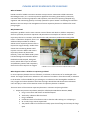
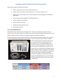
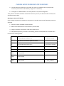
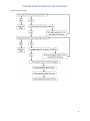
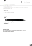
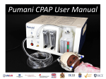
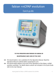
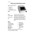
PUMANI bCPAP GUIDELINES FOR CLINICIANS An Overview of the Pumani bCPAP, Indications for bCPAP, and Instructions for Use PUMANI bCPAP GUIDELINES FOR CLINICIANS What is bCPAP? bCPAP stands for bubble Continuous Positive Airway Pressure. Sometimes called Continuous Distending Pressure, it is a constant pressure applied to the airway, generated by continuous, consistent flow of air during inspiration and expiration, with the aim of opening collapsed lung segments and maintaining patency in already opened air spaces. bCPAP is now being introduced in Malawi as the next step in the management of severe respiratory distress in children after nasal prong oxygen. Why bCPAP now? Respiratory problems are the most common cause of illness and death in children. Respiratory distress syndrome, meconium aspiration and pneumonia are examples of common causes of respiratory distress in neonates, while bronchiolitis and pneumonia predominate among older infants. In Malawian hospitals, there is no additional help available for children whose severe respiratory distress fails to respond to oxygen therapy. Studies have shown that introducing bCPAP could improve survival in babies with severe respiratory disease by about 70%. Recently, a low cost bubble CPAP system has been designed and tested at Queen Elizabeth Central Hospital, with good results, which offers hope to neonatal and paediatric patients with respiratory distress who fail to improve on oxygen. A bCPAP in use in the neonatal ward of QECH in March 2012. What happens when a child has a respiratory problem? In most respiratory diseases there is effectively a reduction in the amount of air exchanged in the alveoli. This might result from a reduction in the amount of air taken in from the outside; a reduction in the alveolar surface available for gas exchange or a reduction in the haemoglobin available to carry oxygen and carbon dioxide to and from the lungs. We will describe how these mechanisms come into play in some of the common causes of respiratory distress in children. These are some of the common respiratory diseases in neonates and young children: 1. Respiratory Distress Syndrome, RDS (also called Hyaline Membrane Disease, HMD) a. Lung is not mature and alveoli not well developed b. Thick sticky substance fills the alveoli c. Lung is solidified d. Insufficient levels of surfactant result in alveolar walls sticking in or collapsing in e. As a result, lungs cannot expand well f. The patient needs a lot of effort to initially open the stiff lungs and to keep the lungs open 2 PUMANI bCPAP GUIDELINES FOR CLINICIANS 2. Pneumonia/pneumonitis a. There is an inflammatory process going on in the lungs due to infection or other agents b. Lungs are swollen and firm c. Thick exudate fills the alveoli d. Some surfactant is destroyed during the inflammatory process e. As a result, areas of the lung will not expand well while others will collapse f. Additional effort or pressure is needed to open the lungs and to prevent them from collapsing back 3. Airway Obstruction a. Depends on cause and level of obstruction b. Mostly there is swelling of the lining of the airway, reducing the lumen c. The status in ‘a’ and ‘b’ will result in more effort needed to move air in and out through the airway, depending on the level of the obstruction d. Sometimes thick secretions in airway and/or the alveoli e. Might have segments of collapse, while others might be hyper-expanded f. Either the lung cannot expand, or cannot ventilate The major effect of these pathologies is to reduce the lung surface area available for gaseous exchange, resulting in hypoxia. Respiratory Distress Respiratory distress describes a response to the hypoxia. Initially the body fights to maintain oxygen levels delivered to the brain by increasing the work of the cardiovascular and respiratory systems. Clinically the patient will demonstrate the following: 1. 2. 3. 4. 5. Increased respiratory rate and heart rate Chest in-drawings and use of accessory muscles Grunting and nasal flaring Poor feeding Low oxygen saturations As the hypoxia progresses, the patient might become pale and cyanosed, and the level of consciousness may deteriorate. The aim of treatment in a patient with respiratory distress is to achieve better oxygenation and reverse the hypoxia and therefore improve some of the signs that are mentioned above. How does the bCPAP help? On inspiration, bCPAP drives air with additional pressure into collapsed alveoli and opens them. This is like what happens when you first blow into a collapsed balloon. This process is sometimes called ‘recruitment’. Because of the continuous airflow, this pressure is maintained even when the patient breathes out and therefore the alveoli do not collapse at the end of expiration. This makes the lungs expand more easily and increases the surface area available for gaseous exchange and therefore improves oxygenation. This in turn reduces the need for increased work of breathing. 3 PUMANI bCPAP GUIDELINES FOR CLINICIANS When does my patient need bCPAP? bCPAP is usually used as the next step in respiratory therapy after oxygen in patients who present with respiratory distress. Therefore the majority of the patients who go on bCPAP treatment will be those who remain with severe respiratory distress despite oxygen therapy. However, some patients might require bCPAP right from the beginning judging from the degree of distress and the overall clinical picture. As a guide, patients with the following conditions can benefit from bCPAP: 1. Respiratory distress syndrome 2. Meconium aspiration syndrome 3. Transient tachypnoea of the newborn (TTN) 4. All forms of pneumonia or pneumonitis 5. Bronchiolitis 6. Apnoea of prematurity 7. Lung collapse / atelectasis 8. Upper airway obstruction 9. Pneumocystis Pneumonia (PCP) All other indications should be discussed with senior clinicians in your hospital. In the situation where you have a child with severe respiratory distress and cannot be sure about the differential diagnosis, it will be safer to start the bCPAP and then discuss with more senior people on how to proceed. Always remember that clinicians at the district or central hospitals in your service area should be at your disposal to give advice where you have questions about specific patients. Specific indications for bCPAP Signs of severe respiratory distress as defined by the WHO: 1. Tachypnoea 2. Severe subcostal or intercostal recessions 3. Use of accessory muscles 4. Nasal flaring 5. Grunting; particularly in small babies, the presence of grunting represents a patient who may be struggling to maintain Positive End Expiratory Pressure (PEEP) and therefore may benefit from bCPAP 6. Oxygen saturations below 90% despite 2 L/min of oxygen therapy 7. Cyanosis 8. Agitation / restlessness 9. Breathlessness / exhaustion 4 PUMANI bCPAP GUIDELINES FOR CLINICIANS Patients who might not benefit from bCPAP: 1. Patients who do not have severe respiratory distress 2. Patients whose respiratory distress improves on 2 L/min of oxygen or less 3. Upper airway abnormalities such as choanal atresia, tracheosophageal atresia/fistula, cleft palate 4. Severe cardiovascular instability and impending arrest 5. Absent respiratory drive/effort 6. Profound sepsis and shock 7. Congenital diaphragmatic hernia 8. Severe birth asphyxia The Pumani bCPAP Machine All bCPAP machines consist of four basic elements. The first is a high flow air pump run from a power source. Then there must be a conduit for air from the pump, through a flow valve with an air gauge to regulate airflow and then to a pressure regulator and patient interface. In the Pumani bCPAP machine (pictured below), which is being rolled out in Malawi, two aquarium pumps powered by electrical mains and housed in a metal container provide the flow. The flow valves are attached on the outside front of the unit. A water bottle on the right side of the machine serves as a pressure regulator. The outlet to the patient interface is located on the front of the unit. Additionally, an inlet from an oxygen source is provided for attachment when a patient requires oxygen in addition to the bCPAP flow and pressure. The oxygen flow is regulated either on the oxygen concentrator gauge or through the gauge on the bCPAP unit itself. The oxygen and blended flows are set on the bCPAP using the table below (also found in the Pumani User Manual). 5 PUMANI bCPAP GUIDELINES FOR CLINICIANS Commencing a patient on bCPAP Requirements: 1. Air flow and pressure source (Pumani bCPAP machine) 2. Oxygen source (oxygen concentrator) 3. Power supply (Power cable should be attached from the back of the machine) 4. Bottle tubing, patient tubing 5. Hat, nasal prongs (sizes 1-4, depending on the size of the patient’s nostrils), safety pins, elastics 6. Orogastric tubes 7. Suction machine and catheters 8. Saline drops 9. Pulse oximeter 10. Respiratory Rate timer Setting up a Patient on the Pumani bCPAP Machine Location. The bCPAP machine should be set up in the high care area / corner of your paediatric / neonatal ward to ensure above average monitoring. If possible, this location should be close to the nursing station so that the nursing staff can always easily keep an eye on the patient. It works better if all patients on bCPAP are nursed close together instead of being scattered in different corners of the ward. Setting up the machine. If you have a patient with severe respiratory distress, first of all carry out the necessary ABC assessment. 1. Put the patient on nasal prong oxygen, starting with 1 L/min and assess response. Increase the oxygen flow to 2 L/min if there is no response. If at 2 L/min O2 administration there are still signs of severe distress or saturation below 90%, increase the oxygen flow to the maximum flow rate and, if possible, use face mask oxygen and then start setting up the bCPAP. Face masks should be run with at least 5 liters of oxygen. Make sure you keep an eye on the patient while setting up the machine. 2. Check to ensure that the bCPAP machine is in working order, the power is connected, and that the oxygen concentrator, tubings and attachments are all available 3. Fill the bottle with tap water, up to 6 cm 4. Connect the bottle tubing to the bottle connector (labeled with an picture of a bottle) and the patient tubing to the patient connector (labeled with a picture of a baby) 5. Start all patients on the following flow settings: Total Air flow at 6 L/min 6 PUMANI bCPAP GUIDELINES FOR CLINICIANS Oxygen flow: o Neonates: 3 L/min o Patients over 1 month old: 4 L/min Preparing & inserting the prongs. Do not insert the prongs without the machine on. 1. Select appropriate sizes for the prongs and hat 2. Transfer the patient, while still on oxygen, to the CPAP bed or warming crib 3. Clean the nose with water; gently suction nostrils 4. Insert orogastric tube for feeds and for deflating the abdomen 5. Place hat on the baby 6. Ensure the nostrils are open, apply 2 drops of saline to each nostril to keep the nostrils open and moist 7. Attach the prongs to the hat with the pins and elastics 8. Remove the oxygen tubing immediately before inserting the nasal prongs 9. Put the nasal prongs far enough into the nose to completely fill the nostril, but maintain at least ¼ cm of space between the prongs and septum 10. Check for bubbling water in the bottle. If bubbling does not occur, check that the prongs fill the nostrils completely, that the tubings are well connected and are not kinking. Try increasing the total flow by 1 L at a time, up to 10 L/min. Monitoring patients on bCPAP Patients on bCPAP (as any other very sick patient) require more than average routine monitoring. However, this will depend on the nursing capacity on the ward. To ensure that the aim of providing bCPAP to patients is achieved, the following standard of monitoring should be provided as a minimum: About an hour after putting a patient on bCPAP, the patient should be reviewed and the following recorded in the notes: airway patency, current bCPAP settings (water level, Total flow, Oxygen flow), RR, HR, grunting, nasal flaring, oxygen saturations, recessions, abdominal distention, and general clinical condition. This is useful in terms of reviewing response to bCPAP and in helping to decide whether to change the bCPAP settings or not. If there is no improvement in oxygen saturations: 1. Check the tubings; ensure all connections are correct and no kinking 2. Check the airway; apply saline drops to nose and suction thoroughly 3. Check for abdominal distension; aspirate stomach and leave OGT to openly drain 4. Increase oxygen flow by 0.5 L 7 PUMANI bCPAP GUIDELINES FOR CLINICIANS 5. Then increase the pressure by 1 cm water at a time, to a maximum of 8 cm (any further increase in pressure must be made by a registrar or consultant) 6. Call registrar or Medical Officer to review patient or help with management Then patients on bCPAP should be reviewed at least every 6 hours; the above details recorded, and appropriate steps taken. Weaning a Patient off of bCPAP Start weaning the patient on bCPAP once the patient is clinically stable and the following criteria are achieved: 1. Patient has been on bCPAP at least 24 hours 2. RR less than 60/minute for at least 6 hours (for neonates) 3. Oxygen saturation consistently > 90% for at least 6 hours 4. No significant grunting, recessions, nasal flaring, apnoea or bradycardia for at least 6 hours Procedure for weaning bCPAP: 1. Decrease Water Level by 1 cm Every 6 hours until 5 cm level is reached Wait 6 hours to allow the patient to settle 2. Decrease Oxygen Flow by 0.5 L Every 6 hours until 1 L/min flow is reached Wait 6 hours to allow the patient to settle 3. Remove patient from CPAP and place on 2 L of O2 1 Hour Later Assess patient 6 Hours Later Assess patient 12 Hours Later Assess patient Continue assessing every 12 hours 8 PUMANI bCPAP GUIDELINES FOR CLINICIANS Flow chart for weaning: 9 PUMANI bCPAP GUIDELINES FOR CLINICIANS Complications of bCPAP Blocked nostrils Very common. Prevent and treat by regular saline drops and gentle suctioning. Nasoseptal irritation or necrosis Septal irritation common, necrosis less common. Prevented by careful selection of nasal prong sizes and not inserting the prongs too far into the nostrils. Further prevented by regular application of saline drops to the nostrils to prevent drying and crusting of the nasal mucosa. Distended abdomen This is common but prevented and treated by inserting an orogastric tube. You may need to aspirate stomach contents or leave the OGT on free drainage. Nose bleed Common in the dry season. Prevent and treat by applying regular nasal saline drops. Pneumothorax This is very uncommon. Usually due to excessive pressures applied to small, stiff lungs. Suspect if there is a sudden deterioration in respiration and oxygenation in a patient who was doing well on bCPAP or a patient who seems to be worse after starting bCPAP. Any patient not responding to CPAP of 8 cm water should be reviewed by a senior or trained clinician. Pulmonary haemorrhage This is very uncommon. Also due to very high pressures applied to small, immature lungs. Suspect if there is sudden deterioration in clinical condition, especially if there is associated pallor and significant bloody frothing from mouth or nose. Can be prevented by being cautious with the pressures applied to the baby’s lungs. Any patient not responding to CPAP of 8 cm water should be reviewed by a senior or trained clinician. Restarting bCPAP The criteria and procedure for restarting bCPAP are the same as for the initial commencement as listed above. Restarting bCPAP will also be subject to availability of a bCPAP machine and at the reviewing clinician’s discretion. Queries Further information about the Pumani bCPAP machine, troubleshotting, patient set-up, etc. is available in the Pumani bCPAP User Manual. 10