1
ExoQuick™ Exosome Precipitation Solution
Cat. # EXOQ__A-1
Data acquisition and analysis
These experiments were done in
collaboration with Particle Characterization
Laboratories, Inc (PCL).
2. Magnetic Beads and Exosome Surface
Markers
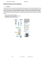
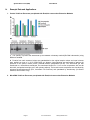
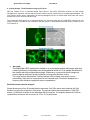
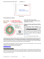
SBI has developed a magnetic streptavidin 9.1 um
Exo-Flow bead system. The 9.1 um diameter of the
beads enables more exosome capture per volume
added when compared to 4 um Dynabeads. This is
significant in that some exosome subpopulations
that are desired may only be present in very low
numbers. The increased surface area enables the
more efficient capture of these rare exosomes.
Bigger is better.
Fig. 1: Exo-Flow streptavidin magnetic beads
Depending upon the specific exosomes you wish to purify, a particular biotinylated antibody may be used to couple to
the Exo-Flow streptavidin beads. The Exo-Flow kits are modular, thus you can select from various pre-validated
capture antibody kits, or utilize your own biotinylated capture antibody corresponding to the exosome surface marker
specific for the exosomes of interest in your model system. SBI has thoroughly tested a variety of capture antibodies
that work quite well to flow-sort exosomes from either serum or cell culture samples.
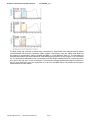
The data below were generated on a BD LSR II instrument and are meant as examples of how to set the gate
settings and perform data analysis using FlowJo software.
Information on FlowJo software can be viewed online.
http://www.flowjo.com/
Exo-Flow software gate settings
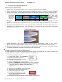
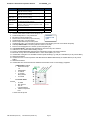
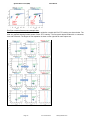
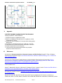
The forward and side scatter data for the 9.1 um Exo-Flow beads are shown below for samples containing no
captured exosomes stained with Exo-FITC (left panel) and then data for serum CD9-captured exosomes stained with
Exo-FITC. Set the gate primarily on the majority bead singlets (outlined in a black oval, red arrow pointing to the gate
setting) prior to full flow analysis.
888-266-5066 (Toll Free)
650-968-2200 (outside US)
Page 9