1
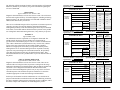
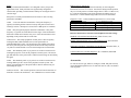
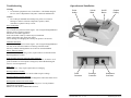
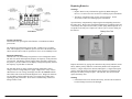
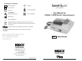
Explanation of Symbols
REF
Reorder Number
SN
Serial Number
Keep Dry
Type B Applied Part
Latex Free
ATTENTION:
See instructions for use.
Class II Equipment
Manufacturer
User Manual for
LifeDop L350R Series (Tabletop Doppler)
®
Date of Manufacture
Symbol indicates that the device should be sent to the special
agencies according to local regulations for separate collection after
its useful life.
Product conforms to the Medical Device Directive 93/42/EEC
Authorized Representative in the European Community.
®
®
LifeDop , Summit DopplerTM and Wallach are trademarks of CooperSurgical, Inc.
®
Medichoice is a registered trademark of Owens & Minor, Inc.
REF L350R, MC350R
®
Clorox is a registered trademark of The Clorox Company.
© 2013 Wallach Surgical Devices
95 Corporate Drive
Trumbull, CT 06611 USA
Phone: 800-243-2463
(203) 799-2000
Fax: (203) 799-2002
[email protected]
www.wallachsurgical.com
Made in the USA
MAN0019-DFU • Rev. A • 4/13
Wallach Surgical Devices
MAN0019-DFU • Rev. A • 4/13
Wallach Surgical Devices
Thank you for choosing LifeDop® L350 products. We believe you have
purchased the finest Doppler on the market today. Your total satisfaction is
our highest priority as we strive to continually improve our products and
services. Please contact us with any suggestions. We look forward to
enjoying a long-term relationship with you!
Wallach Surgical Devices
95 Corporate Drive
Trumbull, CT 06611 USA
Year of manufacture
located on the device.
Here’s how you can reach us…
Phone: 1-800-243-2463
(203) 799-2000
Fax:
(203) 799-2002
e-mail us at: [email protected]
visit our website at: www.wallachsurgical.com
Warranty and Servicing Policy
The warranty on this product is that it will be free from defects in material
and workmanship for 24 months from the original sale of the device.
Product life is specified to be 5 years from manufacture, though the device
may be repairable beyond this timeframe. This includes all parts and labor
required to repair or replace the unit to original specifications and shipping
costs associated with sending the product back to the customer. Customer is
responsible for providing adequate packaging materials and shipping costs to
Wallach Surgical Devices. Products shall be repaired or replaced in a
reasonable amount of time.
Wallach Surgical Devices’ liability for any claim is limited to materials and
labor associated with repair or replacement. In no event shall Wallach
Surgical Devices be liable for incidental or consequential losses or damages
in connection with the purchase of this product.
Wallach Surgical Devices disclaims all express or implied warranties,
agreements or arrangements other than issued in this warranty.
Table of Contents:
Page
Intended Use/Contraindications/Warnings .............................. 1
Description of Product ............................................................ 3
Operation and Installation ........................................................ 5
Obtaining Doppler Signals ....................................................... 8
Maintenance and Cleaning ...................................................... 10
Replacing Batteries ................................................................. 11
Troubleshooting ..................................................................... 12
Clinical References ................................................................ 13
Accessories ............................................................................. 13
Specifications .......................................................................... 14
Warranty and Servicing Policy ............................................... 17
Explanation of Symbols ................................................... Back Cover
Please read the manual carefully and become familiar with the operation,
features and maintenance of your LifeDop L350R prior to using the device
or accessories.
Wallach Surgical Devices is not responsible for damages to the device
that occur as a result of the inadequate packaging on return shipments to
Wallach Surgical Devices, improper maintenance or cleaning as described
in the user manual, misuse, abuse, alteration of the equipment from
its original specifications, or dismantling of the unit (other than by
Wallach Surgical Devices approved service technicians).
Service Returns – To return products:
1. Call Wallach Surgical Devices to obtain a Return Authorization and to
receive any final instructions prior to shipping.
2. Clean the product prior to shipping.
3. Ensure the device is well-packaged and suitable for shipment.
Send the product to:
Repair Department
Wallach Surgical Devices
95 Corporate Drive
Trumbull, CT 06611 USA
For customer service, please call 1-800-243-2463 or (203) 799-2000.
(This mark excludes the
Medichoice® model: MC350R)
MAN0019-DFU • Rev. A • 4/13
i
Wallach Surgical Devices
MAN0019-DFU • Rev. A • 4/13
17
Wallach Surgical Devices
Obstetrical Probe Information:
ISATA (max) (mW/cm2)
Po (mW)
Effective Radiating Area (cm2)
Ultrasound Frequency (MHz)
Pulse Duration
Repetition Freq.
ISATA
ISPTA.3
ISPPA.3
MI
Pr.3
Wo
fc
zsp
x-6, y-6
EBD
Model Number
2 MHz
3 MHz
19.6
17.0
48.0
24.5
2.45
1.57
2.1 MHz
3.2 MHz
CW
CW
CW
CW
the spatial-average temporal-average intensity (mwatts per cm2).
the derated spatial-peak temporal-average intensity (mwatts per cm2).
the derated spatial-peak pulse-average intensity (watts per cm2).
the Mechanical Index.
the peak rarefactional pressure (megapascals) associated with the transmit pattern
giving rise to the value reported for MI.
the total time-average ultrasonic power (mwatts).
the probe center frequency (MHz).
the axial distance at which the reported parameter is measured (cm).
are the –6dB beam dim. in the x-y plane where zsp is found (cm).
the entrance beam dimensions (cm). These dimensions are the same as the
dimensions of the transmit crystal.
Measurement Uncertainties:
Power:
Pressure:
Intensity (Ispta):
Frequency:
+34, -42%
+11, -16%
+23, -26%
± 5%
Vascular (4, 5 and 8 MHz Probes)
This product will be used to detect blood flow in veins and arteries for
assisting in the detection of peripheral vascular disease.
Caution
• U.S. Federal law restricts this device to sale by or on the order of a
licensed practitioner.
Contraindications
Warnings
• The vascular probes (4, 5 and 8 MHz) are not for fetal use.
• The ultrasound probes are not to be used on or near the eyes.
• The device is for use only on intact skin.
Any line-connected accessory must meet applicable safety standards.
The derated peak rarefactional pressure is calculated from the value of measure water (pr) by:
Pr.3 = exp(-0.115*0.3*f*z)*pr
(where pressure is given in megapascals)
Additional Output Reporting Information for IEC 61157
4 MHz: Iob < 91 mW/cm2
5 MHz: Iob < 51 mW/cm2
8 MHz: Iob < 47 mW/cm2
The 2 MHz, 2 MHz WP and 3 MHz obstetrical probes are exempt from the declaration
requirements of IEC61157. These probes meet the conditions: Iob < 20 mW/cm2, Ispta < 100
mW/cm2, and Pr < 1 MPa. Iob is output power divided by beam area.
Note that parameter Zsp in the probe reporting tables is the same parameter as Ip in IEC 61157.
16
Obstetric (2 and 3 MHz Probes)
This product will be used to detect fetal heart beats as an aid for determining
fetal viability.
• Do not plug any part of this device into a telephone or modem system.
Acoustic Output Parameters are measured in water. Derated values, denoted by the subscript
“.3”, take into account a conservative level of attenuation that would be encountered in the
human body. The derated intensity values (I.3) are obtained from water values of intensity (Iw)
at a depth of z calculated by:
I.3 = exp(-0.23*0.3*f*z)*Iw
(where f is the probe frequency in MHz and z is the depth in centimeters)
MAN0019-DFU • Rev. A • 4/13
Intended Use
Wallach Surgical Devices
• This device is not intended for use with HF surgical equipment.
If there are questions or concerns regarding these warnings or contraindications,
please do not hesitate to contact Wallach Surgical Devices for further clarification.
Caution
• Dropping the LifeDop 350, probe or accessories may cause damage to
the housing or electronics.
In order to preserve, protect and improve the quality of the
environment, protect human health and utilize natural resources
prudently and rationally – do not dispose of waste electrical or
electronic equipment (WEEE) as unsorted municipal waste.
Contact local WEEE disposal sites.
Safety of Ultrasound
LifeDop 350s were designed with physician and patient safety in mind. In
early design phases all potential hazards were eliminated or reduced to As
Low As Reasonably Achievable (ALARA) by adhering to good design
practices and industry wide safety standards. Ultrasound procedures should
be performed with the ALARA principle in mind when delivering ultrasound
energy into the body.
MAN0019-DFU • Rev. A • 4/13
1
Wallach Surgical Devices
The following official statements from the American Institute of Ultrasound
Medicine (AIUM) are provided for your general information regarding the
safe use of ultrasound.
Clinical Safety
Approved March 1997, October 1982
Diagnostic ultrasound has been in use since the late 1950s. Given its known
benefits and recognized efficacy for medical diagnosis, including use during
human pregnancy, the American Institute of Ultrasound in Medicine herein
addresses the clinical safety of such use:
There are no confirmed biological effects on patients or instrument operators
caused by exposures from present diagnostic ultrasound instruments.
Although the possibility exists that such biological effects may be identified
in the future, current data indicate that the benefits to patients of the prudent
use of diagnostic ultrasound outweigh the risks, if any, that may be present.
Prudent Use
Approved May 1999
The AIUM advocates the responsible use of diagnostic ultrasound. The
AIUM strongly discourages the non-medical use of ultrasound for
psychosocial or entertainment purposes. The use of either two-dimensional
(2D) or three-dimensional (3D) ultrasound to only view the fetus, obtain a
picture of the fetus or determine the fetal gender without a medical
indication is inappropriate and contrary to responsible medical practice.
Although there are no confirmed biological effects on patients caused by
exposures from present diagnostic ultrasound instruments, the possibility
exists that such biological effects may be identified in the future. Thus
ultrasound should be used in a prudent manner to provide medical benefit to
the patient.
Safety in Training and Research
Approved March 1997, March 1983
Diagnostic ultrasound has been in use since the late 1950s. There are no
confirmed adverse biological effects on patients resulting from this usage.
Although no hazard has been identified that would preclude the prudent and
conservative use of diagnostic ultrasound in education and research,
experience from normal diagnostic practice may or may not be relevant to
extended exposure times and altered exposure conditions. It is therefore
considered appropriate to make the following recommendation:
In those special situations in which examinations are to be carried out for
purposes other than direct medical benefit to the individual being examined,
the subject should be informed of the anticipated exposure conditions, and of
how these compare with conditions for normal diagnostic practice.
MAN0019-DFU • Rev. A • 4/13
2
Wallach Surgical Devices
Transducer Model: LifeDop 4 MHz
Application(s): Peripheral Vascular
Operating Mode: Continuous-Wave (cw)
ACOUSTIC OUTPUT
Global Maximum Value
Pr.3
Associated
Acoustic
Parameter
(mW)
fc
(MHz)
zsp
EBD
(cm)
1.0
Ele. (cm)
1.15
Operating Mode: Continuous-Wave (cw)
MI
0.04
0.09
(Mpa)
(MHz)
zsp
(cm)
ISPTA.3
2
(mW/cm )
223
ISPPA.3
2
(W/cm )
0.22
12.8
0.013
5.3
5.3
5.3
0.85
0.85
0.85
x-6
(cm)
0.4
0.4
y-6
(cm)
0.6
0.6
Az (cm)
0.4
Ele. (cm)
0.8
Operating Mode: Continuous-Wave (cw)
ACOUSTIC OUTPUT
MI
0.03
0.09
(Mpa)
wo
(mW)
fc
(MHz)
zsp
MAN0019-DFU • Rev. A • 4/13
0.5
1.0
(mW)
EBD
1.2
0.5
0.45
fc
Beam Dimensions
4.0
1.2
Az (cm)
Transducer Model: LifeDop 8 MHz
Application(s): Peripheral Vascular
Associated
Acoustic
Parameter
4.0
1.2
(cm)
wo
Global Maximum Value
Pr.3
4.0
y-6
Global Maximum Value
Pr.3
EBD
0.047
(cm)
ACOUSTIC OUTPUT
Beam Dimensions
47.2
x-6
Transducer Model: LifeDop 5 MHz
Application(s): Peripheral Vascular
Associated
Acoustic
Parameter
ISPPA.3
2
(W/cm )
.278
0.05
0.07
(Mpa)
wo
Beam Dimensions
ISPTA.3
2
(mW/cm )
278
MI
(cm)
ISPTA.3
2
(mW/cm )
229
ISPPA.3
2
(W/cm )
0.23
13.9
0.014
8.0
8.0
8.0
0.66
0.66
0.66
x-6
(cm)
0.2
0.2
y-6
(cm)
0.4
0.4
Az (cm)
0.3
Ele. (cm)
0.6
15
Wallach Surgical Devices
Specifications
Description of Product
Degree of protection against electric shock:
The LifeDop 350 is factory configurable to include many different features
and product enhancements. Along with user interchangeable ultrasound
transducers, the LifeDop 350 device is well suited to meet your specific
needs.
Type B Applied part
Class II Equipment
Degree of protection against ingress of water:
IPX4 – extending 2.5 cm from tip
IPX1 – entire probe 2.5 cm from tip, excluding connector
Designed and tested to meet:
IEC601-1, IEC60601-1-2, IEC60601-2-37
Dimensions (h x w x l):
Weight:
200 x 150 x 100 mm
1000 grams
Operating temperature:
10 to 40 °C
Operating humidity:
30 to 75 %
Transport/Storage temperature:
–20 to 50 °C
Transport/Storage humidity:
5 to 90%, non-condensing
(beyond 30 days, battery to be stored between –20 and 30 °C)
Battery life:
Batteries provided with LifeDop 350:
5 hours continuous minimum
1000 1-minute exams typical
Battery type and voltage:
3 – AA NiMH 1.5 volt (rechargeable)
Audio bandwidth and power:
350 Hz – 2 kHz, 0.5 W
Heart rate calculation accuracy:
±3 BPM over range 50 to 220 BPM
Main Unit
The tabletop style main unit is ergonomically designed to be comfortable to
carry and allow easy access to each control feature. Each unit is individually
tested and inspected to ensure the highest quality standards.
SSQ – Superior Sound Quality. Every LifeDop 350 is designed with a state
of the art speaker sound system that produces excellent sound quality and
long-term reliability.
Recharge – We offer the ease of use of a rechargeable system or in some
limited areas a non-rechargeable unit. Either way, the LifeDop 350 battery
system has been designed with your long-term battery life needs in mind.
LCD Display – The LCD display allows you to view the fetal or vascular
heart rate in larger, easy-to-read digits, monitor battery life and battery
recharging, and observe signal strength indicators, and provides multiple
diagnostic indicators that ensure your unit is functioning at peak
performance levels.
Operating Conditions: There are no user controls which affect the
ultrasound output.
Attention: Consult Accompanying Documents
MAN0019-DFU • Rev. A • 4/13
14
Wallach Surgical Devices
MAN0019-DFU • Rev. A • 4/13
3
Wallach Surgical Devices
Probes
LifeDop 350 ultrasound transducers were designed to meet your specific
applications needs. Each probe has been ergonomically designed for
comfort while providing excellent maneuverability for locating the fetus or
vascular target.
Each probe is carefully measured and tested to ensure it meets exacting
performance standards.
2 MHz - Late term obstetrical examination. This probe frequency is
typically used during the last trimester for deep fetal positions associated
with larger women. Waterproof versions of the 2 MHz probe are available.
3 MHz – Early and general-purpose obstetrical examination. This probe
frequency is a general use model ideal for most stages of fetal examination.
Fetal heart sounds can be heard as early as 12 weeks and sometimes sooner
depending on the position and size of the fetus.
Radio Frequency Interference
The LifeDop 350 family was tested for immunity to electromagnetic
interference at a level of 3 V/meter. Interference during normal operation
may occur in the presence of fields stronger than 3 V/meter. If this occurs,
try to increase the distance between the LifeDop 350 and the source of
interference. Contact Wallach Surgical Devices for more information.
Diagnostic Codes – Contact Wallach Surgical Devices’ Service Dept.
1 – Temperature too low
5 – 5 Volt Supply too low
2 – Temperature too high
6 – 5 Volt Supply too high
3 – Reference Voltage too low
7 – Battery Voltage too low
4 – Reference Voltage too high
8 – Battery Voltage too high
Reference materials for Obstetrical and Peripheral Vascular testing:
Handbook of Fetal Heart Rate Monitoring; Julian T. Parer, 1997
4 MHz Broad – This unique peripheral vascular probe is ideal for quickly
locating brachial, radial and ankle arteries in the performance of
Ankle/Brachial Index testing. The broad beam of the 4 MHz probe allows
the user to place the probe over the general location of the artery and with
very little movement find the vessel for fast blood pressure measurements.
Doppler Ultrasound and Its Use In Clinical Measurement; Peter Atkinson
and John P. Woodcock, 1982
5 MHz – This standard “pencil” style probe is an excellent vascular tool for
locating deep specific vessels in the peripheral vascular system. The narrow
grip and small face of the probe make it ideal for maneuvering for maximizing
the signal.
Current Noninvasive Vascular Diagnosis; Ali F. Aburahma, Edward B.
Diethrich, 1988
8 MHz – This standard “pencil” style probe is an excellent vascular tool for
locating shallow specific vessels in the peripheral vascular system. The
narrow grip and small face of the probe make it ideal for maneuvering for
maximizing the signal.
Accessories
Noninvasive Diagnosis of Peripheral Vascular Disease; W. Robert Felix, Jr.,
1988
To order accessories (gel, batteries, rechargers, stands, and probe cleaner),
contact Wallach Surgical Devices at 1-800-243-2463 or (203) 799-2000 to
order by phone.
8 MHz Sterilizable – This vascular probe has a narrow beam for detection of
blood flow in both veins and arteries. It is validated for use in sterile fields.
MAN0019-DFU • Rev. A • 4/13
4
Wallach Surgical Devices
MAN0019-DFU • Rev. A • 4/13
13
Wallach Surgical Devices
Troubleshooting
Operation and Installation
Warning
• Use alternate equipment in case of unit failure. Call Wallach Surgical
Devices’ Service Department if the probe or main unit malfunctions.
Probe
Holder
Volume
Slider
On/Off
Button
Graphic
Display
Caution
• Do not drop or mishandle the LifeDop 350, probes or accessories.
Damage to sensitive electrical components, speakers, cables,
transducers or plastic is likely to occur.
Poor sound quality
Inadequate gel use
Try to relocate the probe for a better signal – refer to Signal Strength Indicator
Improper choice of probe Frequency
Interference from other equipment
Probe coiled cable or battery contacts may be intermittent
Debris in the speaker may cause poor sound
Device damage from dropping the LifeDop 350, probes or accessories
Heart Rate inaccurate
Try to relocate the probe for a better signal – refer to Signal Strength Indicator
For OB, ensure maternal sounds are not mixing with fetal sounds
Ensure by manual counting that the rate is between 50 and 220 BPM
Battery indicator flashing
Consult the Battery Monitoring section; replace batteries as described in the
Replacing Batteries section.
Probe frequency does not match the connected probe
Check probe that is attached to ensure it is the correct one, or check to see if
probe is attached. If correct probe is being used, contact Wallach Surgical
Devices’ Service Department.
Error 5 or 7
Batteries are low. They require replacement or recharging.
Recharge indicator flashing
Recharge cycle is complete or the batteries didn’t require recharge.
Recharge indicator off after charging
Battery level error has occurred – refer to Diagnostic Codes and contact service.
Probe
Connector
Recharge
Connector
Headphone
Connector
Rechargeable unit does not hold a charge
Verify that the correct recharge adaptor is being used - Use only Wallach
Surgical Devices products.
Batteries are old - Refer to Maintenance Section.
MAN0019-DFU • Rev. A • 4/13
12
Wallach Surgical Devices
MAN0019-DFU • Rev. A • 4/13
5
Wallach Surgical Devices
Replacing Batteries
Warnings
• Replace batteries only with batteries supplied by Wallach Surgical
Devices. See the Accessories section for reordering of parts information.
• The battery compartment only accepts AA size batteries. See the
Accessories section for reordering of parts information.
Open the battery compartment by depressing the tab and pulling outward on
the battery door. Remove the existing drained batteries by pushing on the end
of the battery that compresses the battery contact spring and lift upwards. It is
acceptable to carefully use a simple tool, such as a pen, to assist in lifting out
the batteries.
Battery Door Tab
LCD Panel
Turning Unit On/Off
Turn the unit on by pressing the On/Off button. LCD indicators indicate
power status.
The LifeDop 350 automatically shuts off after 3 minutes if it is not being
used. This complete power shutdown preserves the life of the batteries and
ensures the unit will be ready for operation in case it was accidentally left on.
Diagnostic Monitoring
Once the unit is on, the LifeDop 350 performs a series of diagnostic checks.
The unit first checks and temporarily displays the frequency of the probe that
is being used. This display will not reappear unless the probe is changed or
the power is cycled, in which case the display will again temporarily confirm
the frequency of probe that is connected.
The unit then checks for proper internal operating temperature, battery
voltage, reference voltage and power supply voltage levels. If any of these
characteristics are out of range, the display will show the ERROR indicator
and a failure code associated with the diagnostic error. Diagnostic functions
are periodically checked while the unit is on to ensure the LifeDop 350 is
operating at peak performance. Refer to the Troubleshooting section for a
listing of failure codes.
MAN0019-DFU • Rev. A • 4/13
6
Wallach Surgical Devices
Replace the batteries by paying close attention to the polarity indicators on the
battery and the polarity indicators on the battery holder in the compartment.
Positive (+) aligns with positive (button) and negative (-) aligns with negative
(spring). Insert the battery such that the spring contacts are loaded first and
then press the battery firmly into place. After all three have been inserted,
replace the battery door.
Warning
• If the batteries have been inserted incorrectly, the unit will not function
but the LifeDop 350 will not be damaged.
MAN0019-DFU • Rev. A • 4/13
11
Wallach Surgical Devices
Maintenance and Cleaning
Warnings
• The LifeDop 350 is not designed for liquid immersion. Do not soak the
main unit or probes in liquids. Use only spray or wipe cleaners and
disinfectants. Do not use products containing bleach.
• The LifeDop 350 is not designed for sterilization processes such as
autoclaving, gamma radiation or hydrogen gas.
• The LifeDop 350 is not intended to be used on open skin. If there is
evidence of open wound contamination, disinfect the probe before
using again as described below.
The LifeDop 350 requires very little maintenance. However, it is important
to continuing function of the unit and the health of the patients that the unit
is cleaned and examined regularly per the following guidelines:
After every examination:
Excess gel should be wiped off prior to docking the probe. Probes and main
unit should be cleaned with a damp cloth using warm water or presaturated
isopropyl alcohol wipes. In particular, pay close attention to clean the seams
along the plastic lines at the probe face but do not allow water or spray to
enter through the connectors or speaker grill.
Battery Monitoring
All LifeDop 350 units perform continuous battery monitoring and give a
visual indication of battery level. The display shows a multiple level
battery-shaped indicator that indicates the voltage level of the battery.
The battery outline will flash when the battery level is very low, indicating
that the user should change the batteries soon after the current examination
is complete.
Recharging
Warnings
• Do not use any other wall adaptor unit other than that supplied with the
LifeDop 350. Otherwise, major electrical damage is likely to occur.
See the Accessories section for reordering of parts information.
• Do not attempt to recharge alkaline batteries. Otherwise, major
electrical damage is likely to occur.
It is recommended that the LifeDop 350 be recharged prior to first use. To
recharge the unit, plug the recharge jack from the wall adaptor into the unit’s
recharge connector. The battery-shaped indicator will cycle in a repeated
rising pattern to indicate the unit is recharging.
To disinfect unit, use commercially available spray or wipe disinfectants
registered with the EPA. Clorox® Broad Spectrum Quaternary Disinfectant
is the only disinfectant that is Wallach Surgical Devices approved for use
with the 350 Series Products. Follow the manufacturer’s instructions and
wipe unit until it is dry of solutions. Examiners should wash hands and
change gloves after every exam. Refer to local and hospital policies for
cleaning and disinfection policies.
Store unit in a clean area free from dust and debris. Follow temperature and
humidity guidelines as specified at the end of this manual.
Recharge jack from
power supply plugs
into connector
Warning
• If the unit is to be stored for longer than 90 days without use, remove
the batteries prior to storage.
Periodically (at least annually):
Inspect the main unit and probes for signs of cracks or breaks in the
mechanical housing. Inspect cables and connectors for signs of wear or
failure. The user should discontinue use of the unit with any sign of loss of
housing integrity. Contact Wallach Surgical Devices for service.
The recharge cycle will be limited to 14 hours or until the maximum battery
voltage level has been reached. Once the recharge cycle has been discontinued
normally, the LCD indicator will flash at a low rate. The unit will not
overcharge the batteries. If it determines that the batteries do not require
charging, the charge cycle will be interrupted after 30 seconds.
It is recommended that rechargeable batteries be replaced annually.
MAN0019-DFU • Rev. A • 4/13
10
Wallach Surgical Devices
MAN0019-DFU • Rev. A • 4/13
7
Wallach Surgical Devices
Obtaining Doppler Signals
Caution
• Doppler examinations should be performed only by trained individuals.
For any Doppler examination, it is essential that an adequate supply of
coupling gel is used to transmit the ultrasound energy from the probe to the
surface of the skin. Re-apply more gel if it starts to dry out or spread so
thinly that an air gap occurs between the probe and the skin. It is not
necessary to cover the entire surface of the probe, only the probe face.
Applying too much gel makes the unit difficult to clean and does not aid in
the performance of the probe.
Volume Control
The audio level can be adjusted using the Volume Slider. Slide the control
up toward the display to increase the volume. Slide the control back away
from the display to decrease it.
Signal Quality Indicator
An inadequate signal can produce erroneous rates from the heart rate
calculation. The signal level that is being obtained is shown on the Signal
Quality Indicator bars. This indicator provides a visual aid in obtaining a
strong audio signal by showing the pulsatile nature of the signal. A large
difference between the highest and lowest signal bars that are lit confirms
that the quality of the signal is good and thus ensures the heart rate
calculation is operating at peak performance.
The heart rate can be verified manually by counting the audible beats for
20 seconds and multiplying by 3, or for 15 seconds and then multiplying
by 4. Counting for less than 15 seconds is not recommended due to a
decrease in accuracy with the small sample size.
Obstetrical (continued)
Many times when attempting to detect the fetal heart, the maternal vascular
sounds are heard instead of (or in some cases, in addition to) the fetal
sounds. These maternal sounds can come from one of the major arteries,
the placenta or the umbilical cord. The maternal vascular sounds are
typically higher in frequency at a lower rate. The heart rate calculation
will display either the maternal rate or the fetal rate, whichever portion of
the signal is stronger.
If the fetal heart sounds cannot be located using the procedure as described
above, a second exam should be performed using another commercially
available fetal monitor as a repeated test.
5 and 8 MHz Vascular
Peripheral arterial sounds are typically higher in frequency. For the best
sounds, angle the probe approximately 45 degrees from the skin surface over
the general location of the vessel. Slowly move the probe side to side and
vary the angle of the probe until the vascular sounds are heard. Changing
the angle of the probe has an effect on the frequency of the sound. The
steeper the probe angle is, the higher the frequency of the sound.
Peripheral venous sounds are not typically periodic and vary greatly
depending on patient movement and breathing. These sounds are more like
the wind at the ocean and vary in pitch as the patient moves or breathes.
4 MHz Vascular
The use of the 4 MHz vascular probe is the same as the 8 MHz as described
above, except tilting the probe 45 degrees is not necessary since the crystals
are angled inside the probe cap. This allows the user to simply place the
probe flat on the peripheral vascular surface to scan for the flow sounds by
moving the flat probe face across the skin surface above the vessel.
Obstetrical
Fetal heart sounds are quite different from peripheral vascular blood flow
sounds. Fetal sounds are typically much lower in frequency and much
higher in rate. For early term fetal detection, start the probe at the pubic
bone and slowly move along the midline – rocking the probe slowly from
side to side until a heartbeat is heard. For mid to late term fetal detection
the best chance of finding the heart sounds are to start on the fundus and
move toward the navel and from one side of the abdomen to the other,
slowly rocking the probe until the heartbeat is heard. The fetal heart
reminds many people of a galloping horse and can vary in tone from a
distant swishing sound to a hard clopping sound depending on the position
of the baby and probe.
MAN0019-DFU • Rev. A • 4/13
8
Wallach Surgical Devices
Proper alignment of
the 4 MHz vascular
probe with respect
to the vessel
MAN0019-DFU • Rev. A • 4/13
9
Wallach Surgical Devices
Maintenance and Cleaning
Warnings
• The LifeDop 350 is not designed for liquid immersion. Do not soak the
main unit or probes in liquids. Use only spray or wipe cleaners and
disinfectants. Do not use products containing bleach.
• The LifeDop 350 is not designed for sterilization processes such as
autoclaving, gamma radiation or hydrogen gas.
• The LifeDop 350 is not intended to be used on open skin. If there is
evidence of open wound contamination, disinfect the probe before
using again as described below.
The LifeDop 350 requires very little maintenance. However, it is important
to continuing function of the unit and the health of the patients that the unit
is cleaned and examined regularly per the following guidelines:
After every examination:
Excess gel should be wiped off prior to docking the probe. Probes and main
unit should be cleaned with a damp cloth using warm water or presaturated
isopropyl alcohol wipes. In particular, pay close attention to clean the seams
along the plastic lines at the probe face but do not allow water or spray to
enter through the connectors or speaker grill.
Battery Monitoring
All LifeDop 350 units perform continuous battery monitoring and give a
visual indication of battery level. The display shows a multiple level
battery-shaped indicator that indicates the voltage level of the battery.
The battery outline will flash when the battery level is very low, indicating
that the user should change the batteries soon after the current examination
is complete.
Recharging
Warnings
• Do not use any other wall adaptor unit other than that supplied with the
LifeDop 350. Otherwise, major electrical damage is likely to occur.
See the Accessories section for reordering of parts information.
• Do not attempt to recharge alkaline batteries. Otherwise, major
electrical damage is likely to occur.
It is recommended that the LifeDop 350 be recharged prior to first use. To
recharge the unit, plug the recharge jack from the wall adaptor into the unit’s
recharge connector. The battery-shaped indicator will cycle in a repeated
rising pattern to indicate the unit is recharging.
To disinfect unit, use commercially available spray or wipe disinfectants
registered with the EPA. Clorox® Broad Spectrum Quaternary Disinfectant
is the only disinfectant that is Wallach Surgical Devices approved for use
with the 350 Series Products. Follow the manufacturer’s instructions and
wipe unit until it is dry of solutions. Examiners should wash hands and
change gloves after every exam. Refer to local and hospital policies for
cleaning and disinfection policies.
Store unit in a clean area free from dust and debris. Follow temperature and
humidity guidelines as specified at the end of this manual.
Recharge jack from
power supply plugs
into connector
Warning
• If the unit is to be stored for longer than 90 days without use, remove
the batteries prior to storage.
Periodically (at least annually):
Inspect the main unit and probes for signs of cracks or breaks in the
mechanical housing. Inspect cables and connectors for signs of wear or
failure. The user should discontinue use of the unit with any sign of loss of
housing integrity. Contact Wallach Surgical Devices for service.
The recharge cycle will be limited to 14 hours or until the maximum battery
voltage level has been reached. Once the recharge cycle has been discontinued
normally, the LCD indicator will flash at a low rate. The unit will not
overcharge the batteries. If it determines that the batteries do not require
charging, the charge cycle will be interrupted after 30 seconds.
It is recommended that rechargeable batteries be replaced annually.
MAN0019-DFU • Rev. A • 4/13
10
Wallach Surgical Devices
MAN0019-DFU • Rev. A • 4/13
7
Wallach Surgical Devices
Replacing Batteries
Warnings
• Replace batteries only with batteries supplied by Wallach Surgical
Devices. See the Accessories section for reordering of parts information.
• The battery compartment only accepts AA size batteries. See the
Accessories section for reordering of parts information.
Open the battery compartment by depressing the tab and pulling outward on
the battery door. Remove the existing drained batteries by pushing on the end
of the battery that compresses the battery contact spring and lift upwards. It is
acceptable to carefully use a simple tool, such as a pen, to assist in lifting out
the batteries.
Battery Door Tab
LCD Panel
Turning Unit On/Off
Turn the unit on by pressing the On/Off button. LCD indicators indicate
power status.
The LifeDop 350 automatically shuts off after 3 minutes if it is not being
used. This complete power shutdown preserves the life of the batteries and
ensures the unit will be ready for operation in case it was accidentally left on.
Diagnostic Monitoring
Once the unit is on, the LifeDop 350 performs a series of diagnostic checks.
The unit first checks and temporarily displays the frequency of the probe that
is being used. This display will not reappear unless the probe is changed or
the power is cycled, in which case the display will again temporarily confirm
the frequency of probe that is connected.
The unit then checks for proper internal operating temperature, battery
voltage, reference voltage and power supply voltage levels. If any of these
characteristics are out of range, the display will show the ERROR indicator
and a failure code associated with the diagnostic error. Diagnostic functions
are periodically checked while the unit is on to ensure the LifeDop 350 is
operating at peak performance. Refer to the Troubleshooting section for a
listing of failure codes.
MAN0019-DFU • Rev. A • 4/13
6
Wallach Surgical Devices
Replace the batteries by paying close attention to the polarity indicators on the
battery and the polarity indicators on the battery holder in the compartment.
Positive (+) aligns with positive (button) and negative (-) aligns with negative
(spring). Insert the battery such that the spring contacts are loaded first and
then press the battery firmly into place. After all three have been inserted,
replace the battery door.
Warning
• If the batteries have been inserted incorrectly, the unit will not function
but the LifeDop 350 will not be damaged.
MAN0019-DFU • Rev. A • 4/13
11
Wallach Surgical Devices
Troubleshooting
Operation and Installation
Warning
• Use alternate equipment in case of unit failure. Call Wallach Surgical
Devices’ Service Department if the probe or main unit malfunctions.
Probe
Holder
Volume
Slider
On/Off
Button
Graphic
Display
Caution
• Do not drop or mishandle the LifeDop 350, probes or accessories.
Damage to sensitive electrical components, speakers, cables,
transducers or plastic is likely to occur.
Poor sound quality
Inadequate gel use
Try to relocate the probe for a better signal – refer to Signal Strength Indicator
Improper choice of probe Frequency
Interference from other equipment
Probe coiled cable or battery contacts may be intermittent
Debris in the speaker may cause poor sound
Device damage from dropping the LifeDop 350, probes or accessories
Heart Rate inaccurate
Try to relocate the probe for a better signal – refer to Signal Strength Indicator
For OB, ensure maternal sounds are not mixing with fetal sounds
Ensure by manual counting that the rate is between 50 and 220 BPM
Battery indicator flashing
Consult the Battery Monitoring section; replace batteries as described in the
Replacing Batteries section.
Probe frequency does not match the connected probe
Check probe that is attached to ensure it is the correct one, or check to see if
probe is attached. If correct probe is being used, contact Wallach Surgical
Devices’ Service Department.
Error 5 or 7
Batteries are low. They require replacement or recharging.
Recharge indicator flashing
Recharge cycle is complete or the batteries didn’t require recharge.
Recharge indicator off after charging
Battery level error has occurred – refer to Diagnostic Codes and contact service.
Probe
Connector
Recharge
Connector
Headphone
Connector
Rechargeable unit does not hold a charge
Verify that the correct recharge adaptor is being used - Use only Wallach
Surgical Devices products.
Batteries are old - Refer to Maintenance Section.
MAN0019-DFU • Rev. A • 4/13
12
Wallach Surgical Devices
MAN0019-DFU • Rev. A • 4/13
5
Wallach Surgical Devices
Probes
LifeDop 350 ultrasound transducers were designed to meet your specific
applications needs. Each probe has been ergonomically designed for
comfort while providing excellent maneuverability for locating the fetus or
vascular target.
Each probe is carefully measured and tested to ensure it meets exacting
performance standards.
2 MHz - Late term obstetrical examination. This probe frequency is
typically used during the last trimester for deep fetal positions associated
with larger women. Waterproof versions of the 2 MHz probe are available.
3 MHz – Early and general-purpose obstetrical examination. This probe
frequency is a general use model ideal for most stages of fetal examination.
Fetal heart sounds can be heard as early as 12 weeks and sometimes sooner
depending on the position and size of the fetus.
Radio Frequency Interference
The LifeDop 350 family was tested for immunity to electromagnetic
interference at a level of 3 V/meter. Interference during normal operation
may occur in the presence of fields stronger than 3 V/meter. If this occurs,
try to increase the distance between the LifeDop 350 and the source of
interference. Contact Wallach Surgical Devices for more information.
Diagnostic Codes – Contact Wallach Surgical Devices’ Service Dept.
1 – Temperature too low
5 – 5 Volt Supply too low
2 – Temperature too high
6 – 5 Volt Supply too high
3 – Reference Voltage too low
7 – Battery Voltage too low
4 – Reference Voltage too high
8 – Battery Voltage too high
Reference materials for Obstetrical and Peripheral Vascular testing:
Handbook of Fetal Heart Rate Monitoring; Julian T. Parer, 1997
4 MHz Broad – This unique peripheral vascular probe is ideal for quickly
locating brachial, radial and ankle arteries in the performance of
Ankle/Brachial Index testing. The broad beam of the 4 MHz probe allows
the user to place the probe over the general location of the artery and with
very little movement find the vessel for fast blood pressure measurements.
Doppler Ultrasound and Its Use In Clinical Measurement; Peter Atkinson
and John P. Woodcock, 1982
5 MHz – This standard “pencil” style probe is an excellent vascular tool for
locating deep specific vessels in the peripheral vascular system. The narrow
grip and small face of the probe make it ideal for maneuvering for maximizing
the signal.
Current Noninvasive Vascular Diagnosis; Ali F. Aburahma, Edward B.
Diethrich, 1988
8 MHz – This standard “pencil” style probe is an excellent vascular tool for
locating shallow specific vessels in the peripheral vascular system. The
narrow grip and small face of the probe make it ideal for maneuvering for
maximizing the signal.
Accessories
Noninvasive Diagnosis of Peripheral Vascular Disease; W. Robert Felix, Jr.,
1988
To order accessories (gel, batteries, rechargers, stands, and probe cleaner),
contact Wallach Surgical Devices at 1-800-243-2463 or (203) 799-2000 to
order by phone.
8 MHz Sterilizable – This vascular probe has a narrow beam for detection of
blood flow in both veins and arteries. It is validated for use in sterile fields.
MAN0019-DFU • Rev. A • 4/13
4
Wallach Surgical Devices
MAN0019-DFU • Rev. A • 4/13
13
Wallach Surgical Devices
Specifications
Description of Product
Degree of protection against electric shock:
The LifeDop 350 is factory configurable to include many different features
and product enhancements. Along with user interchangeable ultrasound
transducers, the LifeDop 350 device is well suited to meet your specific
needs.
Type B Applied part
Class II Equipment
Degree of protection against ingress of water:
IPX4 – extending 2.5 cm from tip
IPX1 – entire probe 2.5 cm from tip, excluding connector
Designed and tested to meet:
IEC601-1, IEC60601-1-2, IEC60601-2-37
Dimensions (h x w x l):
Weight:
200 x 150 x 100 mm
1000 grams
Operating temperature:
10 to 40 °C
Operating humidity:
30 to 75 %
Transport/Storage temperature:
–20 to 50 °C
Transport/Storage humidity:
5 to 90%, non-condensing
(beyond 30 days, battery to be stored between –20 and 30 °C)
Battery life:
Batteries provided with LifeDop 350:
5 hours continuous minimum
1000 1-minute exams typical
Battery type and voltage:
3 – AA NiMH 1.5 volt (rechargeable)
Audio bandwidth and power:
350 Hz – 2 kHz, 0.5 W
Heart rate calculation accuracy:
±3 BPM over range 50 to 220 BPM
Main Unit
The tabletop style main unit is ergonomically designed to be comfortable to
carry and allow easy access to each control feature. Each unit is individually
tested and inspected to ensure the highest quality standards.
SSQ – Superior Sound Quality. Every LifeDop 350 is designed with a state
of the art speaker sound system that produces excellent sound quality and
long-term reliability.
Recharge – We offer the ease of use of a rechargeable system or in some
limited areas a non-rechargeable unit. Either way, the LifeDop 350 battery
system has been designed with your long-term battery life needs in mind.
LCD Display – The LCD display allows you to view the fetal or vascular
heart rate in larger, easy-to-read digits, monitor battery life and battery
recharging, and observe signal strength indicators, and provides multiple
diagnostic indicators that ensure your unit is functioning at peak
performance levels.
Operating Conditions: There are no user controls which affect the
ultrasound output.
Attention: Consult Accompanying Documents
MAN0019-DFU • Rev. A • 4/13
14
Wallach Surgical Devices
MAN0019-DFU • Rev. A • 4/13
3
Wallach Surgical Devices
The following official statements from the American Institute of Ultrasound
Medicine (AIUM) are provided for your general information regarding the
safe use of ultrasound.
Clinical Safety
Approved March 1997, October 1982
Diagnostic ultrasound has been in use since the late 1950s. Given its known
benefits and recognized efficacy for medical diagnosis, including use during
human pregnancy, the American Institute of Ultrasound in Medicine herein
addresses the clinical safety of such use:
There are no confirmed biological effects on patients or instrument operators
caused by exposures from present diagnostic ultrasound instruments.
Although the possibility exists that such biological effects may be identified
in the future, current data indicate that the benefits to patients of the prudent
use of diagnostic ultrasound outweigh the risks, if any, that may be present.
Prudent Use
Approved May 1999
The AIUM advocates the responsible use of diagnostic ultrasound. The
AIUM strongly discourages the non-medical use of ultrasound for
psychosocial or entertainment purposes. The use of either two-dimensional
(2D) or three-dimensional (3D) ultrasound to only view the fetus, obtain a
picture of the fetus or determine the fetal gender without a medical
indication is inappropriate and contrary to responsible medical practice.
Although there are no confirmed biological effects on patients caused by
exposures from present diagnostic ultrasound instruments, the possibility
exists that such biological effects may be identified in the future. Thus
ultrasound should be used in a prudent manner to provide medical benefit to
the patient.
Safety in Training and Research
Approved March 1997, March 1983
Diagnostic ultrasound has been in use since the late 1950s. There are no
confirmed adverse biological effects on patients resulting from this usage.
Although no hazard has been identified that would preclude the prudent and
conservative use of diagnostic ultrasound in education and research,
experience from normal diagnostic practice may or may not be relevant to
extended exposure times and altered exposure conditions. It is therefore
considered appropriate to make the following recommendation:
In those special situations in which examinations are to be carried out for
purposes other than direct medical benefit to the individual being examined,
the subject should be informed of the anticipated exposure conditions, and of
how these compare with conditions for normal diagnostic practice.
MAN0019-DFU • Rev. A • 4/13
2
Wallach Surgical Devices
Transducer Model: LifeDop 4 MHz
Application(s): Peripheral Vascular
Operating Mode: Continuous-Wave (cw)
ACOUSTIC OUTPUT
Global Maximum Value
Pr.3
Associated
Acoustic
Parameter
(mW)
fc
(MHz)
zsp
EBD
(cm)
1.0
Ele. (cm)
1.15
Operating Mode: Continuous-Wave (cw)
MI
0.04
0.09
(Mpa)
(MHz)
zsp
(cm)
ISPTA.3
2
(mW/cm )
223
ISPPA.3
2
(W/cm )
0.22
12.8
0.013
5.3
5.3
5.3
0.85
0.85
0.85
x-6
(cm)
0.4
0.4
y-6
(cm)
0.6
0.6
Az (cm)
0.4
Ele. (cm)
0.8
Operating Mode: Continuous-Wave (cw)
ACOUSTIC OUTPUT
MI
0.03
0.09
(Mpa)
wo
(mW)
fc
(MHz)
zsp
MAN0019-DFU • Rev. A • 4/13
0.5
1.0
(mW)
EBD
1.2
0.5
0.45
fc
Beam Dimensions
4.0
1.2
Az (cm)
Transducer Model: LifeDop 8 MHz
Application(s): Peripheral Vascular
Associated
Acoustic
Parameter
4.0
1.2
(cm)
wo
Global Maximum Value
Pr.3
4.0
y-6
Global Maximum Value
Pr.3
EBD
0.047
(cm)
ACOUSTIC OUTPUT
Beam Dimensions
47.2
x-6
Transducer Model: LifeDop 5 MHz
Application(s): Peripheral Vascular
Associated
Acoustic
Parameter
ISPPA.3
2
(W/cm )
.278
0.05
0.07
(Mpa)
wo
Beam Dimensions
ISPTA.3
2
(mW/cm )
278
MI
(cm)
ISPTA.3
2
(mW/cm )
229
ISPPA.3
2
(W/cm )
0.23
13.9
0.014
8.0
8.0
8.0
0.66
0.66
0.66
x-6
(cm)
0.2
0.2
y-6
(cm)
0.4
0.4
Az (cm)
0.3
Ele. (cm)
0.6
15
Wallach Surgical Devices
Obstetrical Probe Information:
ISATA (max) (mW/cm2)
Po (mW)
Effective Radiating Area (cm2)
Ultrasound Frequency (MHz)
Pulse Duration
Repetition Freq.
ISATA
ISPTA.3
ISPPA.3
MI
Pr.3
Wo
fc
zsp
x-6, y-6
EBD
Model Number
2 MHz
3 MHz
19.6
17.0
48.0
24.5
2.45
1.57
2.1 MHz
3.2 MHz
CW
CW
CW
CW
the spatial-average temporal-average intensity (mwatts per cm2).
the derated spatial-peak temporal-average intensity (mwatts per cm2).
the derated spatial-peak pulse-average intensity (watts per cm2).
the Mechanical Index.
the peak rarefactional pressure (megapascals) associated with the transmit pattern
giving rise to the value reported for MI.
the total time-average ultrasonic power (mwatts).
the probe center frequency (MHz).
the axial distance at which the reported parameter is measured (cm).
are the –6dB beam dim. in the x-y plane where zsp is found (cm).
the entrance beam dimensions (cm). These dimensions are the same as the
dimensions of the transmit crystal.
Measurement Uncertainties:
Power:
Pressure:
Intensity (Ispta):
Frequency:
+34, -42%
+11, -16%
+23, -26%
± 5%
Vascular (4, 5 and 8 MHz Probes)
This product will be used to detect blood flow in veins and arteries for
assisting in the detection of peripheral vascular disease.
Caution
• U.S. Federal law restricts this device to sale by or on the order of a
licensed practitioner.
Contraindications
Warnings
• The vascular probes (4, 5 and 8 MHz) are not for fetal use.
• The ultrasound probes are not to be used on or near the eyes.
• The device is for use only on intact skin.
Any line-connected accessory must meet applicable safety standards.
The derated peak rarefactional pressure is calculated from the value of measure water (pr) by:
Pr.3 = exp(-0.115*0.3*f*z)*pr
(where pressure is given in megapascals)
Additional Output Reporting Information for IEC 61157
4 MHz: Iob < 91 mW/cm2
5 MHz: Iob < 51 mW/cm2
8 MHz: Iob < 47 mW/cm2
The 2 MHz, 2 MHz WP and 3 MHz obstetrical probes are exempt from the declaration
requirements of IEC61157. These probes meet the conditions: Iob < 20 mW/cm2, Ispta < 100
mW/cm2, and Pr < 1 MPa. Iob is output power divided by beam area.
Note that parameter Zsp in the probe reporting tables is the same parameter as Ip in IEC 61157.
16
Obstetric (2 and 3 MHz Probes)
This product will be used to detect fetal heart beats as an aid for determining
fetal viability.
• Do not plug any part of this device into a telephone or modem system.
Acoustic Output Parameters are measured in water. Derated values, denoted by the subscript
“.3”, take into account a conservative level of attenuation that would be encountered in the
human body. The derated intensity values (I.3) are obtained from water values of intensity (Iw)
at a depth of z calculated by:
I.3 = exp(-0.23*0.3*f*z)*Iw
(where f is the probe frequency in MHz and z is the depth in centimeters)
MAN0019-DFU • Rev. A • 4/13
Intended Use
Wallach Surgical Devices
• This device is not intended for use with HF surgical equipment.
If there are questions or concerns regarding these warnings or contraindications,
please do not hesitate to contact Wallach Surgical Devices for further clarification.
Caution
• Dropping the LifeDop 350, probe or accessories may cause damage to
the housing or electronics.
In order to preserve, protect and improve the quality of the
environment, protect human health and utilize natural resources
prudently and rationally – do not dispose of waste electrical or
electronic equipment (WEEE) as unsorted municipal waste.
Contact local WEEE disposal sites.
Safety of Ultrasound
LifeDop 350s were designed with physician and patient safety in mind. In
early design phases all potential hazards were eliminated or reduced to As
Low As Reasonably Achievable (ALARA) by adhering to good design
practices and industry wide safety standards. Ultrasound procedures should
be performed with the ALARA principle in mind when delivering ultrasound
energy into the body.
MAN0019-DFU • Rev. A • 4/13
1
Wallach Surgical Devices
Thank you for choosing LifeDop® L350 products. We believe you have
purchased the finest Doppler on the market today. Your total satisfaction is
our highest priority as we strive to continually improve our products and
services. Please contact us with any suggestions. We look forward to
enjoying a long-term relationship with you!
Wallach Surgical Devices
95 Corporate Drive
Trumbull, CT 06611 USA
Year of manufacture
located on the device.
Here’s how you can reach us…
Phone: 1-800-243-2463
(203) 799-2000
Fax:
(203) 799-2002
e-mail us at: [email protected]
visit our website at: www.wallachsurgical.com
Warranty and Servicing Policy
The warranty on this product is that it will be free from defects in material
and workmanship for 24 months from the original sale of the device.
Product life is specified to be 5 years from manufacture, though the device
may be repairable beyond this timeframe. This includes all parts and labor
required to repair or replace the unit to original specifications and shipping
costs associated with sending the product back to the customer. Customer is
responsible for providing adequate packaging materials and shipping costs to
Wallach Surgical Devices. Products shall be repaired or replaced in a
reasonable amount of time.
Wallach Surgical Devices’ liability for any claim is limited to materials and
labor associated with repair or replacement. In no event shall Wallach
Surgical Devices be liable for incidental or consequential losses or damages
in connection with the purchase of this product.
Wallach Surgical Devices disclaims all express or implied warranties,
agreements or arrangements other than issued in this warranty.
Table of Contents:
Page
Intended Use/Contraindications/Warnings .............................. 1
Description of Product ............................................................ 3
Operation and Installation ........................................................ 5
Obtaining Doppler Signals ....................................................... 8
Maintenance and Cleaning ...................................................... 10
Replacing Batteries ................................................................. 11
Troubleshooting ..................................................................... 12
Clinical References ................................................................ 13
Accessories ............................................................................. 13
Specifications .......................................................................... 14
Warranty and Servicing Policy ............................................... 17
Explanation of Symbols ................................................... Back Cover
Please read the manual carefully and become familiar with the operation,
features and maintenance of your LifeDop L350R prior to using the device
or accessories.
Wallach Surgical Devices is not responsible for damages to the device
that occur as a result of the inadequate packaging on return shipments to
Wallach Surgical Devices, improper maintenance or cleaning as described
in the user manual, misuse, abuse, alteration of the equipment from
its original specifications, or dismantling of the unit (other than by
Wallach Surgical Devices approved service technicians).
Service Returns – To return products:
1. Call Wallach Surgical Devices to obtain a Return Authorization and to
receive any final instructions prior to shipping.
2. Clean the product prior to shipping.
3. Ensure the device is well-packaged and suitable for shipment.
Send the product to:
Repair Department
Wallach Surgical Devices
95 Corporate Drive
Trumbull, CT 06611 USA
For customer service, please call 1-800-243-2463 or (203) 799-2000.
(This mark excludes the
Medichoice® model: MC350R)
MAN0019-DFU • Rev. A • 4/13
i
Wallach Surgical Devices
MAN0019-DFU • Rev. A • 4/13
17
Wallach Surgical Devices
Explanation of Symbols
REF
Reorder Number
SN
Serial Number
Keep Dry
Type B Applied Part
Latex Free
ATTENTION:
See instructions for use.
Class II Equipment
Manufacturer
User Manual for
LifeDop L350R Series (Tabletop Doppler)
®
Date of Manufacture
Symbol indicates that the device should be sent to the special
agencies according to local regulations for separate collection after
its useful life.
Product conforms to the Medical Device Directive 93/42/EEC
Authorized Representative in the European Community.
®
®
LifeDop , Summit DopplerTM and Wallach are trademarks of CooperSurgical, Inc.
®
Medichoice is a registered trademark of Owens & Minor, Inc.
REF L350R, MC350R
®
Clorox is a registered trademark of The Clorox Company.
© 2013 Wallach Surgical Devices
95 Corporate Drive
Trumbull, CT 06611 USA
Phone: 800-243-2463
(203) 799-2000
Fax: (203) 799-2002
[email protected]
www.wallachsurgical.com
Made in the USA
MAN0019-DFU • Rev. A • 4/13
Wallach Surgical Devices
MAN0019-DFU • Rev. A • 4/13
Wallach Surgical Devices