1
fS 2011
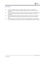
where
I rest is the measured reflected light intensity in the baseline condition, and I test is the reflected
light intensity during the presentation of the stimulus.
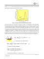
Figure 2 Absorption factors of 3 main chromophores in tissue
Brain tissues have three significant chromophores: water, oxy-hemoglobin (oxy-Hb) and deoxyhemoglobin (deoxy-Hb). In functional brain imaging applications, the main interest is to monitor oxy-Hb
and deoxy-Hb concentration changes. Within the 700-900nm range, or “optical window“, tissue is
relatively transparent because all significant chromophore absorption factors are low. The dominant
chromophores in the optical window are oxy-Hb and deoxy-Hb, and their concentration changes can be
calculated using Equation 3. Absorption factors of the three main chromophores with respect to
wavelength are given in Figure 2. Within the optical window, there is an isosbestic point where the
absorption spectra of oxy-Hb and deoxy-Hb cross. Measurements by choosing two wavelengths, one
below and one above this isosbestic point, could reveal the individual effects of oxy-Hb and deoxy-Hb.
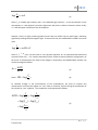
(6)
(7)
7|Page
H. Ayaz (1.0e)