1
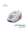
APPY1™ Test Kit - English
Intended Use
The APPY1 Test is a qualitative In Vitro Diagnostic Multivariate Index Assay (IVDMIA) used to aid in the identification of
patients at low risk for acute appendicitis. The APPY1 Test measures the concentrations of the myeloid-related protein
heterocomplex MRP 8/14 (calprotectin) and C-reactive protein (CRP) in EDTA-plasma by lateral flow immunoassay.
The quantitative concentrations of MRP 8/14 and CRP, in addition to an independently-determined white blood cell
(WBC) count, are then computed by a preprogrammed proprietary algorithm to generate an APPY1 Test result.
Indications for Use
The APPY1 Test is indicated for use in pediatric, adolescent, and young adult patients, ages 2 through 20 years old, with
abdominal pain and other physical and clinical signs and symptoms suggestive of acute appendicitis.
For Professional Use Only
Summary and Explanation
Diagnosing patients presenting with abdominal pain remains one of the most common and challenging issues in
emergency medicine. Over 22.2 million patients visited European and U.S. hospital emergency departments (ED) in
2010 with the primary complaint of abdominal pain.1,2
Appendicitis has the highest incidence in pediatric, adolescent and young adult patients 10-19 years of age,3 but
the diagnosis of appendicitis in these patients remains challenging because children present with a wide variety of
atypical clinical features.4 Up to 50% of infants and young children with AA may present with atypical signs and
symptoms,5 which can delay diagnosis and lead to life-threatening complications such as perforated appendix,
abdominal abscess, peritonitis, sepsis, and shock. Therefore, it is important to identify diagnostic tools which aid in
an objective, timely, and accurate diagnosis and risk stratification of patients with suspected acute appendicitis (AA).
At present, the ability to accurately diagnose appendicitis is limited to a collection of physical and clinical signs and
symptoms, and various diagnostic modalities. Multiple clinical prediction tools using classic symptoms as data points
have been developed to aid in the diagnosis of appendicitis. The performance of these scoring systems is inconsistent
and highly variable.5,6 Moreover, these systems are not utilized routinely by emergency departments.7 A WBC count
is routinely performed, but has demonstrated limited utility as a single marker, even when it is combined with signs
and symptoms suggestive of AA.8 Depending on the physician’s initial patient risk-assessment, diagnostic imaging,
including abdominal ultrasound (US) or computed tomography (CT) with or without contrast, may be ordered in cases
where the clinical presentation is equivocal and more information is needed. While CT is accurate in the diagnosis
of acute appendicitis when the appendix can be clearly visualized, it carries significant risk from increased exposure
to ionizing radiation thereby increasing the reliance on less harmful tests.9-12 Recent reports have shown that the use
of CT in the pediatric population has reached a plateau; however, the concern over CT exposure, especially in young
patients, has not diminished.12,13
The combination of three biomarkers in the APPY1 Test, rather than use of any individual marker, improves the ability
to identify patients at low risk.7 The APPY1 Test, used in conjunction with physical and clinical findings, provides
additional objective information to help guide clinical decision-making in the risk stratification of patients presenting
with abdominal pain suggestive of acute appendicitis. Patients identified as low risk by the APPY1 Test may be
managed more conservatively, e.g., with minimal or no additional testing or imaging, observation only, or earlier
discharge.
Principles of the Test
The APPY1 System is comprised of the APPYReader™ Instrument (the reader), a single-use APPY1 Test Cassette, a
single-use vial of dilution buffer, and a single-use vial of lyophilized antibodies conjugated to fluorescently-labeled
microparticles. The test procedure calls for the addition of patient plasma to the dilution buffer, followed by
reconstitution of the lyophilized conjugate with the diluted plasma/buffer. Available MRP 8/14 and CRP complexes
in the diluted patient sample are bound by the reconstituted antibody conjugate. An aliquot of this mixture is
then applied to the test cassette, where the now fluorescently-labeled MRP 8/14 and CRP complexes bind to the
corresponding capture zone antibodies on the nitrocellulose membrane. The test cassette is immediately inserted
into the reader which measures the concentration of each analyte present in the sample based on the fluorescence
intensity of each test line capture zone. The concentration of MRP 8/14 and CRP present in the sample is calculated
based preprogrammed calibration curves on RFID tags embedded within each individual test cassette. The
concentrations of the two markers, in combination with WBC values obtained from a hospital’s hematology analyzer
and entered into the dedicated reader, are used to generate the patient’s APPY1 Test result utilizing a preprogrammed
APPY1 Test Instructions for Use • L10003EN 04 • English • © 2014 Venaxis, Inc.
Page 2
proprietary algorithm. The APPY1 System also includes electronic (dry) and procedural (liquid) quality controls to
ensure the integrity of the results. The total time required for sample processing and testing is approximately 20
minutes.
MRP 8/14 (also known as S100A8/A9 or calprotectin) is a calcium-binding protein complex present in the cytoplasm
of neutrophils that is associated with nonspecific acute inflammatory conditions. MRP 8/14 has been found to be
differentially elevated in the appendix tissue and peripheral blood in patients with acute appendicitis.15-18
C-reactive protein (CRP) is produced by the liver and is associated with inflammation. CRP is an acute-phase protein
that is elevated in various diseases and conditions associated with nonspecific acute inflammation. Elevated
concentrations of CRP in peripheral blood have been correlated with acute appendicitis.19-21 Analytical performance
characteristics of the APPY1 Test CRP assay is consistent with that of a high sensitivity CRP assay (hsCRP), as described
below.
Elevated total WBC counts are associated with acute appendicitis when symptoms are present for 24 hours or longer.22
Leukocytosis is nonspecific for acute appendicitis as it is also seen in various other infectious and inflammatory
disorders. Studies suggest that a normal WBC count, in conjunction with low levels of MRP 8/14 and CRP and other
biomarkers, decreases the probability that acute appendicitis is present.18
Reagents and Materials
F10000
• APPY1 Test Kit, 25 tests
»» APPY1 Test Cassette, pouched, x 25
• Nitrocellulose membrane striped with monoclonal antibodies specific for CRP and MRP 8/14, and an
unrelated polyclonal goat anti-chicken IgY as an internal control.
»» APPY1 Test Buffer, 990 µL, x 25
»» APPY1 Test Conjugate, lyophilized cake, x 25
»» Instructions for Use, x 1
Materials Required but Not Provided
• APPYReader Kit
F10008
• APPYReader QC Cassette, pouched, x 1
F10004
• APPY1 Control Kit, 10 vials
F10005
•
•
•
•
•
»» APPY1 Control Level 1, 40 µL, x 5
»» APPY1 Control Level 2, 40 µL, x 5
Centrifuge for plasma processing, fixed angle rotor ≥ 45 degrees or swing bucket rotor, capable of achieving
1,300 x g ± 100 (Relative Centrifugal Force, RCF)
Calibrated micropipettes and disposable micropipette tips capable of dispensing 10 µL, 70 µL, and 150 µL
Vortex mixer
Mini-centrifuge (for pulse spins of Controls if necessary)
Personal protective equipment and biohazardous waste disposal containers
Storage and Stability
• Upon receipt, store the APPY1 Test Kit at 2º-8º C (35.6º-46.4º F) away from direct light. The kit components
should be stored in the original packaging, unopened, until ready to use. The APPY1 Test Kit is stable until the
expiration date printed on the kit box and test components when stored as recommended.
• The APPYReader Quality Control (QC) Cassette is stored at 15°-30°C (59°-86°F) in the original packaging,
unopened, until the first use. The QC cassette is reusable and should be resealed in the original pouch
immediately after each use. Do not discard. Protect from exposure to direct light.
• The APPY1 Control Kit is stored in its original packaging at ≤ -20°C (-4°F). Do not store in a frost-free freezer.
Only the number of required control vials should be thawed just prior to use and then discarded after testing.
The APPY1 Control 1 and Control 2 vials should not be refrozen after thawing.
APPY1 Test Instructions for Use • L10003EN 04 • English • © 2014 Venaxis, Inc.
Page 3
Precautions
General Precautions
• For professional use only.
• Each test cassette is intended for single use with one patient specimen. Test cassettes cannot be reused.
• Do not mix components from different test kit lots, as components are matched within each kit lot for
optimum performance.
• Do not remove test cassettes from their pouch until ready to use. Do not remove test conjugate from the kit
packaging until ready to use.
• Store the APPYReader QC Cassette in its original, light-protective, resealable pouch between each use. After
each use, return the QC cassette to the original resealable pouch. Do not discard.
• Do not use APPY1 Control vials that have been thawed and refrozen.
• Do not puncture or damage the test cassette during handling prior to use. If punctured or damaged, discard
and use a new test cassette.
• Do not use kit components beyond the printed expiration date on the kit box and component labels.
• After loading the test sample into the sample port of the test cassette, close the cassette drawer immediately
to initiate test run. Delay may result in an invalid test result.
• A confirmed patient WBC value must be entered within 90 minutes of initiating test run to obtain a valid result.
• All materials that have been in contact with human blood or its derivatives should be discarded as
biohazardous waste.
• The APPY1 Control Kit contains derivatives of pooled human source plasma. Each individual donor unit has
been tested and found negative for HBsAg, HCV, HIV-1, HIV-2, HIV-1Ag, HIV-1-NAT, ALT, and syphilis by FDAapproved methods. Because no test method can offer complete assurance that infectious agents are absent,
these reagents should be handled as potentially infectious human plasma. The use of Universal Precautions
and Blood-borne Pathogen Safety Rules is recommended. Each laboratory should follow their established
institutional guidelines for laboratory safety practices.
• The test buffer contains 0.095% sodium azide as a preservative. In case of skin contact, flush with copious
amounts of water. Sodium azide may react with lead and/or copper plumbing to form explosive metal azides;
flush with excess water upon disposal.
• All patient specimens should be handled as though capable of transmitting disease. The use of Universal
Precautions and Blood-borne Pathogen Safety Rules is recommended. Each laboratory should follow their
established institutional guidelines for laboratory safety practices.
Specimen Collection and Storage
Patient blood should be drawn through routine venipuncture or an established IV-line (with flush) into a K2-EDTA
(lavender top) tube containing 1.8 mg of EDTA/cc of blood. After blood collection, centrifuge the tube for 10 minutes
at 1300 x g ± 100 (RCF) at ambient room temperature. Centrifugation must start within one (1) hour of specimen
collection time. The centrifuge must have either a fixed rotor angle greater than 45 degrees or a swinging bucket
rotor. To calculate the conversion of RPM to RCF, refer to the user manual for the centrifuge model, or contact Venaxis®
Technical Support.
Test Procedure
Reader Setup
• Follow reader setup as described in the APPYReader User Manual and/or Start-Up Guide.
• Log into the reader using your assigned Operator ID.
• Allow the reader to warm up for at least 10 minutes before running any patient samples.
Prepare QC and Test Reagents
• If performing liquid QC, remove required number of Control 1 and Control 2 vials from the APPY1 Control Kit
stored at ≤ -20° C. Thaw at ambient room temperature while reader is warming up. See APPY1 Control Kit
Instructions For Use for more information.
• Remove sufficient test cassettes, test buffer vials, and test conjugate vials from an APPY1 Test Kit stored at
2-8° C necessary to complete all testing (QC and patient samples). Allow components to reach ambient room
temperature while reader is warming up.
APPY1 Test Instructions for Use • L10003EN 04 • English • © 2014 Venaxis, Inc.
Page 4
Run the QC Cassette
• Perform each day of patient testing and/or before running Controls 1 and 2. Patient samples cannot be tested
unless the QC cassette has a valid (PASS) result within the prior 24 hours.
• IMPORTANT: To maintain the expected useful life of the QC cassette, avoid light exposure. Store and reseal
the QC cassette in its original foil pouch immediately after each use. Do not discard the QC cassette until
expiration date.
• See APPYReader QC Cassette Instructions For Use for more information.
1. Log in as an operator or change to operator mode.
2. Press
to display the Main Menu screen
3. Use
and
4. Use
and
to highlight Maintenance and press Select.
to highlight Check APPYReader and press Select.
5. Use
and
to highlight APPYReader QC Cassette and press Select.
The reader drawer will open and the Insert Cassette screen will be displayed.
The APPYReader QC Cassette screen is displayed when the test finishes.
6. To print the QC cassette result:
a. Press Options.
and
to highlight Print Result and press Select.
b. Use
7. To send the result to a Laboratory Information System, refer to the APPYReader User Manual.
8. Press Back to return to the Check APPYReader screen or press
to return to the Main Menu screen.
• Expected Values
a. If the relative fluorescence unit (RFU) values detected on the QC cassette are within the acceptable
ranges programmed on the QC cassette RFID chip, the test result is reported as PASS.
b. If the RFU values detected on the QC cassette are outside the acceptable ranges programmed on
the QC cassette RFID chip, the test result is reported as FAIL and the operator is locked-out from
running patient samples. Repeat the test with a different QC cassette.
c. If the result of the second QC cassette is PASS, proceed to testing patient samples.
d. If a different QC cassette is not available, or if the result of the second QC cassette is FAIL, the reader
will remain locked. Contact Venaxis Technical Support at +1-303-794-2000 or your local distributor
for assistance.
Testing Patient Samples
• Prepare APPY1 Test Cassette
1. Log in as an operator or change to operator mode.
2. Press
3. Use
to display the operator Main Menu screen.
and
to highlight Run Test and press Select.
4. Enter the patient ID using one of the following methods:
• Manually, use the numeric keypad to enter the patient ID.
a.
Press Keypad to enter letters, spaces, and punctuation.
b. When the keypad is displayed on the reader’s screen, use
to highlight the desired character.
c.
,
,
, and
Press Confirm & Next to add the highlighted character to the patient ID.
d. Repeat Steps a and b as necessary.
e.
When the patient ID is complete, press Back or select Done to exit the Keypad feature.
• Using the optional external barcode reader, scan the patient’s barcode.
5. On the Enter Patient ID screen, press Confirm.
6. If the patient WBC result is available, enter it and press Confirm. Otherwise press Skip.
If you press Skip, you will be asked to enter the patient WBC after the test.
7. Remove test cassette from pouch and place it into the cassette drawer.
APPY1 Test Instructions for Use • L10003EN 04 • English • © 2014 Venaxis, Inc.
Page 5
• Prepare Patient Plasma Specimen for Testing
1. Set up test buffer and test conjugate vials.
2. Use a new disposable pipette tip for each transfer.
3. Remove 10 µL of patient plasma specimen from the centrifuged K2-EDTA tube, being careful not to
disturb the buffy coat layer. Transfer to the test buffer vial. Cap and mix well by vortex or manually.
4. Transfer 150 µL of the diluted patient plasma sample to the test conjugate vial. Mix well by pipetting up
and down 8-10 times, ensuring all conjugate is resuspended. Avoid introducing air to the mixture, which
will create foam.
• Initiate Run
1. Immediately add 70 µL of the diluted patient sample/antibody conjugate mixture to the test cassette
sample port.
2. Gently close the drawer. The test will start automatically.
3. The reader displays the status of the test on the Run Test screen.
4. When the test is done, you will be asked to enter the WBC or to confirm the WBC entered prior to the
test. The test cassette must remain in the reader during this time.
5. Re-enter the patient WBC and press Confirm. Test Result screen will then be displayed.
6. Press Next to display the Confirm Patient ID screen.
7. Confirm that the patient ID displayed on this screen is correct.
• View and Print Results
1. If the patient ID is correct: press Confirm to view the test result on the Result List screen. If the reader’s
automatic printing feature is enabled, the result is printed automatically.
2. If the patient ID is incorrect:
• Enter the appropriate patient ID and press Confirm to display the Patient ID does not match
screen.
• Re-enter the patient ID and press Confirm to view the test result on the Result List screen.
3. If the reader’s automatic printing feature is enabled, the result is printed automatically.
to eject the drawer. Remove the test cassette and dispose of in biohazardous waste container.
4. Press
Interpretation of Results
APPY1 TEST RESULT
INTERPRETATION
NEGATIVE/Low Risk For Appendicitis
Below clinical decision point for the test.
Patient is at low risk for acute appendicitis.
INCONCLUSIVE For Appendicitis
Above clinical decision point for the test.
Inconclusive risk for acute appendicitis. See Limitations below.
INVALID RESULT OR ERROR MESSAGE
Repeat test. If retest still results in Invalid Result or Error Message,
consult the APPYReader User Manual for troubleshooting instructions
or contact Technical Support.
Quality Control
Integrated Control Features
• The reader software includes an initialization self-check that calibrates the optical motors, checks the cassette
transport system, checks the optics receiver system, and performs an integrity check of the reader settings.
Failure to meet pre-programmed specifications will result in an Invalid Result or Error Message.
External Controls
• APPYReader QC Cassette
The APPYReader QC Cassette provides an optical and system suitability check for proper reader operation, and
should be run daily before running liquid QC reagents or patient specimens. The QC cassette must provide
passing results in order to proceed with testing patient samples.
APPY1 Test Instructions for Use • L10003EN 04 • English • © 2014 Venaxis, Inc.
Page 6
• APPY1 Control Kit
Liquid quality controls are available as a procedural control for the APPY1 Test. The established ranges for
each control are provided with each kit lot. Follow the Instructions for Use that come with each kit to run the
controls as necessary. Controls should be tested:
»»
»»
»»
»»
»»
At least once a month
With each new lot and new shipment of APPY1 Test Kits
If there is any question regarding system integrity, reagent storage conditions, or the reliability of any
test result
In accordance with national, country, or local guidelines
In accordance with your laboratory’s QC procedures
Limitations
1.A Negative/Low Risk for Appendicitis APPY1 Test result does not preclude a diagnosis of acute appendicitis.
In the early stages of the acute phase response (<24 hours from onset of symptoms) or in focal appendicitis,
plasma levels of MRP 8/14 and CRP may not have reached clinically significant levels, resulting in a false
negative test result.
2.An Inconclusive for Appendicitis APPY1 Test result should not be used as a diagnostic test for acute appendicitis,
or interpreted as “positive” as other inflammatory disorders or infectious disease may demonstrate increased
plasma levels of MRP 8/14 and CRP and elevated WBC count.
3.The APPY1 Test result is based on a score calculated by a proprietary algorithm using multivariate inputs. The
score is then compared to a pre-determined clinical decision point to arrive at the APPY1 Test result.
4. MRP 8/14, CRP and WBC are up-regulated nonspecifically in inflammatory conditions such as acute infections,
acute tissue injury, and other inflammatory disorders. Therefore, a diagnosis of acute appendicitis should be
made in the context of patient history, and physical and clinical findings.
5. Intra-individual variations in CRP levels may range from 30-60%. Variability in MRP 8/14 levels is also well
characterized.
6. Errors in specimen processing may result in false negative or false positive results.
Expected Results
The algorithm and the clinical decision point/cut-off value were derived from a pilot clinical study of 503 patients.
Based on Receiver Operating Curve (ROC) analysis and clinical agreement with discharge diagnosis, the clinical
decision point for the APPY1 Test was established. Patient test results below the cut-off are considered NEGATIVE/Low
Risk for Acute Appendicitis, and results above the cut-off are considered INCONCLUSIVE Risk for Acute Appendicitis.
Clinical Performance
The clinical validation study of the APPY1 System was conducted on archived frozen plasma samples. Samples were
collected during a previous multisite, prospective, open label observational study of the evaluation, assessment,
triage, and disposition of patients 2 – 20 years old presenting to emergency departments with symptoms suggestive
of acute appendicitis. Patients enrolled in the study had blood drawn, processed to plasma, and then frozen within
2 hours. Specimens were stored on site at ≤ -70ºC until shipment to Venaxis, where they were archived and stored
at ≤ -70ºC until testing. Blinded sample testing was conducted in Venaxis’ laboratory using the APPY1 System. The
APPY1 Test results were compared to the original discharge diagnosis (Negative for acute appendicitis, AA-) or
histopathology (Positive for acute appendicitis, AA+).
A total of 465 patient samples collected during previous pilot clinical studies and having sufficient volume for the
complete analysis were used in the validation study. Subjects ranged in age from 2-20 years old, mean age 12.0 ± 4.3
years. Approximately 49% were male, 52% female. Caucasians were the predominant race at 70%, Hispanic/Latino
19%, and Other 11%. There was no significant difference in mean age between the AA+ cohort (12.5±3.8), and the
AA- cohort (11.9±4.5).
APPY1 Test Instructions for Use • L10003EN 04 • English • © 2014 Venaxis, Inc.
Page 7
APPY1 Test Results Compared to Clinical Diagnosis
APPY1 Test Result N=465
AA-
AA+
Below cut-off: NEGATIVE/Low Risk for AA
154
6
160
Above cut-off: INCONCLUSIVE for AA
179
126
305
333
132
465
Clinical Performance Data
Measure
Estimate (95% CI)
AA Prevalence
28.4% (24.5 – 32.6)
Sensitivity
95.5% (90.4 – 97.9)
Specificity
46.2% (41.0 – 51.6)
NPV
96.3% (92.1 – 98.3)
PPV
41.3% (35.9 – 46.9)
Positive Likelihood Ratio
1.78 (1.60 – 1.99)
Negative Likelihood Ratio
0.098 (.045 - .217)
Analytical Performance
Limit of Detection
The Limit of Blank (LOB), Limit of Detection (LOD) and Limit of Quantitation (LOQ) were determined by 2 operators
testing 3 lots of APPY1Test Kits against 6 plasma samples and blank samples of APPY1Test Buffer over 3 days, with the
samples randomized each day.
LOD, LOB and LOQ
Limit
CRP, µg/mL
MRP, µg/mL
Blank
0.201
0.030
Detection
0.254
0.045
Quantitation
0.341
0.064
Linearity
For each analyte, linearity was demonstrated for an interval from approximately the LOD/LOQ to above the upper
truncation value of the assay. The assay is linear for both analytes in the range of the clinical cut-off value. MRP assay
is linear from 0.052 to 0.933 µg/mL; CRP assay is linear from 0.187 to 51.4 µg/mL. Using CRP LOD as the low value, the
CRP assay is linear from 0.254 µg/mL to 51.4 µg/mL.
Measured MRP 8/14 µg/ml
MRP 8/14 Linearity
1.2
y = 0.9998x - 0.0062
R² = 0.9959
1.0
0.8
0.6
0.4
0.2
0.0
0.0
0.2
0.4
0.6
0.8
1.0
Estimated µg/ml
APPY1 Test Instructions for Use • L10003EN 04 • English
2014 Venaxis, Inc.
CRP• ©
Linearity
70
Page 8
0.0
0.0
0.2
0.4
0.6
0.8
1.0
Estimated µg/ml
Measured CRP µg/mL
CRP Linearity
70
60
50
40
30
20
10
0
-10 0
y = 1.0545x - 0.451
R² = 0.9915
10
20
30
40
60
50
Estimated µg/mL
Precision
Precision performance of the APPY1 Test was evaluated in accordance with the CLSI guideline EP-5A2, “Evaluation of
Precision Performance of Quantitative Measurement Methods; Approved Guideline.” Five plasma specimens between
LOQ and the cut-off (APPY1 Test score of 4.0), at or close to the cut-off, and at intervals above the cut-off were tested
over 20 days, 2 runs per day, 2 replicates per run, 1 operator. Samples were randomized across 5 readers on each
test day. To complete the APPY1 Test score calculation, a fixed WBC was assigned to each specimen. Total percent
coefficient of variation (% CV) ranged from 0.0 to 2.8% for the APPY1 Test score, 6.5 to 12.0 % for MRP 8/14, and 4.8 to
15.1% for CRP. Near the established cut-off, % CVs for MRP 8/14 and CRP were ≤ 10.6%.
Precision Testing: APPY1 Test Score with Fixed WBC
Sample
1
2
3
4
5
N
80
80
80
80
80
APPY1
Test score
(Mean)
3.2
3.9
4.0
4.1
5.2
Within-run
SD
0.000
0.000
0.050
0.037
0.143
Between-run
Total
%CV
0.0
0.0
1.2
0.9
2.8
SD
0.000
0.000
0.000
0.027
0.000
%CV
0.0
0.0
0.0
0.7
0.0
SD
0.000
0.000
0.050
0.046
0.148
%CV
0.0
0.0
1.2
1.1
2.8
1
2
Sample
3
4
5
0.10
0.006
6.3
0.000
0.0
0.006
6.5
0.15
0.009
6.1
0.007
4.5
0.011
7.1
0.24
0.024
10.0
0.002
0.7
0.025
10.6
0.31
0.016
5.1
0.012
3.8
0.021
6.9
0.52
0.058
11.2
0.000
0.0
0.062
12.0
1
2
Sample
3
4
5
0.58
0.027
4.6
0.013
2.3
0.028
4.8
2.31
0.126
5.5
0.097
4.2
0.149
6.5
5.06
0.341
6.7
0.280
5.5
0.453
8.6
5.25
0.364
6.9
0.262
5.0
0.453
8.6
18.88
2.953
15.6
0.000
0.0
2.851
15.1
Precision Testing: MRP 8/14 Concentration
Parameters
MRP 8/14 µg/mL
Mean
Repeatability (within run)
Between run
Total
SD
%CV
SD
%CV
SD
%CV
Precision Testing: CRP Concentration
Parameters
CRP µg/mL
Mean
Repeatability (within run)
Between run
Total
SD
%CV
SD
%CV
SD
%CV
APPY1 Test Instructions for Use • L10003EN 04 • English • © 2014 Venaxis, Inc.
Page 9
Reproducibility
Reproducibility was evaluated in Venaxis’ laboratory by 3 operators, each with a dedicated reader, blinded to the other
operators’ results. An operator + reader combination was designated as a separate “site” for a total of 3 sites. Each
operator tested 5 plasma specimens plus 2 controls for 3 days, 2 runs per day, 2 replicates per run. The sample order
was varied in each test run. To complete the APPY1 Test score calculation, a fixed WBC was assigned to each specimen.
For APPY1 Test scores, the % CV for within-run (repeatability) was ≤ 3.4%, the % CV for between-run was ≤ 0.9%, the %
CV for between-day was ≤ 0.7%, and the % CV between-operator/site was ≤ 0.5%. Total imprecision (reproducibility)
was ≤ 3.4%. The results for APPY1 Test score, MRP 8/14 and CRP are shown below:
Reproducibility Testing: APPY1 Test Score with Fixed WBC
Parameters
APPY1 Test Score
Mean
Repeatability (within run)
Between run
Between day
Between operator
Reproducibility (total)
SD
%CV
SD
%CV
SD
%CV
SD
%CV
SD
%CV
1
2
Sample
3
4
5
3.2
0.014
0.4
0.004
0.1
0.005
0.1
0.003
0.1
0.015
0.5
3.9
0.019
0.5
0.010
0.3
0.014
0.4
0.005
0.1
0.026
0.7
4.0
0.048
1.2
0.019
0.5
0.010
0.2
0.011
0.3
0.054
1.3
4.2
0.049
1.2
0.017
0.4
0.012
0.3
0.010
0.3
0.054
1.3
5.4
0.168
3.1
0.049
0.9
0.036
0.7
0.030
0.5
0.182
3.4
1
2
Sample
3
4
5
0.10
0.003
2.9
0.002
1.9
0.003
2.6
0.010
9.6
0.009
8.6
0.15
0.010
6.6
0.005
3.1
0.007
4.7
0.002
1.6
0.013
8.8
0.25
0.029
11.5
0.009
3.5
0.006
2.4
0.006
2.5
0.031
12.6
0.34
0.026
7.7
0.008
2.3
0.007
2.0
0.006
1.7
0.029
8.4
0.59
0.656
11.1
0.013
2.3
0.017
3.0
0.015
2.5
0.071
12.0
1
2
3
4
5
0.63
0.039
6.1
0.016
2.5
0.012
1.9
0.009
1.4
0.044
7.0
2.52
0.166
6.6
0.130
5.2
0.116
4.6
0.048
1.9
0.245
9.7
5.55
0.309
5.6
0.281
5.1
0.065
1.2
0.099
1.8
0.434
7.8
5.94
0.568
9.6
0.228
3.8
0.132
2.2
0.121
2.0
0.638
10.7
24.34
3.375
13.9
1.226
5.0
0.743
3.1
0.902
3.7
3.776
15.5
Reproducibility Testing: MRP 8/14 Concentration
Parameters
MRP 8/14 µg/mL
Mean
Repeatability (within run)
Between run
Between day
Between operator
Reproducibility (total)
SD
%CV
SD
%CV
SD
%CV
SD
%CV
SD
%CV
Reproducibility Testing: CRP Concentration
Sample
Parameters
CRP µg/mL
Mean
Repeatability (within run)
Between run
Between day
Between operator
Reproducibility (total)
SD
%CV
SD
%CV
SD
%CV
SD
%CV
SD
%CV
APPY1 Test Instructions for Use • L10003EN 04 • English • © 2014 Venaxis, Inc.
Page 10
Interference
Pooled plasma samples containing low , medium , and high concentrations of MRP 8/14 and CRP were evaluated
for interference by hemoglobin, bilirubin (conjugated and unconjugated), lecithin, human anti-mouse antibodies
(HAMA), Rheumatoid Factor (RF), and Immunoglobulin G (IgG). Control values for each plasma pool (L, M, H) were
established using the mean of 3 replicate measurements. The effect of each interfering substance was assessed using
the mean of 3 replicate measurements on each plasma pool. Each plasma pool was assigned a fixed WBC value in
order to calculate the APPY1 Test score. The low plasma pool was assigned a WBC value of 4, the medium pool was
assigned a WBC value of 7 and the high plasma pool was assigned a WBC value of 10, giving APPY1 Test scores of 3.1,
3.6 and 4.6, respectively. No significant interference was observed on the APPY1 Test score for any of the interferents,
all differences were below 10%. However, there was a potential interference detected at low levels of MRP 8/14 when
challenged with unconjugated bilirubin at these test levels. As demonstrated in the graph below, this interference
has very little effect on the APPY1 Test score. Additionally, there was interference detected at low levels of CRP when
comparing the RF positive plasma sample with its control. However, low levels of CRP were detected in this sample
by immunoblot, independent of the test pools. If a cleaner source of RF is tested the % difference shown below will
be reduced. A similar result was obtained with all levels of MRP 8/14 when testing the IgG interferent. It was shown
that the purified IgG is contaminated with MRP 8/14 and that if a cleaner source material is obtained the interference
effects shown below should also be reduced.
Interference Testing: APPY1Test
Potential Interferent
Concentration
Medium Pool
High Pool APPY1
APPY1 Test Score
Test Score = 4.6
= 3.6
% Difference from Control Sample or NSD
0.69
-0.79
-0.08
Low Pool APPY1
Test Score = 3.1
Hemoglobin
2 g/L
Bilirubin, unconjugated
342 µmol/L
1.37
0.56
1.78
Bilirubin, conjugated
342 µmol/L
-0.28
-1.52
-1.80
Lecithin
11.1 mmol/L
0.38
-0.09
0.17
HAMA
153.3 ng/mL
0.24
-0.93
-2.18
Rheumatoid Factor
60 IU/mL
0.06
0.42
-1.99
IgG
52.8 mg/mL
5.55
7.06
4.30
APPY1 Test Instructions for Use • L10003EN 04 • English • © 2014 Venaxis, Inc.
Page 11
REFERENCES
1.
EU Market Study by Venaxis, Inc. (Benelux, France, Italy, Germany, UK), 2012.
2.
Venaxis analysis of data from: National Hospital Ambulatory Medical Care Survey, 2010. Available at: http://www.cdc.gov/nchs/ahcd/
ahcd_questionnaires.htm#public_use. Accessed November 2012.
3.
Addiss D, et al. The Epidemiology of Appendicitis and Appendectomy in the United States. American Journal of Epidemiology.
1990;132:910-25.
4.
Becker, et al. Atypical Clinical Features of Pediatric Appendicitis. Academic Emergency Medicine. 2007;14:124-129.
5.
Brennan G. Pediatric Appendicitis: Pathophysiology and appropriate use of diagnostic imaging. Canadian Journal of Emergency
Medicine. 2006;8:425-432.
6.
Kharbanda, et al. A Clinical Decision Rule to Identify Children at Low Risk for Appendicitis. Pediatrics. 2005;116:709-716.
7.
Huckins DS, et al. A novel biomarker panel to rule out acute appendicitis in pediatric patients with abdominal pain,
American Journal of Emergency Medicine. (in press), http://dx.doi.org/10.1016/j.ajem.2013.06.016.
8.
Cardall, et al. Clinical Value of the Total White Blood Cell Count and Temperature in the Evaluation of Patients with Suspected
Appendicitis. Academic Emergency Medicine. 2004;11:1021-1027.
9.
Hennelly K and Bachur R. Appendicitis Update. Current Opinion Pediatrics. 2011;23:1-5.
10. Wan M, et al. Acute Appendicitis in Young Children: Cost Effectiveness of US versus CT in Diagnosis – A Markov Decision Analytical
Model. Radiology. 2009;250:378-386.
11. Glatter R. What role do imaging studies play in diagnosing pediatric appendicitis in the ED? MedScape Emergency Medicine, Web
MD. June 18, 2010. Available at: http://www.medscape.com/viewarticle/723549. Accessed April 9, 2012.
12. Stoker J, et al. Imaging patients with acute abdominal pain. Radiology. 2009;253:31-46.
13. Bachur R, et al. Advanced Radiologic Imaging for Pediatric Appendicitis: 2005-2009 Trends and Outcomes. Journal of Pediatrics. 2011.
14. Menoch M, et al. Trends in Computed Tomography Utilization in the Pediatric Emergency Department. Pediatrics. 2012:129:e690-e697.
15. Bealer J and Colgin M. S100A8/A9: A potential New diagnostic Aid for Acute Appendicitis. Academic Emergency Medicine.
2010:17:333-336.
16. Mills A, et al. Diagnostic Characteristics of S100A8/A9 in a Multicenter Study with Patients With Acute Right Lower Quadrant
Abdominal Pain. Academic Emergency Medicine. 2012;19:48-55.
17. Kharbanda A, et al. Novel Serum and Urine Markers for Pediatric Appendicitis. Academic Emergency Medicine. 2012;19:56-62.
18. Thuijl G, et al. A Pilot study on potential new plasma markers for diagnosis of acute appendicitis. 2010.
19. Shindoh J, et al. Diagnostic Power of Inflammatory Markers in Predicting Severity of Appendicitis. Hepato-Gastroenterology.
2011;58:2003-2006.
20. Cole M, Maldonado N. Evidence-Based Management of Suspected Appendicitis in the Emergency Department. Emergency Medicine
Practice. 2011;13:1-32.
21. Siddique K, et al. Diagnostic accuracy of white cell count and C-reactive protein for assessing the severity of paediatric appendicitis.
Journal of the Royal Society of Medicine. 2011;2:59.
22. Doraiswamy NW. Progress of acute appendicitis: a study in children. British Journal of Surgery 1978;65:877-9.
Symbol Key: Symbolschlüssel: Leyenda de los símbolos: Légende des symboles: Descrizione dei simboli:
Verklaring van symbolen:
Symbol
Symbol
Símbolo
Symbole
Simbolo
Symbool
EN
DE
ES
FR
IT
NL
Used for
Verwendet für
Significa
Utilisé pour
Spiegazione
Gebruikt voor
CE Mark
CE-Zeichen
Marca CE
Marque CE
Marcatura CE
CE-markering
Contents
Inhalt
Contenido
Contenu
Contenuto
Inhoud
Venaxis, Inc.
Emergo Europe
1585 South Perry Street
Castle Rock, CO 80104 USA
Molenstraat 15
2513 BH, The Hague,
The Netherlands
www.venaxis.com
Tel: +1-303-794-2000
Fax: +1-303-798-8332
The APPY1 Test Kit is for U.S. Export Only.
Venaxis is a registered trademark of Venaxis, Inc. APPY1 and APPYReader are trademarks of Venaxis, Inc.
APPY1 Test Instructions for Use • L10003 04 • © 2014 Venaxis, Inc.
Page 67