1
PRECICE® Enzymes & Kits
User Manual
Ref: # K0307-01
PRECICE® dCK Screening Assay Kit
For research use only. Not for use in diagnostic procedures
I. Introduction
dCK Assay Kit was specially designed to follow the enzymatic activity of purified deoxycytidine kinase (dCK, EC 2.7.1.74) in
vitro. This user manual gives the instructions for standard assays in 96-well plate.
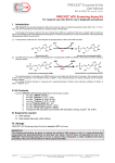
The principle of the assay is based on the use of deoxyinosine (dIR) as a substrate of dCK and a coupled reaction involving a
highly active IMPDH (Inosine Monophosphate Dehydrogenase, bacterial recombinant) for a direct measurement of the deoxyinosine
monophosphate (dIMP) formed by dCK.
(1)
In the presence of dIR and ATP, dCK catalyses the phosphorylation of dIR to form dIMP and ADP:
N
O
HO
ATP
ADP
O
N
N
N
O
O
HO P O
NH
dCK
Deoxyinosine (dIR)
HO
Deoxyinosine monophosphate (dIMP)
The dIMP formed is then oxidized to deoxyxanthosine monophosphate (dXMP) by IMPDH
NADH2 formation.
NAD
NADH2
N
O
O
O
N
HO
HO P O
NH
N
IMPDH
OH
HO
Deoxyinosine monophosphate (dIMP)
NH
N
OH
HO
(2)
O
N
in the presence of NAD, leading to
N
O
O
P O
OH
HO
O
N
N
H
NH
O
Deoxyxanthosine monophosphate (dXMP)
This coupling reaction is immediate when IMPDH activity is much higher than dCK activity in the assay. The enzymatic activity
of dCK, which corresponds to the formation kinetics of dIMP, is then stoichiometrically and directly monitored by the formation
kinetics of NADH2.
The velocity of NADH2 formation is measured with a spectrophotometer at 340nm (molar extinction coefficient of NADH 2 at
340nm = 6220 M-1.cm-1)
II. Kit Contents
A standard PRECICE® dCK Screening Assay Kit (one 96-well plate) contains:
1.
2.
3.
4.
5.
6.
7.
8.
one tube " IMPDH"
one tube "Cofactor 1”
one tube "Cofactor 2”
one tube “Cofactor 3”
one vial "Reaction Buffer 5x "
one tube "Deoxyinosine"
one tube "Human dCK enzyme", 100mU
®
Transparent 96-well plate (round-bottom 96-well plate Corning, Costar , ref. 3797)
III. Equipments required
1) Plate agitator
2) Plate reader fitted with a filter 340nm.
IV. Storage
®
PRECICE dCK Screening Assay Kit must be stored at -20°C until used.
IMPORTANT:
The following instructions are given to measure the activity of dCK enzyme in vitro, in a range allowing this
measurement by spectrophotometry as described here below. NovoCIB does not guarantee the use of its
®
PRECICE dCK Screening Assay Kit or of one or several of its components, in other conditions than those
described in this user manual and/or for other purpose than R&D.
Page 1/3 NovoCIB SAS, 115 avenue Lacassagne, 69003 Lyon, France 69003 Lyon, France
[email protected]
Tel / Fax +33 (0)478536395
#K0307-01-UM. v Version 150924LB
www.novocib.com
PRECICE® Enzymes & Kits
User Manual
Ref: # K0307-01
V. Experimental Protocol I - for non-nucleoside dCK inhibitors
Important: Spin all tubes before solubilizing.
V.1. Reconstitute IMPDH enzyme
Add 250µL of deionized water to “IMPDH” tube. Agitate gently until complete dissolution of the powder.
V.2. Preparation of standard reaction buffer (1x)
i)
Add the content of "Reaction Buffer 5x" tube (4ml) to 16ml of deionized water to prepare “Reaction Buffer 1x”.
ii) Transfer quantitatively the content of 2 tubes with "Cofactor 1" and “Cofactor 2” to the tube with “Reaction buffer 1x”.
To do so:
iii) –Add 1ml of “Reaction buffer 1x” to each tube with cofactors and mix them by inverting or pipeting up and down until
the powder dissolved.
iv) - transfer by pipetting the content of two tubes back into a vial “Reaction buffer 1x”;
v) - repeat to be sure that all reagent and enzymes of the small tubes and vial are recovered. Mix by gently inverting until
complete dissolution. Avoid bubbles.
vi) Transfer quantitatively the content of “IMPDH” tube to “Reaction buffer 1x” with co-factors.
vii) Add 200µL of deionized water to “Cofactor 3” tube (ATP). Agitate until dissolved. Take off 20µL of thus prepared ATP
solution to “Reaction buffer 1x” already containing Cofactor 1, Cofactor 2, and IMPDH.
viii) Solubilize the content of Human dCK enzyme" tube by adding 1ml of complete “Reaction buffer 1x” with cofactors and IMPDH, transfer by pipeting the content of the tube back into a vial “Reaction buffer 1x”.
Composition of complete reaction buffer: 100mM Tris-HCl, 250mM KCl, 10mM MgCl2, BSA 0.5mg/ml, 5mM DTT, 5mM NAD, 250µM
ATP, IMPDH 50mU/ml, human recombinant dCK 5mU/ml.
V.3. Preparation of deoxyinosine solution for starting the reaction
Add 960µl of deionized water to "Deoxyinosine" tube and mix by inverting. 1ml of 20mM dIR solution is obtained.
VI. Following dCK activity in vitro
VI.1. Pre-incubation phase (15')
i)
Program the plate-reader in a kinetics mode with the measurements done every 1 minutes, absorbance at 340 nm, 37°C,
agitation before the kinetics for 1 min, duration time 15min.
ii) Add 200µL of standard reaction buffer per well.
iii) Agitate and measure absorbance at 340nm (A340). Record this first set of data.
5.2.2. Start the reaction and incubate (40')
i)
Eject the plate from the plate-reader.
ii) Program the plate-reader in a kinetics mode with the measurements done every 1 minutes, absorbance at 340 nm, 37°C,
agitation before the kinetics for 1 min, duration time 30-40min.
iii) Start the reaction by adding 10µL of 20mM of "Deoxyinosine" per well (1mM dIR final concentration).
iv) Place the plate in the plate-reader and start the measurements. Record second set of data.
VII. Experimental Protocol II; for nucleoside analogues dCK inhibitors
Important: Spin all tubes before solubilizing.
VII.1. Reconstitute IMPDH enzyme
Add 250µL of deionized water to “IMPDH” tube. Agitate gently until complete dissolution of the powder.
VII.2. Preparation of standard reaction buffer (1x)
i) Add the content of "Reaction Buffer 5x" tube (4ml) to 16ml of deionized water to prepare “Reaction Buffer 1x”.
ii) Transfer quantitatively the content of 2 tubes with "Cofactor 1", “Cofactor 2" to the tube with “Reaction buffer 1x”.
Important: Do not add "Cofactor 3" (ATP)!
Page 2/3 NovoCIB SAS, 115 avenue Lacassagne, 69003 Lyon, France 69003 Lyon, France
[email protected]
Tel / Fax +33 (0)478536395
#K0307-01-UM. v Version 150924LB
www.novocib.com
PRECICE® Enzymes & Kits
User Manual
Ref: # K0307-01
To do so:
iii) -pipet 1ml of “Reaction buffer 1x” to each tube with cofactors and mix them by inverting or pipeting up and down until
the powder dissolved.
iv) - transfer by pipeting the content of two tubes back into a vial “Reaction buffer 1x”;
v) - repeat to be sure that all reagent and enzymes of the small tubes and vial are recovered. Mix by gently inverting until
complete dissolution. Avoid bubbles.
vi) Transfer quantitatively the content of “IMPDH” tube to “Reaction buffer 1x” with co-factors.
vii) Add 1ml of “Reaction buffer 1x” to "Deoxyinosine" tube and transfer the solution back to “Reaction buffer 1x”
already containing Cofactor 1, Cofactor 2, and IMPDH.
viii) Solubilize the content of "Human dCK enzyme" tube by adding 1ml of complete “Reaction buffer 1x” with cofactors and IMPDH, transfer by pipeting the content of the tube back into a vial “Reaction buffer 1x”.
Composition of complete reaction buffer: 100mM Tris-HCl, 250mM KCl, 10mM MgCl2, BSA 0.5mg/ml, 5mM DTT, 5mM NAD, 1mM
deoxyinosine, IMPDH 50mU/ml; human recombinant dCK 5mU/ml.
VII.3. Preparation of ATP solution for starting the reaction
Add 1ml of deionized water to the tube "Cofactor 3" containing 30mg of ATP powder. Mix until dissolved. 50mM ATP solution is
obtained.
VIII. Following dCK activity in vitro
VI.1. Pre-incubation phase (15')
i) Program the plate-reader in a kinetics mode with measurements done every 1 minutes, absorbance at 340 nm, 37°C,
agitation before the kinetics for 1 min, duration time 15min.
ii) Add 200µL of standard reaction buffer per well.
iii) Agitate and measure absorbance at 340nm (A340). Record this first set of data.
5.2.2. Start the reaction and incubate (40')
i) Eject the plate from the plate-reader
ii) Program the plate-reader in a kinetics mode with shaking for 1min and the measurements done every 1 minutes,
absorbance at 340 nm, 37°C, agitation before the kinetics for 1 min, duration time 60min.
iii) Start the reaction by adding 10µL of 50mM ATP per well (2,5mM ATP final concentration).
iv) Place the plate in the plate-reader and start the measurements. Record second set of data.
Assay validation:
Specific competitive inhibition of deoxyinosine phosphorylation by dCK
by deoxycytidine and gemcitabine in comparison to other ribo- and
deoxyribonucleosides and ribavirine.
0.75
0.7
Absorbance at 340nm
0.65
control
0.6
deoxyguanosine1mM
uridine 1mM
deoxyuridine 1mM
thymidine 1mM
gemcitabine 0.5mM
cytidine 1mM
deoxycytidine 1mM
ribavirine
0.55
0.5
0.45
0.4
0.35
0.3
0.25
0
5
10
15
20
25
30
35
Figure 1.
Nucleosides and nucleoside analogues
(2µL of 100mM solution) were added to
96-well plate, followed by the addition
of 200µL of standard reaction mix
containing DTT, NAD, IMPDH and dCK.
10 min later, the reaction was started
by the addition of ATP solution (10µL
per well) and the increase in
absorbance at 340nm was followed for
30min.
Results:
Of 8 deoxy- and ribonucleosides
tested,
only
deoxycytidine
and
gemcitabine
competitively
inhibit
deoxyinosine phosphorylation by dCK
which
totally
consistent
with
previously published data on their
substrate properties.
Time after ATP addition (second incubation), min
Page 3/3 NovoCIB SAS, 115 avenue Lacassagne, 69003 Lyon, France 69003 Lyon, France
[email protected]
Tel / Fax +33 (0)478536395
#K0307-01-UM. v Version 150924LB
www.novocib.com