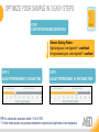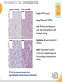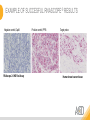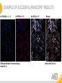
1
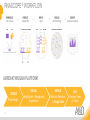
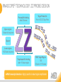
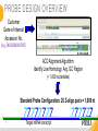
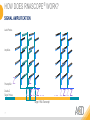
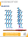
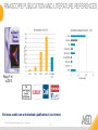
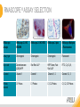
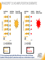
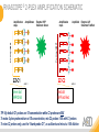
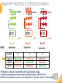
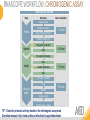
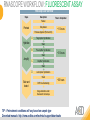
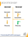
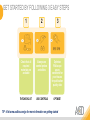
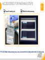
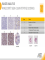
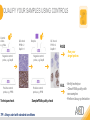
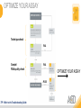
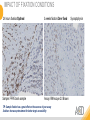
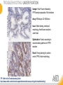
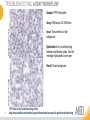
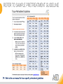
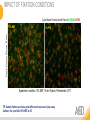
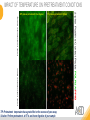
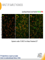
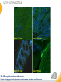
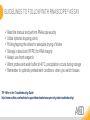
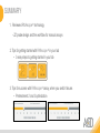
READY, SET, GO! GETTING STARTED WITH RNASCOPE® Presented by: Jacqueline Akech, Ph.D. May 12th, 2015 Senior Scientist Advanced Cell Diagnostics ©2014 Advanced Cell Diagnostics, Inc. | Confidential and Proprietary | For Research Use Only (RUO), not intended for diagnosis. TOPICS • How Does RNAscope® Work? • Getting Started with RNAscope® in your Laboratory • Tips and Tricks on Running the Assay • Frequently Asked Questions • Time for Q&A 2 RNASCOPE® OVERVIEW 3 RNASCOPE ® WORKFLOW A BREAKTHROUGH PLATFORM UNIQUE Probe Design 4 SIGNAL Amplification + Background Suppression SINGLE Molecule Detection in Single Cells ANY Genome, Gene or Tissue RNASCOPE® TECHNOLOGY: ZZ PROBE DESIGN Preamplifer binding site: 28-bases Target Probe Set: Pool of 20 ZZ Oligo Pairs Upper region: 14-base tail sequence Spacer Lower region: ~8-25-base sequence Target-specific binding site: ~50-base region RNA Target Region 1KB 20 pairs of ~50 bases mRNA transcript detection: Highly specific & robust signal amplification 5 PROBE DESIGN OVERVIEW Customer Gene-of-interest Accession No. (e.g.,ENSG00000097007) ACD Alignment Algorithm: Identify Low Homology, Avg. GC Region (~ 1,000 nucleotides) Standard Probe Configuration: 20 Z-oligo pairs = 1,000 nt 6 ZTarget ZZmRNA ZZtranscript ZZZ… …ZZZZZZ HOW DOES RNASCOPE ® WORK? SIGNAL AMPLIFICATION Label Probes Amplifiers Preamplifier Double Z Target Probes Z Z ZZZZ… Target RNA Transcript 7 … ZZZZ HOW DOES RNASCOPE ® WORK? SIGNAL AMPLIFICATION ZZ Z Z Z Z … X … ZZ Z Z Z Non-specific Target 8 20 Z pairs x 20 Amplifiers x 20 Labels 8000 labelled molecules per 1kb region HOW DOES RNASCOPE ® WORK? Specific Signal Amplification 9 RNASCOPE PUBLICATION AND LITERATURE REFERENCES Wang, F. et al.2012 Visit www. acdbio.com and download a publication of your interest ©2014 Advanced Cell Diagnostics, Inc acdbio.com 10 RNASCOPE ® WORKFLOW 11 RNASCOPE ® ASSAY SELECTION RNAscope Assays RNAscope 2.0 HD BROWN RNAscope 2.0 HD RED RNAscope 2-plex RNAscope Multiplex – Fluoroscence Assay type Chromogenic Chromogenic Chromogenic Fluorescent Dye used Diaminobenzene (DAB)-HRP Fast Red -ALP HRP-Green, Fast Red -ALP FITC, Cy3, Cy5, Channel Channel 1 Channel 1 Channel 1, 2 Channel 1, 2, 3 Probes channel C1 Probes C1 Probes C1, C2 Probes C1, C2, C3 Probes RNASCOPE ® 2.0 HD AMPLIFICATION SCHEMATIC Enzyme: HRP Substrate: DAB Amplification steps Amp6-Red Label probe Pre Amp/AMP1 Amp6-Brown Label probe Pre Amp/AMP1 Amplification steps ZZ mRNA 1 2.0 HD BROWN Brown dot HRP/DAB ZZ mRNA 1 2.0 HD RED Red dot AP/Fast Red TIP : Do not interchange reagents within Brown/Red assays or across similar 2.0 HD Assays By default 2.0 HD assays require C1 probes that are ready to use, no further dilution is required Enzyme: ALP Substrate: FastRed RNASCOPE ® 2-PLEX AMPLIFICATION SCHEMATIC Enzyme: HRP Substrate: Green Amplification steps Amp6-Red Pre Amp/AMP1 Amp6-Brown Pre Amp/AMP1 Amplification steps ZZ-C1 Green dot HRP/DAB mRNA 1 ZZ-C2 mRNA 2 Red dot AP/Fast Red TIP: By default C1 probes are 1X concentration while C2 probes are 50X To make 2-plex probe mixture at 1X concentration, mix C2 probes 1:50 with C1 probes To view C2 probes only, use the “blank-probe-C1”, as a diluent and mix at a 1:50 dilution Enzyme: ALP Substrate: FastRed RNASCOPE ® MULTIPLEX FLUORESCENT SCHEMATIC Amp3-C2 Amp3-C1 Alexa 488 Amp3-C3 Atto 550 Amp1-C1 ZZ-C1 ZZ-C2 Alexa-488 495nm/520nm ZZ-C3 mRNA 3 mRNA 2 mRNA 1 Ex/Em Amp4-Alt A Amp1-C3 Amp4-Alt A Amp1-C2 Amp4-Alt A Atto 647 Atto-550 Atto-647 555nm/575nm 645nm/670m Color Module C1 C2 C3 Amp4 Alt A GREEN - Alexa 488 ORANGE--Atto 550 FAR RED--Atto 647 Amp4 Alt B ORANGE--Atto 550 GREEN - Alexa 488 FAR RED--Atto 647 Amp4 Alt C ORANGE--Atto 550 FAR RED--Atto 647 GREEN - Alexa 488 TIP: By default C1 probes are 1X concentration while C2 and C3 probes are 50X To make 3-plex probe mixture at 1X concentration, mix C2 and C3 probes 1:50 with C1 probe If C2 and C3 are all at 50X concentration, use the “blank-probe-C1” as a diluent and mix at a 1:50 dilution RNASCOPE WORKFLOW: CHROMOGENIC ASSAY RNAscope user workflow Description Deparaffinization Steps H2O2 block (Pretreat 1) Pretreat Time to Completion ~1.5 hours Epitope retrieval (Pretreat 2) Protease (Pretreat 3 or 4) A Hybridize Target probe hybridization Wash B ~2.5 hours Pre-amplifier hybridization Wash Amplify C Amplifier hybridization ~1.5 hours Wash D Label probe hybridization Wash Stain and detect ~1.5 hours Detection Hematoxylin stain Image detection under standard light microscope / scanner TIP : Detection protocols will vary based on the chromogenic assay used Download manuals: http://www.acdbio.com/technical-support/downloads RNASCOPE WORKFLOW: FLUORESCENT ASSAY RNAscope user workflow Steps Description Fixation Pretreat Dehydration Time to Completion ~1.5 hours Protease digestion (Pretreat 3/4) A Hybridize Target probe hybridization Wash B Pre-amplifier hybridization Wash Amplify C ~3.5 hours Amplifier hybridization Wash D Label probe hybridization Wash Stain and detect ~0.5 hours DAPI Counterstaining Image detection under fluorescent microscope TIP : Pretreatment conditions will vary based on sample type Download manuals: http://www.acdbio.com/technical-support/downloads ONE DAY OR TWO DAY ASSAY? ONE DAY ASSAY TWO DAY ASSAY Sample preparation Sample preparation DAY 1 Sample pretreatment Sample pretreatment DAY 2 RNAscope assay RNAscope assay TIP : Review the User Manuals PART 1 and PART 2 for optional stopping points GETTING STARTED WITH RNASCOPE IN YOUR LAB 19 GET STARTED BY FOLLOWING 3 EASY STEPS 1 2 3 Check that all required materials are available Always use control probes and slides Optimize RNAscope assay conditions for your tissues for publication quality data THE CHECKLIST USE CONTROLS OPTIMIZE TIP : Visit www.acdbio.com/go for more information on getting started THE CHECKLIST: WHAT YOU NEED RNAscope target probes (single-plex and multiplex) Positive and Negative control probe Hot–Plate for pretreatment/ target retrieval step RNAscope reagent kit ( Detection Kit, Pretreatment kits & Wash Buffer) HybEZ Hybridization system EZ Batch slide processing system or Tissue-Tek system Ecomount for 2.0 HD Red & 2-plex chromogenic assay RNAscope control slides Immedge hydrophobic barrier pen User supplied reagents (refer to user manual) Read the user manual (Part 1 – Sample Prep & Part 2 – Detection Assay) TIP : Visit www. acdbio.com/go for more information on getting started. Checklist is available on the website and in the manual USING A HOT PLATE Hotplate for retrieval/boiling TIP : When using a hot plate for pre-treatment step – pay close attention to the TIME and boiling TEMPERATURE RNASCOPE ® REAGENT KIT CONTENTS OLD NEW Contents of the reagent kit 1. Pretreatment reagents 2. RNAscope detection kit 3. Wash buffer TIP : Warm probes at 40 ºC for 10 minutes before use TIP :Warm 50x wash buffer at 40 ºC for 20 minutes if you notice a precipitation USING A HYBEZ HYBRIDIZATION OVEN HyBEZ hybdrization system TIP: HybEZ oven is required as it provides both temperature and humidity control, necessary to obtain optimal RNAscope results ACCESSORIES FOR WASHING STEPS Tissue Tek washing tray EZ Batch for slide processing TIP: ACD EZ Batch slide processing tray is easy and convenient for loading multiple slides for washing steps. QUALIFY YOUR SAMPLES USING CONTROLS 26 IMAGE ANALYSIS RNASCOPE® SEMI- QUANTITATIVE SCORING QUALIFY YOUR SAMPLES USING CONTROLS Control slides e.g. Hela Negative control probe, e.g. DapB Positive control probe, e.g. PPIB Technique check QC check PPIB > 2 DapB < 1 Sample QC check PPIB > 2 DapB < 1 PASS Run your target probes Negative control probe, e.g. DapB Positive control probe, e.g. PPIB Sample/RNA quality check TIP : Always start with standard conditions FAIL •Verify technique • Check RNA quality with new samples •Perform Assay optimization OPTIMIZE YOUR ASSAY Technique check FAIL Sample/ RNA quality check FAIL OPTIMIZE YOUR ASSAY PASS TIP : Refer to the Troubleshooting Guide OPTIMIZE YOUR ASSAY 30 OPTIMIZE YOUR SAMPLE IN 3 EASY STEPS STEP 1 START WITH STANDARD CONDITIONS Observe Staining Pattern High background, over-digested? = underfixed No signal/weak signal, under-digested? = overfixed STEP 2 ADJUST PRETREATMENT 2, BOILING TIME STEP 3 ADJUST PRETREATMENT 3/4, PROTEASE TIME* TIP: For cultured cells, protease is diluted 1:15 in 1X PBS * For fresh frozen samples, only protease pretreatment is required and is performed at room temperature 32 TROUBLESHOOTING: OVERDIGESTION 8 min Pretreat 2 , 30 min Pretreat 3 15 min Pretreat 2 , 30 min Pretreat 3 Negative control dapB Positive control PPIB Sample: FFPE Xenograft Assay: RNAscope 2.0 HD Red Issue: Destroyed morphology, ghost nuclei, high nuclear background, weak hematoxylin staining Optimization: Decrease pretreatment 2 conditions. Result: Strong staining for positive controls with no/negligible background, intact morphology, strong hematoxylin staining TIP: Visit http://www.acdbio.com/technicalsupport/downloads/rnascope-ish-guide-troubleshooting/ EXAMPLE OF SUCCESFUL RNASCOPE ® RESULTS Negative control, DapB RNAscope 2.0 HD Red Assay Positive control, PPIB Target probe Human breast cancer tissue EXAMPLE OF SUCCESFUL RNASCOPE ® RESULTS Hs POLR2A/Alexa 488 Hs PPIB/Atto 550 RNAscope Multiplex Fluorescent Assay Amp 4 ALT A Hs UBC/Atto 647 Merged Human Hela Cell Line TROUBLESHOOTING TIPS CHROMOGENIC ASSAYS 35 FACTORS AFFECTING RNASCOPE® ASSAY PERFORMANCE Fixation conditions are not optimal RNA is degraded Hybridization conditions not optimal Samples drying during assay THE SOLUTIONS Fix samples as recommended. E.g., for FFPE use 10% NBF RT, 16-32 hrs Acquire new samples and assess RNA quality Use the HybEZ hybridization oven only Use Immedge pen and add adequate reagents to avoid drying NBF: Neutral Buffered Formalin IMPACT OF FIXATION CONDITIONS 24 hours fixation/Optimal Sample: FFPE brain sample 3 weeks fixation/Over fixed Assay: RNAscope 2.0 Brown TIP: Sample fixation has a great effect on the success of your assay Solution: Increase pretreatment for better target accessibility Synaptophysin TROUBLESHOOTING: UNDER FIXATION Positive control, Rn PPIB Sample: Flash Frozen followed by FFPE sample preparation, Rat intestines Assay: RNAscope 2.0 HD Brown Issue: Weak staining, destroyed morphology, fresh frozen sample is under fixed Positive control, Rn PPIB Optimization: Fixation according to recommended guidelines for FFPE samples Result: Strong staining for positive control, PPIB, intact morphology 38 TIP : Refer to the Troubleshooting Guide http://www.acdbio.com/technical-support/downloads/rnascope-ish-guide-troubleshooting/ TROUBLESHOOTING: ASSAY WORKFLOW Sample: FFPE Hela pellet Negative control, dapB Assay: RNAscope 2.0 HD Brown Issue: Tissue dried out, high background Optimization: Do not allow drying between amplification steps. Use the Immedge hydrophobic barrier pen Negative control, dapB Result: Clean background 39 TIP: Refer to the Troubleshooting Guide; http://www.acdbio.com/technical-support/downloads/rnascope-ish-guide-troubleshooting/ REFER TO SAMPLE PRETREATMENT GUIDELINE TIP : Refer to the user manual for tissue specific pretreatment guidelines TROUBLESHOOTING TIPS MULTIPLEX FLUORESCENT ASSAY 41 IMPACT OF FIXATION CONDITIONS 2-plex Mouse Positive Control Probe Mm POLR2A/PPIB 4% PFA Fresh Frozen Mouse Brain 10% NBF Experiment condition: 10% NBF, 15 min Fixation, Pretreatment 4, RT TIP: Sample fixation can have a great effect on the success of your assay Solution: Use prechilled 10% NBF at 4ºC IMPACT OF TEMPERATURE ON PRETREATMENT CONDITIONS Fresh Frozen Mouse Brain Fresh Frozen Mouse Kidney RT protease pretreatment 4 /Optimal TIP: Pretreatment temperature has a great effect on the success of your assay Solution: Perform pretreatment at RT to avoid over digestion of your sample 2-plex Positive Control Probe POLR2A/PPIB 40oC protease pretreatment 4 /Over digested IMPACT OF SAMPLE THICKNESS 2-plex Mouse Positive Control Probe Mm POLR2A/PPIB 15 um 20 um Fresh Frozen Mouse Brain 10um Experiment condition: 10% NBF, 15 min Fixation, Pretreatment 4, RT TIP: Sample thickness can signal in your samples Solution: Use recommended sample thickness, 10-20um AUTOFLUORESCENCE Mouse FFPE Kidney Mouse FFPE Intestine Mouse FFPE Colon Mouse FFPE Brain TIP: FFPE sample have inherent autofluorescence Solution: Use appropriate background correction software to reduce autofluorescence MULTIPLEX FLUORESCENT ASSAY 101—PROBLEMS AND SOLUTIONS SOURCE ISSUE Microscopy No/weak signal Nonspecific signal Sample No/weak signal PROBLEM SOLUTION 1. Wrong filter setting/longer emission cut off 2. Wrong exposure 3. Inappropriate imaging enhancing with software 1. 2. 1. 2. 1. 2. Compromised RNA quality Sample preparation (high autofluorescence background on the sample 3. 3. 4. Use correct filter settings Do not use using autoexposure at first, verify signal with naked eye Use known image enhancing software e.g. Nuance Use new sample with proven RNA quality Follow the pretreatment guideline recommended Always perform assay with 3-plex positive control and 3-plex negative probes to assess RNA quality Always check signal with naked eye under objective lens first MULTIPLEX FLUORESCENT ASSAY 101—TIPS AND TRICKS • Be aware of the suggested filter settings for your microscope • Use the suggested pretreatment condition • Use the sample preparation protocol (PART 1) for your samples for optimal results • Always run a 3-plex positive control and negative control to assess RNA quality and to verify microscope setting are appropriate • Always evaluate the results by eye first before capturing images FREQUENTLY ASKED QUESTIONS 48 FREQUENTLY ASKED QUESTIONS • RNAscope assay compatibility with different tissues RNAscope manual assay can be used with FFPE, fresh-frozen, fixed-frozen and cultured cells. RNAscope automated assays are primarily supported with the FFPE tissue. Please refer to the User Manual Selection Guide: http://www.acdbio.com/technicalsupport/downloads • Key differences between RNAscope ISH assay and IHC No cooling is required during Epitope retrieval, users should directly put the slides in water at room temperature, dehydrate and proceed to Pretreatment 3 step as per the manual Part 1 TIP: Visit www.acdbio.com/support for additional FAQs 49 GUIDELINES TO FOLLOW WITH RNASCOPE ® ASSAY Read the manual and perform RNAscope exactly Utilize optional stopping points Flicking/tapping the slides for adequate drying of slides Storage in desiccant (FFPE) for RNA integrity Always use fresh reagents Warm probes and wash buffer at 40°C, precipitation occurs during storage Remember to optimize pretreatment conditions, when you switch tissues TIP: Refer to the Troubleshooting Guide http://www.acdbio.com/technical-support/downloads/rnascope-ish-guide-troubleshooting/ SUMMARY 1. Reviewed RNAscope ® technology - ZZ probe design and the workflow for manual assays 2. Tips for getting started with RNAscope ® in your lab – 3 easy steps to getting started in your lab 3. Tips for success with RNAscope ® assay, when you switch tissues – Pretreatment 2 and 3 optimization VISIT THE SUPPORT PAGE TO LEARN MORE Support tab TIP: Visit www.acdbio.com/technical-support/support-overview CONTACT ACD SUPPORT Support via email –[email protected] Support via phone-1-877-376-3636, option 3 Time 8:00am-6:00pm PST Support Resources available on website www.acdbio.com QUESTIONS? 54 Jacqueline Akech ACD Technical Support Advanced Cell Diagnostics, Inc. 3960 Point Eden Way, Hayward, CA 94545 Advanced Cell Diagnostics ©2013 Advanced Cell Diagnostics, Inc. | Confidential and Proprietary