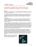
1
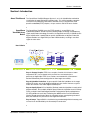
QuantiGene
ViewRNA
For FFPE Samples
User Manual
P/N 17400 Rev A 090708
DRAFT July 8, 2009 10:47 pm
QGViewRNATtile.fm
®
Panomics, Inc.
QuantiGene ViewRNA For FFPE Samples User Manual
Copyright
© Copyright 2009, Panomics, Inc. All rights reserved.
Trademarks
QuantiGene is a registered trademark exclusively licensed to Panomics, Inc. All other trademarks belong to their respective owners.
Citing QuantiGene ViewRNA in Publications
When describing a procedure for publication using this product, please refer to it as the QuantiGene ViewRNA assay.
If a paper cites a QuantiGene ViewRNA product and is published in a research journal, the lead author(s) may receive a travel stipend for use at a
technology conference or tradeshow by sending a copy of the paper to our technical support group at [email protected] or via fax at (510)
818-2610.
Disclaimer
Panomics, Inc. reserves the right to change its products and services at any time to incorporate technological developments. This manual is subject to
change without notice.
Although this manual has been prepared with every precaution to ensure accuracy, Panomics, Inc. assumes no liability for any errors or omissions, nor
for any damages resulting from the application or use of this information.
Contacting Panomics
U.S Corporate Headquarters
Panomics, Inc.(now a part of Affymetrix)
6519 Dumbarton Circle
Fremont, CA 94555
Toll Free: 877 PANOMICS (1.877.726.6642)
Direct: 1.510.818.2600
Fax: 1.510.818.2610
Email: [email protected]
Email: [email protected]
Email: [email protected]
European Headquarters
Panomics Srl
Via Sardegna 1
20060 Vignate-Milano (Italy)
Tel: +39.02.95.360.250
Fax: +39.02.360.992
Email: [email protected]
Email: [email protected]
Email: [email protected]
Asia Pacific Headquarters
Panomics, Inc.
16F Gemdale Plaza Tower A, No. 91
Jiango Road, Beijing 100022
P.R. China
Tel: +86.10.59208157
Fax: +86.10.59208111
Email: [email protected]
Email: [email protected]
Email: [email protected]
DRAFT July 8, 2009 10:47 pm
QGViewRNATtile.fm
Contents
Section I: Introduction . . . . . . . . . . . . . . . . . . . . . . . . . . . . . . . . . . . . . . . . . . . . . . . . . . 1
About This Manual . . . . . . . . . . . . . . . . . . . . . . . . . . . . . . . . . . . . . . . . . . . . . . . . 1
QuantiGene ViewRNA Assay Basics . . . . . . . . . . . . . . . . . . . . . . . . . . . . . . . . . . 1
How It Works . . . . . . . . . . . . . . . . . . . . . . . . . . . . . . . . . . . . . . . . . . . . . . . . . . . . 1
Section II: Required Materials . . . . . . . . . . . . . . . . . . . . . . . . . . . . . . . . . . . . . . . . . . . . 2
QuantiGene ViewRNA FFPE Sample Optimization Kit (Optional) . . . . . . . . . . . . 2
QuantiGene ViewRNA Assay Kit for FFPE Samples . . . . . . . . . . . . . . . . . . . . . . 2
QuantiGene ViewRNA Chromogenic Signal Amplification Kit . . . . . . . . . . . . . . . 2
QuantiGene ViewRNA Assay Probe Sets . . . . . . . . . . . . . . . . . . . . . . . . . . . . . . 3
Optional QuantiGene ViewRNA FFPE Positive Control Kit (Rat Kidney). . . . . . . 3
Required Materials Not Provided . . . . . . . . . . . . . . . . . . . . . . . . . . . . . . . . . . . . . 3
Section III: Recommendations for Experimental Design and Assay Optimization . . . . . 5
Overview . . . . . . . . . . . . . . . . . . . . . . . . . . . . . . . . . . . . . . . . . . . . . . . . . . . . . . . 5
Optimization experiment . . . . . . . . . . . . . . . . . . . . . . . . . . . . . . . . . . . . . . . . . . . 5
Expected Results . . . . . . . . . . . . . . . . . . . . . . . . . . . . . . . . . . . . . . . . . . . . . . . . . 5
Section IV: Assay Workflow . . . . . . . . . . . . . . . . . . . . . . . . . . . . . . . . . . . . . . . . . . . . . . 6
Section V: Sample Pretreatment . . . . . . . . . . . . . . . . . . . . . . . . . . . . . . . . . . . . . . . . . . 7
Overview . . . . . . . . . . . . . . . . . . . . . . . . . . . . . . . . . . . . . . . . . . . . . . . . . . . . . . . 7
Important Procedural Note . . . . . . . . . . . . . . . . . . . . . . . . . . . . . . . . . . . . . . . . . . 7
Before You Start . . . . . . . . . . . . . . . . . . . . . . . . . . . . . . . . . . . . . . . . . . . . . . . . . . 7
Procedure. . . . . . . . . . . . . . . . . . . . . . . . . . . . . . . . . . . . . . . . . . . . . . . . . . . . . . . 8
Section VI: In Situ Hybridization Procedure . . . . . . . . . . . . . . . . . . . . . . . . . . . . . . . . . 10
Overview . . . . . . . . . . . . . . . . . . . . . . . . . . . . . . . . . . . . . . . . . . . . . . . . . . . . . . 10
Important Procedural Notes . . . . . . . . . . . . . . . . . . . . . . . . . . . . . . . . . . . . . . . . 10
Before You Start . . . . . . . . . . . . . . . . . . . . . . . . . . . . . . . . . . . . . . . . . . . . . . . . . 10
Procedure. . . . . . . . . . . . . . . . . . . . . . . . . . . . . . . . . . . . . . . . . . . . . . . . . . . . . . .11
Section VII: Troubleshooting . . . . . . . . . . . . . . . . . . . . . . . . . . . . . . . . . . . . . . . . . . . . 19
Troubleshooting No or Weak Signals . . . . . . . . . . . . . . . . . . . . . . . . . . . . . . . . . 19
High Background on the Tissue Section . . . . . . . . . . . . . . . . . . . . . . . . . . . . . . 19
High Background on Slide . . . . . . . . . . . . . . . . . . . . . . . . . . . . . . . . . . . . . . . . . 20
Hematoxylin Staining is too Strong. . . . . . . . . . . . . . . . . . . . . . . . . . . . . . . . . . . 20
Section VIII: Rat Kidney FFPE Tissue Example . . . . . . . . . . . . . . . . . . . . . . . . . . . . . 21
QuantiGene ViewRNA FFPE User Manual
iii
Table of Contents
iv
QuantiGene ViewRNA FFPE User Manual
Section I: Introduction
Section I: Introduction
About This Manual The QuantiGene ViewRNA Reagent System is an in situ hybridization method for
visualization of target RNA within individual cells. This manual provides complete
instructions for performing QuantiGene ViewRNA assays using formalin-fixed
paraffin-embedded (FFPE) samples, 4-6 µm sections and 16-24 hours fixation.
QuantiGene The QuantiGene ViewRNA assay for FFPE samples is a novel RNA in situ
ViewRNA Assay hybridization product, based on patent-pending Probe Set design and proprietary
Basics signal amplification technology, that offers 10 copy RNA sensitivity in individual cells.
Signal amplification is predicated on specific hybridization of adjacent Probe Set
oligonucleotides to a target RNA (see How It Works below), resulting in excellent
signal-to-noise ratios.
How It Works
PreAmp1
FFPE tissue section
Amp1
LP-AP
Target
Step 1: Prepare Sample. FFPE tissue samples, fixed for 16-24 hours at room
temperature (RT), are sectioned to 4-6 µm thickness and attached to a
positively-charged slide. FFPE tissue sections are treated with a Pretreatment
Solution followed by Protease digestion to allow target accessibility.
Step 2: Hybridize Probe Sets. A gene-specific Probe Set hybridizes to the target
mRNA. For clarity, only single oligonucleotide pairs are shown. However, a typical
Probe Set contains 10 or more oligonucleotide pairs.
Step 3: Amplify Signal. A Pre-Amplifier (PreAmp) molecule hybridizes to each pair of
oligonucleotides, then multiple Amplifier (Amp) molecules hybridize to each PreAmp.
Finally, multiple Label Probe oligonucleotides conjugated to alkaline phosphatase
(LP-AP) hybridize to each Amp. Following the addition of the Fast Red Substrate,
alkaline phosphatase breaks down the substrate to form a precipitate (punctated
dots) that indicates the presence of the target RNA molecule.
Step 4: Image. Target mRNA is visualized using standard brightfield microscopy and
cell nuclei can be identified by the Hematoxylin counterstain.
QuantiGene ViewRNA FFPE User Manual
1
Section II: Required Materials
Section II: Required Materials
QuantiGene
ViewRNA FFPE
Sample
Optimization Kit
(Optional)
For initial optimization of a new tissue, this optional, cost-effective, all-inclusive kit
enables you to optimize assay conditions for up to 2 specific tissues. Once you have
optimized the conditions, those conditions can be used to evaluate targets of interest
using the QuantiGene ViewRNA Assay Kit for FFPE samples, Chromogenic Signal
Amplification Kit, and QuantiGene ViewRNA Probe Sets. This kit is available for
human (QV0100), mouse (QV0101), or rat (QV0102) samples only. Refer to the
Product Insert for quantities of individual components supplied. Kits have a shelf life
of 6 months from the date of delivery.
QuantiGene
ViewRNA Assay
Kit for FFPE
Samples
The components of the QuantiGene ViewRNA Assay Kit for FFPE samples and their
recommended storage conditions are listed below. QuantiGene ViewRNA Assay Kits
for FFPE samples are available in two sizes: QV0050 and QV0051, sufficient for 24 or
96 assays, respectively. Each kit is configured for processing a minimum of 6 slides
per experiment. Refer to the product insert for quantities of individual components
supplied. Kits have a shelf life of 6 months from date of delivery.
Component
Description
Storage
100X Pretreatment
Solution
Aqueous buffered solution
2-8 °C
Proteasea
Aqueous buffered solution
2-8 °C
Hybridization Buffer A
(Hyb A)
Aqueous solution containing formamide and
detergent
2-8 °C
Hybridization Buffer B
(Hyb B)
Aqueous solution containing formamide and
detergent
2-8 °C
Hybridization Buffer C
(Hyb C)
Aqueous solution containing detergent
2-8 °C
Wash Buffer Component 1
(Wash Comp 1)
Aqueous solution containing detergent
15-30 °C
Wash Buffer Component 2
(Wash Comp 2)
Aqueous buffered solution
15-30 °C
a. IMPORTANT! Do not freeze.
QuantiGene
ViewRNA
Chromogenic
Signal
Amplification Kit
2
The components of the QuantiGene ViewRNA Chromogenic Signal Amplification Kit
and their recommended storage conditions are listed below. This kit is available in 2
sizes: QV0200 and QV0201, sufficient for 24 or 96 assays, respectively. Each kit is
configured for processing a minimum of 6 slides per experiment. Refer to the Product
Insert for quantity of individual components supplied. Kits have a shelf life of 6
months from date of delivery.
Component
Description
Storage
PreAmplifier 1 (PreAmp1)
DNA in aqueous buffered solution
–20 °C
Amplifier 1 (Amp1)
DNA in aqueous buffered solution
–20 °C
AP Enhancer Solution
Aqueous buffered solution
2-8 °C
QuantiGene ViewRNA FFPE User Manual
Section II: Required Materials
Fast Red Tablets
Red precipitating substrate for the detection of
alkaline phosphatase activity
2-8 °C
Naphthol Buffer
Buffer required for preparation of Fast Red
Substrate
2-8 °C
Label Probe-AP (LP-AP)
Alkaline Phosphatase-conjugated
oligonucleotide in aqueous buffered solution
2-8 °C
QuantiGene In addition to the Assay Kit for FFPE Samples and the Chromogenic Signal
ViewRNA Assay Amplification Kit, QuantiGene ViewRNA TYPE 1 Probe Sets specific to your targets of
Probe Sets interest must be purchased separately. Probe Sets are available in multiple sizes and
should be stored at –20 °C. Refer to the Product Insert for more information.
IMPORTANT QuantiGene ViewRNA TYPE 1 Probe Sets hybridize to the
PreAmp1/Amp1/LP-AP signal amplification system, and can be used to visualize 10 RNA
copies/cell in FFPE samples.
Accessory Product
Description
Storage
QuantiGene ViewRNA TYPE 1
Probe Set
RNA-specific oligonucleotides for use with
PreAmp1/Amp1/LP-AP
–20 °C
Visit www.panomics.com for the most up-to-date listing of available QuantiGene
ViewRNA Probe Sets. If we do not yet offer a QuantiGene ViewRNA Probe Set for
your RNA of interest, please submit the accession number (include version or provide
gi number) or RNA sequence with your order, and we will design and synthesize it for
you in approximately 2 weeks.
Optional
QuantiGene
ViewRNA FFPE
Positive Control
Kit (Rat Kidney)
The QuantiGene ViewRNA FFPE Positive Control Kit is designed for use with
QuantiGene ViewRNA Assay Kit for FFPE Samples and QuantiGene ViewRNA
Chromogenic Signal Amplification Kit. The kit includes 8, 4-6 µm-thick rat kidney
FFPE sections attached to positively-charged slides as well as TYPE 1 Probe Set for
rat Ubc. Refer to the Product Insert for the optimized conditions and
recommendations for use.
Required Materials Other materials required to perform the QuantiGene ViewRNA assay for FFPE
Not Provided Samples that are not included in the Assay Kit for FFPE Samples or the Chromogenic
Signal Amplification Kit are listed here. When specified, do not use alternate
materials or suppliers.
Table 1
All Sample Types
Required Material
Supplier
Deionized Water (ddH2O)
MLSa
95% Ethanol
VWR
89015-512
10X PBS, pH 7.2-7.4
Bio-Rad Laboratories or
Invitrogen
161-0780
70013-032
Gill's Hematoxylin I
American Master Tech
Scientific
HXGHE1LT
HistoClear
National Diagnostics
HS-202
QuantiGene ViewRNA FFPE User Manual
Part Number
3
Section II: Required Materials
Table 1
All Sample Types (continued)
37% Formaldehydeb
Hydroxidec
Fisher Scientific
F79-1
VWR
JT9726-5
20X SSC (used only for optional stop
point in the procedure)
Ambion
AM9763
ImmEdge Hydrophobic Barrier Pen
Vector Laboratories
H4000
UltraMount
DAKO
S1964
Tissue Tek Staining Dish (clear color), 3
required
American Master Tech
Scientific
LWT4457EA
Tissue Tek Clearing Agent Dish (green
color), 1 required
American Master Tech
Scientific
LWT4456EA
Tissue Tek Vertical 24 Slide Rack, 1
required
American Master Tech
Scientific
LWSRA24
Coplin Jar, two 40-50 mL size
MLS
1000 mL Glass Beaker
MLS
Cover Glass, 22 mm x 30 mm (not
required if using optional tissue culture
incubator)
Fisher Scientific
12-530A
ThermoBrite Hybridization Chamber,
(holds up to 12 slides)
Abbot Molecular
07J91-010 (110V)
Humidifying Strips
Abbot Molecular
30-144115
Isotemp Hot Plate
Fisher Scientific
11-300-49SHP
(120V)
27-30% Ammonium
07J91-020 (240V)
11-302-49SHP
(230V)
Temperature/humidity meter (optional if
using tissue culture incubator)
Fisher-Scientific
11-661-19
Optional. Tissue culture incubator, 40 °C
without CO2 and 80-90% humidity for
processing 12 or more slides per
experiment.
Napco
51201082
VWR
9150860
Optional. Aluminum slide rack for use in
tissue culture incubator.
VWR
100493380
Optional. Incubator or oven utilizing
horizontal airflow and capable of
maintaining 80 °C.
Panomics
QS0700, QS0701
(120V) or QS0710,
QS0711 (220V)
Bright Field Microscope
MLS
Water Bath capable of maintaining
40 +/- 1 °C
MLS
a. MLS = Major Laboratory Supplier
b. WARNING! Formaldehyde is a poison and irritant. Avoid contact with skin and mucous membranes.
c. WARNING! Highly volatile. Use in fume hood.
4
QuantiGene ViewRNA FFPE User Manual
Section III: Recommendations for Experimental Design and Assay Optimization
Section III: Recommendations for Experimental Design and Assay
Optimization
Overview In this section, we provide recommendations for identifying optimal pretreatment
condition for the FFPE tissue in situ hybridization.
One optimization experiment is required for each tissue type of interest. Because the
quality of FFPE blocks vary, depending on their source, optimization is required. We
recommend preparing the FFPE blocks the same way you prepare the tissue
sections you plan to use.
Optimization You will need to prepare ten, 4-6 µm thick FFPE tissue sections from a block which
experiment was prepared in the same way (fixation time, section thickness and tissue type) as
the FFPE tissue of your interest. The optimization experiment will enable you to
identify the optimal condition for in situ hybridization.
Each slide will be treated with a different pretreatment condition and probed with the
same housekeeping gene, for example Ubc. Once an optimal condition for in situ
hybridization is determined, you can apply this pretreatment condition to tissue
sections in combination with the target probe of your interest.
The following table shows the optimization experiment set up:
Protease
Incubation
Time (min)
Pretreatment Boiling Time (min)
5
10
20
10
Slide #2
Slide #5
Slide #8
20
Slide #3
Slide #6
Slide #9
40
Slide #4
Slide #7
Slide #10
0
0
Slide #1
Expected Results For section treated with 0 min pretreatment boiling + 0 min protease incubation (Slide
#1), expect to see no signal.
For sections treated with 5, 10 or 20 min pretreatment boiling and 10, 20 or 40 min
protease incubation (Slide #2-#10), expect to see at least one slide showing strong
signal with good morphology. Once a condition has been optimized, any Probe Set
can be used with similarly treated samples.
See “Section VIII: Rat Kidney FFPE Tissue Example” on page 21 for an example of an
expected result.
QuantiGene ViewRNA FFPE User Manual
5
Section IV: Assay Workflow
Section IV: Assay Workflow
Step
1
See
Page...
Task
FFPE sample preparation (approximately 1.5 h)
a. Baking slides at 60 °C
7
b. Fixing with 10% formaldehyde
Note
2
Sample de-paraffinization (approximately 15 min)
8
3
Sample pre-treatment (approximately 5-20 min)
9
Note
4
Possible stopping point (store slides in 1X PBS at RT)
Protease digestion (approximately 5-40 min)
a. Protease treatment
11
b. Post-fixation in 4% formaldehyde
5
12
b. Wash 3 times
Note
6
13
Possible stopping point (store slides in 6X SSC at RT)
PreAmp hybridization (approximately 25 min)
a. Incubate with PreAmp
14
b. Wash 3 times
7
14
Amp hybridization (approximately 15 min)
a. Incubate with Amp
15
b. Wash 3 times
8
15
LP-AP hybridization (approximately 15 min)
a. Incubate with LP-AP
16
b. Wash 3 times
9
11
Target probe hybridization (approximately 3 h)
a. Incubate with target probe
17
FastRed substrate incubation (approximately 30 min)
a. Incubate with FastRed substrate
b. Rinse off substrate and fix in 4% formaldehyde
6
8
Possible stopping point (dry slides and store at RT)
18
18
10
Counterstain with Gills Hematoxylin
18
11
Mount samples and observe under brightfield microscope
18
QuantiGene ViewRNA FFPE User Manual
Section V: Sample Pretreatment
Section V: Sample Pretreatment
Overview Here we provide recommendations for preparation of samples prior to in situ hybridization.
For a successful in situ hybridization, prepare the FFPE samples as follows:
♦
Fix in 10% neutral buffer formalin for 16-24 hours as soon as the tissues are
extracted.
♦
Section FFPE blocks into 4-6 µm thickness and attach to positively charged
slides (Superfrost Plus, Fisher-Scientific P/N 12-550-15).
♦
Make sure FFPE tissue sections on each slide are smaller than 22 mm x 22 mm in
size. If larger than 22 mm x 22 mm in size, you will require twice as much
prepared working reagents per slide, reducing the total number of assays in the
kit.
Important All reagent volumes shown are based on hydrophobic barrier size of 22 mm x 25 mm
Procedural Note area.
Before You Start
Step
Action
1
Set one Thermobrite to 60 °C or a dry incubator to 60 °C.
2
Bake the slides in Thermobrite with lid open at 60 °C for 30 minutes. Alternatively,
bake slides in a 60 °C dry oven for 30 min. This will increase the tissue attachment
to the slide.
3
Prepare one Coplin jar with 10% formaldehyde in 1X PBS (add 10 mL of 37%
formaldehyde to 27 mL 1X PBS).
4
Prepare 350 mL of 1X PBS.
5
Prepare a second Coplin jar with 1X PBS.
6
Prepare 1X Pretreatment Solution by diluting 8 mL of 100X Pretreatment Solution
to 792 mL ddH2O.
7
Dispense 200 mL of 95% EtOH into a clean Tissue Tek Staining Dish.
8
Ensure 400 mL 95% Ethanol and ddH2O are available.
9
Add 200 mL HistoClear to Tissue Tek Clearing Agent Dish (green).
QuantiGene ViewRNA FFPE User Manual
7
Section V: Sample Pretreatment
Procedure To pretreat samples:
Step
1
Action
Incubate the sections with 10% formaldehyde (in a fume hood) for 1 hour at room
temperature:
c. Submerge the slide sections into the Coplin Jar containing 10%
formaldehyde solution and incubate for 1 hour at room temperature.
d. Move the slides to the Coplin jar containing 1X PBS, replace with fresh1X
PBS.
e. Remove slides from 1X PBS and entirely remove the 1X PBS from the slides
by placing them on edge on a laboratory wipe. Lightly tap until all liquid is
removed and allow to air dry.
2
Remove the paraffin from the samples:
a. Place the FFPE slides on 80 °C Thermobrite (with lid open) or in a dry
incubator at 80 °C for 3 min. The paraffin should be melted as soon as the
slides are on the heat block.
b. Immediately load the slides into a Tissue Tek Vertical Slide Rack and
submerge the slides into a Tissue Tek Clearing Agent Dish (green)
containing 200 mL HistoClear.
c. Incubate the slides in HistoClear (in a fume hood) at room temperature (RT)
for 10 min with frequent agitation.
d. Remove the slides from the HistoClear solution by lifting the slide rack.
Immediately transfer the slides into a Tissue Tek Staining Dish with 95%
EtOH.
e. Rinse off the residual HistoClear by moving the slide rack up and down
several times.
f. Decant the 95% EtOH and rinse the slides with fresh 95% EtOH.
g. Lift the slides out of the 95% EtOH and let the slides air dry on a paper
towel for 5 min at RT.
IMPORTANT Make sure the slide has dried completely before proceeding to step
2h.
h. Create a 22 mm x 25 mm hydrophobic rectangle with the tissue section at
the center using the ImmEdge Hydrophobic Barrier Pen and allow to air dry.
3
8
(Optional) Stopping point. Dry slides and store at RT.
QuantiGene ViewRNA FFPE User Manual
Section V: Sample Pretreatment
To pretreat samples: (continued)
Step
4
Action
Treat sample with 1X Pretreatment Solution:
a. Fill a 1000 mL beaker with 800 mL of 1X Pretreatment Solution and bring
the solution to boil (100-104 °C) by heating it on a hot plate. As soon as the
solution reaches boiling, cover the beaker and turn down the heat to
maintain 100 °C while preventing evaporation.
IMPORTANT The pretreatment solution should completely cover tissues on the
slides.
IMPORTANT To prevent boil over, make sure the pretreatment solution is not
vigorously boiling while submerging the slides.
b. Submerge the slide rack loaded with slides into the boiling pretreatment
solution, cover the beaker and incubate for the time as determined by using
the optimization procedure in “Section III: Recommendations for
Experimental Design and Assay Optimization” on page 5.
c. At the end of time point, remove the slide rack with slides in it and
submerge the entire rack with slides into a Tissue Tek Staining Dish
containing ddH2O.
d. If you are performing the optimization assay, remove the slides at the
appropriate time points and insert the slides in a rack submerged in Tissue
Tek Staining Dish containing ddH2O. Wait until you collect all of the slides
with various time points for pretreatment before proceeding.
e. Rinse the slides once more by decanting the ddH2O and refill with fresh
ddH2O.
5
Optional. If you want to stop at this point, you can transfer the slide rack with
slides into a Tissue Tek Staining Dish containing 200 mL 1X PBS at RT overnight.
QuantiGene ViewRNA FFPE User Manual
9
Section VI: In Situ Hybridization Procedure
Section VI: In Situ Hybridization Procedure
Overview The QuantiGene ViewRNA in situ hybridization protocol can be completed in approximately 8 hours (including pretreatment procedure). The instructions provided here
are for processing 1-24 sections, 1 section per slide. If you process less than 6 slides
at one time, there will be reagent shortages.
Important ♦
Procedural Notes
Use the following procedure to fully remove solutions from slides: drain solution
from the slide surface by placing the slide on its edge on a laboratory wipe and
lightly tap until liquid is removed from the hydrophobic barrier.
♦
Before addition of Working Reagents to samples, verify that hydrophobic barrier
is dry. If necessary, use the corner of a laboratory wipe to wick away remaining
solution.
♦
Always use freshly prepared solutions in the Tissue Tek Staining and Clearing
Agent dishes. Do not save or reuse solutions unless directed to do so (for
example with the 4% formaldehyde solution).
♦
Before opening reagents supplied in the microfuge tubes, briefly centrifuge to
collect contents at the bottom of the tube.
♦
Do not allow tissue sections to dry. Drying can result in low or no signal.
Before You Start
Step
Action
1
Set one Thermobrite to 40 °C and wet the humidifying strips, or use humidifying
oven without CO2.
2
Prepare 1000 mL 1X PBS.
3
Prepare 4% formaldehyde in 1X PBS in one Tissue Tek Staining Dish (add 27 mL of
37% formaldehyde to 223 mL 1X PBS).
4
Pre-warm Hyb A, Hyb B and Hyb C buffers to 40 °C.
5
Thaw Probe Set, PreAmplifier and Amplifier. Place on ice until use.
Note
If using the Optimization Kit, the Probe Set is not required.
6
Fill one Tissue Tek Staining Dish with 200 mL Gill's Hematoxylin.
7
Prepare 3000 mL Wash Buffer by adding components to a 3 L capacity container
in the following order and then mixing well:
♦ 2.5 L ddH20
♦ 27 mL Wash Comp 1
♦ 7.5 mL Wash Comp 2
♦ ddH20 to 3 L
Note Adding the components in the order listed above to prevent precipitates
from forming that would occur when adding Wash Comp 1 and Wash Comp 2
directly together.
10
QuantiGene ViewRNA FFPE User Manual
Section VI: In Situ Hybridization Procedure
Step
8
Action
Prepare 1000 mL of 0.01% ammonium hydroxide in ddH2O by adding 0.33 mL of
30% ammonium hydroxide into 999.67 mL ddH2O and mix well.
! WARNING ! 30% ammonium hydroxide is highly volatile. Use in a fume
hood.
Procedure
Step
1
Action
Perform Protease digestion:
a. Prepare Working Protease Solution by diluting the Protease 1:100 in
1X PBS and mixing well. Place at 40 °C until ready to use.
b. Pre-warm the section by putting the slides on Thermobrite set at 40 °C and
add 200 µL, 1X PBS to each section (make sure the buffer does not dry up
on the sections).
c. Decant the 1X PBS from the slides, one at a time, followed by placing the
slide back onto the Thermobrite and immediately add 200 µL of Working
Protease Solution onto the tissue section. Use a pipette tip to spread the
protease and make sure the entire tissue section is covered.
d. Close the lid and incubate for the amount of time determined in the
optimization experiment (see “Section III: Recommendations for
Experimental Design and Assay Optimization” on page 5).
e. Remove the slides from Thermobrite, decant the Protease Solution, and
place slides in a Tissue Tek Slide Rack.
f. If you are performing the optimization assay, remove the slides from the
Thermobrite, decant the protease solution, and insert the slides in a Tissue
Tek Slide Rack submerged in Tissue Tek Staining Dish containing 200 mL
1X PBS. Wait until you collect all of the slides before proceeding.
g. Submerge the slides into a Tissue Tek Staining Dish containing 200 mL
1X PBS.
h. Decant the 1X PBS and replace with fresh 1X PBS. Move the slide rack up
and down several times.
i.
Move the slides (using a slide rack) into a Tissue Tek Staining Dish
containing 4% formaldehyde and incubate for 5 min at RT.
j.
Fill another clean Tissue Tek Staining Dish with 1X PBS, while waiting for
the incubation.
k. Rinse the slide by moving the slide rack from the 4% formaldehyde solution
to 1X PBS solution. Keep the 4% formaldehyde solution for later use.
l.
Move the slide rack up and down several times to make sure the
formaldehyde is rinsed off.
QuantiGene ViewRNA FFPE User Manual
11
Section VI: In Situ Hybridization Procedure
Step
2
Action
Hybridize the Probe Set:
Note If you are using the Sample Optimization Kit, skip the preparation of
Working Probe Set and instead use species-specific prewarmed Hyb A.
a. Prepare Working Probe Set by diluting the Probe Set 1:50 in pre-warmed
Hyb A and mixing well.
b. Place at 40 °C until use. Scale reagents according to the number of assays
to be run. Include 10% overage. Use the table below as a guide.
Component
1 slide
10 slides
Hyb A (prewarmed)
215.6 µL
2156 µL
QuantiGene ViewRNA TYPE 1
Probe Set
4.4 µL
44 µL
Total Volume
220 µL
2200 µL
\
c. Continue the hybridization according to the method of incubation.
If using...
Thermobrite
oven
Then do this...
a. Before moving tissue sections to the Thermobrite, add
200 µL Working Probe Set to each tissue section.
IMPORTANT The hydrophobic barrier must be drawn at
22 mm (W) x 25 mm (L) to accommodate this volume once
the coverslip is put in place.
b. Gently add a 30 x 22 mm coverslip on top of each
section. Make sure not to introduce any bubbles in
between the section and the coverslip.
c. Move slides to the 40 °C Thermobrite, close the lid
and incubate for 3 hours.
Tissue
culture
incubator
a. Place slides on the aluminum slide rack.
b. Add 200 µL Working Probe Set to each tissue section
(addition of cover slip is not necessary).
c. Put slides in a 40 °C humidified incubator for 3 hours.
12
QuantiGene ViewRNA FFPE User Manual
Section VI: In Situ Hybridization Procedure
Step
3
Action
Wash the samples 4 times (for Thermobrite) with Wash Buffer:
If using...
Thermobrite
oven
Then do this...
a. Remove slides from the 40 °C Thermobrite, and
submerge the slides in a Tissue Tek Staining Dish
(without rack inside) containing 200 mL of Wash
Buffer. The coverslips should fall off the section.
b. As soon as the coverslips fall off, transfer the slides
into a Tissue Tek Slide Rack submerged in another
Tissue Tek Staining Dish containing 200 mL of Wash
Buffer and incubate at RT for 2 min. Move the slide
rack up and down several times to wash the sections.
c. Decant the Wash Buffer and add fresh Wash Buffer.
Incubate the slides in Wash Buffer at RT for 2 min and
move the slide rack up and down several times.
d. Repeat step 3c.
Tissue
culture
incubator
a. Remove slides from the 40 °C incubator, decant the
Hyb A solution from slides, and transfer slides to a
Tissue Tek Slide Rack submerged in a Tissue Tek
Staining Dish containing 200 mL of Wash Buffer and
incubate at RT for 2 min. Move the slide rack up and
down several times to wash the sections.
b. Decant the Wash Buffer and add fresh Wash Buffer.
Incubate the slides in Wash Buffer at RT for 2 min and
move the slide rack up and down several times.
c. Repeat step 3b.
4
Optional stopping point. Prepare 6X SSC. Submerge your slides, in a slide rack,
into a Tissue Tek Staining Dish containing 200 mL 6X SSC. Slides can be left
overnight at RT.
QuantiGene ViewRNA FFPE User Manual
13
Section VI: In Situ Hybridization Procedure
Step
5
Action
Hybridize Pre-Amplifier:
a. Prepare Working PreAmp1 by diluting PreAmp1 1:100 in pre-warmed Hyb B
and mixing well.
b. Place at 40 °C until use. Scale reagents according to the number of assays
to be run. Include 10% overage. Use the table below as a guide.
Component
1 slide
10 slides
Hyb B (prewarmed)
197.8 µL
2178 µL
PreAmp1
2.2 µL
22 µL
Total Volume
220 µL
2200 µL
c. Remove the slides from the staining dish and decant the Wash Buffer from
the slides. Make sure the section is wet and the hydrophobic rectangle
is dry. Always use the corner of a laboratory tissue to wipe the
hydrophobic barrier dry.
If using...
Thermobrite
oven
Then do this...
a. Before moving the slides to the Thermobrite, add
200 µL of Working PreAmp1 to each slide.
b. Move the slides to the 40 °C Thermobrite, close the lid
to incubate for 25 min.
Tissue
culture
incubator
6
a. Place slides on an aluminum slide rack and add
200 µL of Working PreAmp 1 to each slide.
b. Put the slide rack into the 40 °C humidified tissue
culture incubator for 25 min.
Wash the samples 3 times with Wash Buffer:
If using...
Thermobrite
oven
Then do this...
a. Remove slides from the 40 °C Thermobrite, decant
the Hyb B solution from slides, and submerge the
slides in a Tissue Tek Staining Dish containing 200 mL
of Wash Buffer. Incubate at RT for 2 minutes. Move
the slide rack up and down several times to wash the
sections.
b. Decant the Wash Buffer and add fresh Wash Buffer.
Incubate the slides in Wash Buffer at RT for 2 min and
move the slide rack up and down several times.
c. Repeat step 6b.
Tissue
culture
incubator
a. Remove slides from the 40 °C incubator, decant the
Hyb B solution from slides, and transfer slides to a
Tissue Tek Slide Rack submerged in a Tissue Tek
Staining Dish containing 200 mL of Wash Buffer and
incubate at RT for 2 min. Move the slide rack up and
down several times to wash the sections.
b. Decant the Wash Buffer and add fresh Wash Buffer.
Incubate the slides in Wash Buffer at RT for 2 min and
move the slide rack up and down several times.
c. Repeat step 6b.
14
QuantiGene ViewRNA FFPE User Manual
Section VI: In Situ Hybridization Procedure
Step
7
Action
Hybridize Amplifier:
a. Prepare Working Amp1 by diluting Amp1 1:100 in pre-warmed Hyb B and
mixing well.
b. Place at 40 °C until use. Scale reagents according to the number of assays
to be run. Include 10% overage. Use the table below as a guide.
Component
1 slide
10 slides
Hyb B (prewarmed)
197.8 µL
1978 µL
Amp1
2.2 µL
22 µL
Total Volume
220 µL
2200 µL
c. Remove the slides from the staining dish and decant the Wash Buffer from
the slides. Make sure the section is wet and the hydrophobic rectangle
is dry. Always use the corner of a laboratory tissue to wipe the
hydrophobic barrier dry.
If using...
Thermobrite
oven
Then do this...
a. Before moving the slides to the Thermobrite, add
200 µL of Working Amp1 to each slide.
b. Move the slides to the 40 °C Thermobrite, close the lid
to incubate for 15 min.
Tissue
culture
incubator
8
a. Place slides on an aluminum slide rack and add
200 µL of Working Amp1 to each slide.
b. Put the slide rack into the 40 °C humidified tissue
culture incubator for 15 min.
Wash the samples 3 times with Wash Buffer:
If using...
Thermobrite
oven
Then do this...
a. Remove slides from the 40 °C Thermobrite, decant
the Hyb B solution from slides, and put the slides into
a Tissue Tek rack submerged in Tissue Tek Staining
Dish containing 200 mL of Wash Buffer and incubate
at RT for 2 min. Move the slide rack up and down
several times to wash the sections.
b. Decant the Wash Buffer and add fresh Wash Buffer.
Incubate the slides in Wash Buffer at RT for 2 min and
move the slide rack up and down several times.
c. Repeat step 8b.
Tissue
culture
incubator
a. Remove slides from the 40 °C incubator, decant the
Hyb B solution from slides, and transfer slides to a
Tissue Tek Slide Rack submerged in a Tissue Tek
Staining Dish containing 200 mL of Wash Buffer and
incubate at RT for 2 min. Move the slide rack up and
down several times to wash the sections.
b. Decant the Wash Buffer and add fresh Wash Buffer.
Incubate the slides in Wash Buffer at RT for 2 min and
move the slide rack up and down several times.
c. Repeat step 8b.
QuantiGene ViewRNA FFPE User Manual
15
Section VI: In Situ Hybridization Procedure
Step
9
Action
Hybridize Label Probe:
a. Prepare Working LP-AP by first diluting LP-AP 1:10 in prewarmed
Hyb C and mix well, followed by making a 1:100 dilution, yielding a 1:1000
final dilution.
b. Place at 40 °C until use. Scale reagents according to the number of assays
to be run. Include 10% overage. Use the table below as a guide.
Component
1 slide
10 slides
Hyb C (prewarmed)
197.8 µL
1978 µL
LP-AP (1:10 diluted)
2.2 µL
22 µL
Total Volume
220 µL
2200 µL
c. Remove the slides from the staining dish and decant the Wash Buffer from
the slides. Make sure the section is wet and the hydrophobic rectangle
is dry. Always use the corner of a laboratory tissue to wipe the
hydrophobic barrier dry.
If using...
Thermobrite
oven
Then do this...
a. Before moving the slides to the Thermobrite, add
200 µL of Working LP-AP to each slide.
b. Move the slides to the 40 °C Thermobrite, close the lid
to incubate for 15 min.
Tissue
culture
incubator
16
a. Place slides on an aluminum slide rack and add
200 µL of Working LP-AP to each slide.
b. Put the slide rack into the 40 °C humidified tissue
culture incubator for 15 min.
QuantiGene ViewRNA FFPE User Manual
Section VI: In Situ Hybridization Procedure
Step
10
Action
Wash the samples 3 times with Wash Buffer:
If using...
Thermobrite
oven
Then do this...
a. Remove slides from the 40 °C Thermobrite, decant
the Hyb C solution from slides, and put the slides into
a Tissue Tek rack submerged in Tissue Tek Staining
Dish containing 200 mL of Wash Buffer and incubate
at RT for 3 min. Move the slide rack up and down
several times to wash the sections.
b. Decant the Wash Buffer and add fresh Wash Buffer.
Incubate the slides in Wash Buffer at RT for 3 min and
move the slide rack up and down several times.
c. Repeat step 10b.
Tissue
culture
incubator
a. Remove slides from the 40 °C incubator, decant the
Hyb C solution from slides, and transfer slides to a
Tissue Tek Slide Rack submerged in a Tissue Tek
Staining Dish containing 200 mL of Wash Buffer and
incubate at RT for 3 min. Move the slide rack up and
down several times to wash the sections.
b. Decant the Wash Buffer and add fresh Wash Buffer.
Incubate the slides in Wash Buffer at RT for 3 min and
move the slide rack up and down several times.
c. Repeat step 10b.
11
Move the slides to a dry paper towel on the bench. Add 200 µL of the
AP-Enhancer Solution and incubate at RT for 5-10 minutes while preparing the
Fast Red Substrate.
12
Prepare the Fast Red Substrate:
a. Remove the required number of Fast Red tablets from the refrigerator. Allow
tablets to reach RT.
b. Drop one Fast Red tablet into 5 mL of Naphthol Buffer. Vortex to dissolve
tablet.
Note
For best results, use the prepared Fast Red Substrate within 1 hour.
QuantiGene ViewRNA FFPE User Manual
17
Section VI: In Situ Hybridization Procedure
Step
13
Action
Apply the Fast Red Substrate:
a. Decant the AP-Enhancer Solution and place the slides on a dry paper towel
with tissue section on top.
If using...
Thermobrite
oven
Then do this...
a. Add 200 µL Fast Red Substrate, move slides to
Thermobrite hybridization station, close the lid, and
incubate at 40 °C for 30 min in dark.
b. Decant the Fast Red Substrate from slides, place
slides in a Tissue Tek Slide Rack and submerge the
slides in a Tissue Tek Staining Dish containing 200 mL
of 1X PBS.
c. Move the slide rack up and down several times to
rinse off the Fast Red Substrate.
d. Move the slide rack into the Tissue-Tek staining dish
containing 4% formaldehyde (used earlier in the
procedure) and incubate for 5 min at RT.
e. Decant the 4% formaldehyde and replace with
1X PBS.
f. Move the slide rack up and down several times to
rinse off the formaldehyde.
Tissue
culture
incubator
a. Place slides on aluminum slide rack and add 200 µL
Fast Red Substrate to each slide. Put tray into the
40 °C humidified tissue culture incubator for 30 min in
the dark.
b. Remove the Fast Red Substrate from slides, place
slides in a Tissue Tek Slide Rack, and submerge the
slides in a Tissue Tek Staining Dish containing 200 mL
of 1X PBS.
c. Repeat steps 13c-13f from above.
14
Counter stain with Gill's Hematoxylin and add cover glass:
a. Move the slide rack into the Tissue-Tek staining dish containing Gill's
Hematoxylin and incubate for 5 min at RT, or until the nuclei are properly
stained. If they don’t stain, repeat staining.
b. Move the slide rack into another Tissue-Tek staining dish containing
ddH2O.
c. Decant the ddH2O and fill the staining dish with fresh ddH2O.
d. Decant the ddH2O and fill the staining dish with 0.01% Ammonium
Hydroxide and move the rack up and down several times to create a much
more blue color nuclear stain.
e. Remove the slides from the rack and remove the solution on the slide as
much as possible. Allow slides to dry.
f. Place 1-2 drops of the mounting medium on top of the tissue without
making any bubbles.
g. Gently put a cover glass over the sample and avoid making bubbles. Leave
the samples at room temperature for several hours for the mounting
medium to dry.
15
18
View slides under a brightfield microscope.
QuantiGene ViewRNA FFPE User Manual
Section VII: Troubleshooting
Section VII: Troubleshooting
Troubleshooting Troubleshooting no or weak signals:
No or Weak
Recommended Actions
Signals Probable Cause
Tissue drys up during
Working Probe Set
hybridization step
Make sure to place a cover glass over the tissue sample when
using Thermobrite hybridization station at the target probe
hybridization step.
Make sure to wet the strips inside the Thermobrite before
starting hybridization.
Make sure the Thermobrite is placed on a leveled bench.
Tissue drys up during
Protease digestion
Make sure enough Protease solution is added to each tissue for
protease digestion.
Make sure to close the Thermobrite lid during digestion.
Make sure to wet the strips inside the Thermobrite before
starting hybridization.
Fast Red substrate drys
up during incubation
Make sure enough Fast Red Substrate is added to each section.
Make sure to close the Thermobrite lid during digestion.
Make sure to wet the strips inside the Thermobrite before
starting Fast Red reaction.
Tissue over-fixed after
protease digestion
Make sure the tissue sections are not fixed for more than 5 min
in 4% formaldehyde after protease digestion.
Placing the slide on the
40 °C Thermobrite
before adding
Hybridization Buffer,
Enhancer Solution, or
FastRed Solution and
causing the Probe to
dissociate from the
target.
Add the Hybridization Buffer, Enhancer Solution, or FastRed
Solution to slides before moving them to the 40 °C Thermobrite.
High Background Troubleshooting high background on the tissue section:
on the Tissue
Recommended Actions
Section Probable Cause
Insufficient washing
Increase the length of wash by 5 min for each wash step.
Slide drys up during
Working Probe Set
hybridization step
Make sure to place a cover glass over the tissue sample when
using Thermobrite hybridization station at the target probe
hybridization step.
Make sure to wet the strips inside the Thermobrite before
starting hybridization.
Make sure the Thermobrite is placed on a leveled bench.
QuantiGene ViewRNA FFPE User Manual
19
Section VII: Troubleshooting
Fast Red substrate drys
up during incubation
Make sure enough Fast Red substrate is added to each section.
Make sure to close the Thermobrite lid during digestion.
Make sure to wet the strips inside the Thermobrite before
starting Fast Red reaction.
High Background Troubleshooting high background on glass slide:
on Slide
Probable Cause
Recommended Actions
Over protease digestion
of the sample
Optimize the protease digestion time as indicated in the
“Section III: Recommendations for Experimental Design and
Assay Optimization” on page 5.
Insufficient washing
Increase the length of wash by 5 min for each wash step.
Slide drys up during
Working Probe Set
hybridization step
Make sure to place a cover glass over the tissue sample when
using Thermobrite hybridization station at the target probe
hybridization step.
Make sure to wet the strips inside the Thermobrite before
starting hybridization.
Make sure the Thermobrite is placed on a leveled bench
Fast Red substrate drys
up during incubation
Make sure enough Fast Red substrate is added to each section.
Make sure to close the Thermobrite lid during digestion.
Make sure to wet the strips inside the Thermobrite before
starting Fast Red reaction.
Hematoxylin Troubleshooting Hematoxylin staining:
Staining is too
Recommended Actions
Strong Probable Cause
Slides are incubated in
Gill’s Hematoxylin
solution for too long
20
Wash slides several times in ddH2O to reduce blue stain.
Reduce the time the slides are in Gill’s Hematoxylin solution.
QuantiGene ViewRNA FFPE User Manual
Section VIII: Rat Kidney FFPE Tissue Example
Section VIII: Rat Kidney FFPE Tissue Example
QuantiGene ViewRNA FFPE User Manual
21