1
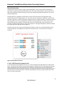
Expresso® Solubility and Expression Screening System Note: Two different storage temperatures required. Vectors, Protease, Polymerase Competent Cells FOR RESEARCH USE ONLY. NOT FOR HUMAN OR DIAGNOSTIC USE. Lucigen Corporation 2905 Parmenter St, Middleton, WI 53562 USA Toll Free: (888) 575-9695 | (608) 831-9011 | FAX: (608) 831-9012 [email protected] www.lucigen.com MA158 Rev B Expresso® Solubility and Expression Screening System Table of Contents Technical Support............................................................................................................................................ 3 Product Designations .......................................................................................................................................... 3 Introduction......................................................................................................................................................... 5 Special Materials and Equipment Needed ........................................................................................................ 5 Process Workflow ............................................................................................................................................... 6 System Details ..................................................................................................................................................... 7 The pSol Vector Suite ................................................................................................................................... 8 E. cloni® 10G Chemically Competent Cells ................................................................................................ 8 Cloning Strategy........................................................................................................................................... 9 pSol His Control Vector ............................................................................................................................... 9 GH1 Control Insert ...................................................................................................................................... 9 Colony Screening ......................................................................................................................................... 9 Protein Expression ....................................................................................................................................... 9 Protein Purification...................................................................................................................................... 9 SelecTEV™ Protease ................................................................................................................................. 10 Detailed Protocol ............................................................................................................................................... 11 1. Preparation of Insert DNA ........................................................................................................... 11 2. Enzyme-free Cloning with the pSol Vectors ................................................................................. 13 3. Heat Shock Transformation of E. cloni® 10G Chemically Competent Cells ............................... 13 4. Colony PCR Screening for Recombinants .................................................................................... 15 5. DNA Purification and Sequencing................................................................................................ 16 6. Induction of Protein Expression ................................................................................................... 16 7. Evaluation of Target Protein Expression and Solubility .............................................................. 17 8. Affinity Purification of 6xHis tagged proteins .............................................................................. 19 9. Cleavage of Fusion Tag with SelecTEV™ Protease .................................................................... 19 10. Removing SelecTEV Protease after Cleavage .............................................................................. 19 11. SelecTEV Cleavage During Dialysis ............................................................................................ 20 Control Information ......................................................................................................................................... 21 References .......................................................................................................................................................... 21 Appendix A: Media Recipes............................................................................................................................. 23 Appendix B: Internal sequencing Primers ..................................................................................................... 23 Appendix C: Colony PCR Screening .............................................................................................................. 24 Appendix D: Vector Map and Sequencing Primers....................................................................................... 25 Appendix E: Cloning Troubleshooting Guide ................................................................................................ 26 Appendix F: Expression/Purification Troubleshooting Guide ..................................................................... 27 Appendix G: Characteristics of Fusion Tags in the pSol Vector Suite ........................................................ 28 2 MA158 Rev B Expresso® Solubility and Expression Screening System Technical Support Lucigen is dedicated to the success and satisfaction of our customers. Our products are tested to assure they perform as specified when used according to our recommendations. It is imperative that the reagents supplied by the user are of the highest quality. Please follow the instructions carefully and contact our technical service representatives if additional information is necessary. We encourage you to contact us with your comments regarding the performance of our products in your applications. Thank you. Lucigen Technical Support Email: [email protected] Phone: (888) 575-9695 Product Guarantee: Lucigen guarantees that this product will perform as specified for one year from the date of shipment. Please avoid using reagents after one year from date of receipt. Product Designations The Expresso Solubility and Expression Screening System contains the pre-processed Expresso pSol Vector suite, E. cloni® 10G Chemically Competent Cells for cloning and protein expression, a cloning control insert, PCR primers for clone verification, recovery medium for transformation, and solutions of L-Rhamnose and D-Glucose for small-scale induction of protein expression. The kit can also be purchased with SelecTEV™ Protease for fusion protein cleavage and/or Accura® High-Fidelity Polymerase to amplify your gene of interest. The system catalog numbers are listed below. Expresso Solubility and Expression Screening System Product Description Expresso Solubility and Expression Screening System Expresso Solubility and Expression Screening System + SelecTEV™ Protease Kit Size 24 reactions Catalog number 49060-1 24 reactions 49062-1 Expresso Solubility and Expression Screening System + Accura High-Fidelity Polymerase 24 reactions 49064-1 Expresso Solubility and Expression Screening System + SelecTEV™ Protease and Accura High-Fidelity Polymerase 24 reactions 49066-1 Part Number(s) A943302-1 A96352-2 A943302-1 A96352-2 A933167-1 A943302-1 A96352-2 A932528-0 A943302-1 A96352-2 A933167-1 A932528-0 Components & Storage Conditions The Expresso Solubility and Expression Screening System may consist of up to four separate packages (depending on catalog number ordered). 3 MA158 Rev B Expresso® Solubility and Expression Screening System Package 1, 2, and 3 Must be stored at -20C. Package 4, containing Competent Cells Must be stored at -80C. Package 1: Expresso® Solubility and Expression Screening Suite Component Concentration Expresso® pSol Vectors (3 reactions each) 12.5 ng/µL 1. pSol AFV 2. pSol SlyD 3. pSol Tsf 4. pSol SUMO 5. pSol Bla 6. pSol MBP 7. pSol GST 8. pSol His Control GH1 Control Insert (Positive Control) 50 ng/µL Primers for PCR screening and sequencing pRham™ Forward Primer 50 pmol/µL ® pETite Reverse Primer 50 pmol/µL Rhamnose Solution 20% w/v Glucose Solution 15% w/v Package 2: SelecTEV™ Protease Component SelecTEV™ Protease, 1000 Units SelecTEV™ 20X Buffer DTT Package 3: Accura® High-Fidelity Polymerase Component Accura® High-Fidelity Polymerase Accura® 2X HF Reaction Buffer Accura® 10X GC Reaction Buffer Betaine Concentration 10 U/µL 20X 100 mM Concentration 2 U/µL 2X 10X 5M Volume 6 µL 10 µL 100 µL 100 µL 1.25 mL 1.25 mL Store at -20C Part # F843211-1 F843212-1 F843213-1 F843214-1 F843215-1 F843216-1 F843217-1 F843218-1 F823219-1 F81887-1 F91710-1 F88889-1 F88890-1 Amount 100 µL 1.0 mL 500 µL Store at -20C Part # F833167 F883093-1 F853091-1 Amount 25 U 1.25 mL 600 µL 1.0 mL Store at -20C Part # F832528-0 F882522-1 F882521-1 F881901-1 Package 4: E. cloni 10G Chemically Competent Cells. Component 24 Reaction Kit E. cloni 10G Chemically Competent Cells 24 X 40 µL Transformation Control pUC19 DNA (10 pg/µL) 20 µL 2 X 12 mL Recovery Medium (Store at -20C or -80C) Competent cells will thaw if stored at -20°C and should be discarded. Store at -80C Part # F96419 F92078-1 F98226-1 4 MA158 Rev B Expresso® Solubility and Expression Screening System Introduction The Expresso® Solubility Screening System enables rapid cloning and expression of a protein of interest fused to a diverse panel of powerful and cleavable fusion tags. This system includes one unique and six literature-validated fusion tags engineered into a suite of cloning-ready pSol vectors for rapid evaluation. Expressioneering® technology allows a single PCR amplicon to be cloned directly into all of the pSol expression vectors without restriction enzymes, ligase, or DNA purification. Each vector contains the rhaPBAD promoter for stable cloning and strong tunable expression within a single host strain. The vectors also encode a 6xHis tag on the N-terminus of each fusion tag to facilitate affinity column purification. The fusion tags can be easily cleaved from the protein of interest using the TEV Protease cleavage site located between the N-terminal fusion tag and the protein of interest. The Expresso Solubility and Expression Screening System may be purchased with SelecTEV™ Protease, a highly site-specific and very active variant of native TEV protease. SelecTEV™ protease and the fusion tags contain N-terminal His tags and can be removed from TEV digestion reactions by IMAC. Important Note: The Expresso Solubility and Expression Screening System is intended to improve the chance of obtaining soluble and functional protein for research purposes. However, there will be proteins whose expression and/or solubility will not improve upon using this kit. For example, Lucigen does NOT recommend using this system for GPCRs, ion channels, and other proteins that are either membrane integrated or associated. Special Materials and Equipment Needed The Expresso Solubility and Expression Screening System supplies many of the items needed to efficiently generate and express recombinant clones. Less common items that must be supplied by the user include: Custom Primers for target gene amplification Sterile polypropylene 17 x 100 mm culture tubes Sonicator equipped with a microtip Resin and columns for immobilized metal affinity chromatography SDS-PAGE equipment. 5 MA158 Rev B Expresso® Solubility and Expression Screening System Process Workflow An example of an Expresso Solubility and Expression Screening System workflow is provided below. Workflow Step Preparation of Insert DNA Expresso Cloning using pSol Vector(s) Colony PCR Screening for Recombinants Grow overnight cultures and split into three aliquots Evaluate Target Protein Expression and Solubility Affinity Purification of 6xHis tagged proteins Cleavage of Fusion Tag with SelecTEV™ Protease Removal of SelecTEV Protease and Solubility Tag Workflow Details Amplify the desired coding sequence by PCR. If the PCR yields a single robust product, proceed directly to step 2: Enzyme-free Cloning with the pSol Vectors. Add PCR product and pSol vector(s) to Chemically Competent E. cloni® 10G Cells. Perform transformation. Incubate the transformation plates overnight at 37C. Perform colony PCR screen. Analyze PCR products by agarose gel electrophoresis. Start overnight cultures of PCR-positive colonies. (1) Isolate plasmid DNA from 1.5 mL of culture. (2) Inoculate cultures for protein expression. (3) Prepare glycerol stocks (optional). Harvest induced cultures. Lyse cells by sonication. Fractionate lysates by centrifugation. Analyze total, soluble, and insoluble protein fractions by SDS-PAGE. Bind cleared lysate to IMAC resin. Wash column and elute fusion proteins. Dialyze overnight to remove imidazole. Incubate the reaction at 30°C for at least 1 hour. Incubation at lower temperatures can be performed but reaction time will need to be increased. Bind the SelecTEV™ Protease digest reaction to an IMAC resin. Collect the Flow-Through, containing the untagged protein of interest. The Histidine tagged SelecTEV™ Protease and Histidine tagged pSol partner will bind to such resins. Reference in Detailed Protocol 1.1 Primer Design 1.2 Amplification of the target gene 3.2 Transformation Protocol 4.1 Colony PCR Screening 5.1 Colony Growth 5.2 DNA Purification 6 Induction of Protein Expression 7 Evaluate Target Protein Expression and Solubility 8 Affinity Purification of 6xHis tagged proteins 9 Cleavage of Fusion Tag with SelecTEV™ Protease 10 Removing SelecTEV Protease after Cleavage 6 MA158 Rev B Expresso® Solubility and Expression Screening System System Details The Expresso Solubility and Expression Screening System uses Expressioneering™ Technology for rapid cloning and expression of solubility-tagged fusion proteins in E. coli. Expressioneering is an in vivo recombinational cloning strategy whereby PCR products can be cloned instantly, with no enzymatic treatment (Figure 1). The target gene is PCR amplified, mixed with the pSol vector(s), and transformed directly into chemically competent cells. Recombination within the host cells seamlessly joins the insert to the vector. Though several solubility tags are available from a variety of vendors, choosing the best tag for a given target protein can only be determined empirically. An important feature of this system is the standardized design of the pSol vectors, which allows a single PCR product to be cloned and tested in all 8 vectors in parallel for selection of the best tag for a given target protein. Figure 1. Expressioneering Technology. A PCR product that contains short homology to the ends of the pSol Expresso vectors is mixed with any of the pre-processed vectors and transformed directly into the chemically competent cells provided. Expression of the fusion protein in the pSol vectors is under control of the rhaPBAD promoter, which is inducible by L-rhamnose. This promoter is recognized by the E. coli RNA polymerase; therefore, a single host strain can be used for both clone construction and protein expression. This single-host strategy allows a much more streamlined workflow than systems requiring separate hosts for cloning and expression. The rhaPBAD promoter is tightly controlled for protein expression. In the absence of rhamnose, the transcriptional activity of rhaPBAD is very low, allowing stable clone construction, even for potentially toxic gene products (1). Transcription is positively controlled by two activators, RhaR and RhaS, which bind rhamnose (2). RhaR activates its own transcription as well as that of RhaS, which in turn activates transcription from rhaPBAD. This regulatory cascade makes transcription from rhaPBAD responsive to variable concentrations of rhamnose, allowing “tunable” control of the target gene expression (3). For proteins that are potentially toxic to the host cells, or that are difficult to express in soluble form, this tuning capability enables adjustment of expression levels for optimal yield of soluble protein. Transcription from the rhaPBAD promoter is also subject to catabolite repression. In the presence of glucose, transcription from rhaPBAD remains inactive even when rhamnose is available. This repression allows the use of “autoinduction” procedures for protein expression, in which cells are inoculated directly into medium containing rhamnose and a small amount of glucose (4, 5). While glucose is present, the cells grow without expression from the rhaPBAD promoter. Expression of the protein of interest occurs only late in the culture, when the glucose is exhausted. 7 MA158 Rev B Expresso® Solubility and Expression Screening System The pSol Vector Suite The pSol Vector Suite is based on Lucigen’s patented pSMART® vectors, which feature transcriptional terminators to prevent unwanted transcription into or out of the cloned sequence. The small size of the pSol Vector backbone facilitates cloning of large inserts and performing DNA manipulations, such as sitedirected mutagenesis. The pSol Vectors are supplied pre-linearized for instant, directional insertion of target genes using Expressioneering™ Technology (Figures 1-3). The vectors contain all signals for expression, which include the rhaPBAD promoter, an efficient ribosome binding site from the T7 gene 10 leader, and translational start and stop codons. Each vector is designed for expression of the target protein as a fusion with an aminoterminal 6xHis-Sol tag. In addition, all of the tags in the kit are positioned for precise removal by TEV protease to produce target protein of nearly native sequence. The single PCR product produced is suitable for cloning into all 8 of the pSol Vectors (Figure 2). The pSol vectors do not contain the lacZ alpha gene fragment, so they do not enable blue/white colony screening. However, the background of empty vector is typically <5%, so minimal colony screening is necessary. Figure 2. pSol Expression Vectors. E. cloni® 10G Chemically Competent Cells E. cloni 10G Chemically Competent Cells are an E. coli strain optimized for high efficiency transformation. The E. cloni 10G cells are ideal for cloning and propagation of plasmid clones, and give high yield and high quality plasmid DNA due to the endA1 and recA1 mutations. The 10G cell strain is also well-suited for protein expression with pSol expression plasmids. This system eliminates the need to shuttle vectors into a separate strain for protein expression. If desired, confirmed pSol constructs may be transferred to other strains for protein expression. 8 MA158 Rev B Expresso® Solubility and Expression Screening System E. cloni 10G Genotype: F- mcrA Δ(mrr-hsdRMS-mcrBC) endA1 recA1 Φ80dlacZΔM15 ΔlacX74 araD139 Δ(ara,leu)7697galU galK rpsL nupG λ- tonA (StrR) E. cloni 10G Chemically Competent Cells produce ≥ 1 x 10⁹ cfu/µg supercoiled pUC19 DNA. As a control for transformation, E. cloni® 10G Competent Cells are provided with supercoiled pUC19 DNA at a concentration of 10 pg/µL. Use 1 µL (10 pg) for transformation. Select pUC19 transformants on plates containing Ampicillin or Carbenicillin (100 µg/mL). Cloning Strategy The pSol vectors are provided in a linearized form, ready for co-transformation with a PCR product containing the gene of interest. The coding sequence is amplified with user-supplied primers that include 18 nucleotides of overlap with the ends of the vector. The forward primer contains sequence corresponding to the TEV cleavage recognition site, and the reverse primer includes stop codons and vector sequence. Recombination between the vector and insert occurs within the host strain, seamlessly fusing the gene of interest to the vector. The method is similar to cloning by homologous recombination (6). It does not require single-stranded ends on the vector or the insert, as in “PIPE” cloning (7). pSol His Control Vector The pSol His Control Vector included with the kit is intended to enable determination of baseline expression and solubility levels that may be achieved with the vector-host system in the absence of a solubility-enhancing tag. GH1 Control Insert The GH1 Control Insert included with the kit is a 0.6 kb PCR fragment that encodes human Growth Hormone 1 (GH1). It is flanked by sequences for Expresso cloning directly into the pSol Vectors. It serves as a positive control for monitoring cloning efficiency, expression, solubility, and TEV protease cleavage. Colony Screening Empty-vector background with the pSol Vectors is typically very low (<5%), so minimal screening is necessary. Colony PCR may be used to verify the presence of inserts. Primers included with the kit are suitable for screening by colony PCR and for sequencing of plasmid DNA. Lucigen recommends sequence analysis to confirm the junctions of the insert with the vector as well as the predicted coding sequence. See Appendix B Appendix B: Internal sequencing Primers for primer sequences that can be used to sequence from within the fusion tag toward the fusion junction. Protein Expression Recombinant plasmids are constructed in the E. cloni 10G host strain and expressed in the same host. After clones have been verified by colony PCR, individual colonies are grown in liquid culture, and protein expression is induced by addition of rhamnose. Expression of Sol-tagged fusion proteins is evaluated by SDS-PAGE analysis. Protein Purification Materials for protein purification are not provided with the Expresso Solubility and Expression Screening System. However, 6xHis tagged proteins produced with this system may be purified by Immobilized Metal Affinity Chromatography (IMAC). Various IMAC reagents are available, from a number of vendors. Follow the resin manufacturer’s guidelines for purification. 9 MA158 Rev B Expresso® Solubility and Expression Screening System SelecTEV™ Protease The Expresso Solubility and Expression Screening System may be purchased with SelecTEV™ Protease (See Product Designations on pg. 3 for ordering information). SelecTEV™ Protease is an enhanced form of Tobacco Etch Virus (TEV) protease that is highly site-specific, more active, and more stable than native TEV protease. SelecTEV™ Protease recognizes the seven amino acid sequence Glu-Asn-Leu-Tyr-Phe-Gln-Gly and cleaves between Gln and Gly with high specificity. The protease can be used to cleave the N-His Sol tag from the protein of interest. SelecTEV™ Protease has an amino-terminal 6xHis tag. Following cleavage of a target protein, the protease can be removed from the cleavage reaction by IMAC. 10 MA158 Rev B Expresso® Solubility and Expression Screening System Detailed Protocol 1. Preparation of Insert DNA 1.1. Primer Design To clone with Expressioneering™ Technology, the target DNA must first be amplified with primers that add flanking sequence identical to those found at the ends of linear pSol vectors (Figure 3). Forward Primer Features The forward primer should be of the general structure: 5’-AAT CTG TAC TTC CAG GGT XXX XXX XXX XXX XXX XXX …-3’ The required sequence at the 5’ end of the forward primer includes 6 of the TEV recognition site codons. This sequence is also found at one end of all of the pSol vectors. The 3’ end of the forward primer anneals to the bottom strand of the target gene, usually beginning with the second codon. The length of complementarity to the target gene will depend on the sequence. Because each Expresso pSol vector contains an ATG initiation codon immediately preceding the 6xHis codons, the ATG initiation codon from the target gene is not needed. Reverse Primer Features The reverse primer should be of the general structure: 5’-GTG GCG GCC GCT CTA TTA XXX XXX XXX XXX XXX XXX …-3’ The required sequence at the 5’ end of the reverse primer matches 18 bases of the downstream end of the pSol vectors. The 3’ end of the reverse primer anneals to the top strand of the target gene, usually including the last codons prior to the stop codon of the coding region. The length of complementarity to the target gene will depend on the sequence. Because the pSol vectors include in-frame stop codons, it is not necessary to include the stop codon of the target gene. Factors affecting the length of the target-specific portion of the primers include: GC content, Tm, and potential for formation of hairpins or primer-dimers. We recommend that targetspecific portion of each primer be designed with a Tm of ~60°C. The annealing temperature used in amplification may be adjusted to accommodate primers with higher or lower Tm. Figure 3. Primers for cloning into the Expresso pSol Vectors. 11 MA158 Rev B Expresso® Solubility and Expression Screening System 1.2. Amplification of the target gene Amplify the desired coding sequence by PCR, using primers designed as described in section 1.1 and a proofreading PCR polymerase. Notes: The performance of the Expresso Solubility and Expression Screening System has been verified with PCR products from various proofreading polymerases, including Accura® High-Fidelity Polymerase. If not using Accura High-Fidelity Polymerase, follow the manufacturer’s recommendations during amplification of the target gene. The use of non-proofreading polymerases (e.g. Taq) is strongly discouraged. 1.2.1. Amplification protocol using Accura High-Fidelity Polymerase: For a 50 µL reaction, assemble the following on ice: Volume, Component µL 25 2X HF Reaction Buffer 4 dNTPs (2.5 mM each) X Forward Primer X Reverse Primer X 0.5 X Template DNA Accura High-Fidelity Polymerase, 2 U/µL Nuclease-free H2O 50 Total volume Cycle the PCR reaction according to the table below. Step Temperature 1. Initial Denaturation 94˚C 2. Denaturation 94˚C 3. Annealing 60˚C 4. Extension 72˚C 5. Repeat steps 2-4 6. Final extension 72˚C 7. Hold 4˚C Final Concentration or Quantity 1X 200 µM 1 µM 1 µM 100 pg – 30 ng plasmid 1U --- Time 30 seconds 15 seconds 15 seconds 60 sec /kb 20 – 30 total cycles 10 minutes ∞ Refer to the Accura® High-Fidelity Polymerase user manual (MA151) for additional PCR guidance (http://lucigen.com/docs/manuals/MA151-Accura-High-FidelityPolymerase.pdf). 12 MA158 Rev B Expresso® Solubility and Expression Screening System 1.2.2. Analysis of Amplified DNA Confirm the size and quality of the amplified DNA by agarose gel electrophoresis. If the reaction yields a single product at a concentration of 10 ng/µL or higher, you can proceed directly to Step 2: Expresso Cloning using pSol Vectors. Note: If the amplification template is an intact circular plasmid encoding kanamycin resistance, high background is likely. Methods to avoid this background include: Gel purification of the PCR product prior to Expresso cloning. DpnI treatment of the PCR product prior to Expresso cloning. For best results, purify PCR product following DpnI treatment. Linearization of template by restriction digest. Purify linear template prior to amplification by PCR. 2. Enzyme-free Cloning with the pSol Vectors Following PCR product verification by agarose gel electrophoresis, the PCR product (1-3 µL or 25-100 ng) is mixed with 25 ng of pSol Vector and transformed directly into competent E. cloni® 10G cells. To ensure optimal cloning results, we strongly recommend the use of Lucigen’s E. cloni 10G Chemically Competent Cells, which are included with the kit. Proceed to Step 3 to perform the transformation. 3. Heat Shock Transformation of E. cloni® 10G Chemically Competent Cells E. cloni 10G Chemically Competent Cells are provided in 40 μL aliquots, sufficient for a single transformation. Transformation is performed by incubation on ice followed by heat shock at 42°C. Note: For maximal transformation efficiency, the heat shock is performed in 15 mL disposable polypropylene culture tubes (17 x 100 mm). The use of other types of tubes may dramatically reduce the transformation efficiency. 3.1. Preparation for Transformation Bring the Recovery Medium to room temperature. Pre-chill a 15 mL disposable polypropylene culture tube (17 x 100 mm) on ice for each transformation. Thaw the E. cloni 10G cells completely on ice (5-10 minutes). 3.2. Transformation Protocol Add vector and insert DNAs to the tubes containing the E. cloni 10G Chemically Competent Cells on ice as indicated below. Reagent Rxn 1 Rxn 2 Rxn 3 Rxn 4 Rxn 5 Rxn 6 Rxn 7 13 MA158 Rev B Rxn 8 Expresso® Solubility and Expression Screening System 12.5 ng/µL pSol AFV Vector 12.5 ng/µL pSol SlyD Vector 12.5 ng/µL pSol Tsf Vector 12.5 ng/µL pSol SUMO Vector 12.5 ng/µL pSol Bla Vector 12.5 ng/µL pSol MBP Vector 12.5 ng/µL pSol GST Vector 12.5 ng/µL pSol Control Vector Amplified Target Fragment 2 µL 2 µL 2 µL 2 µL 2 µL 2 µL 2 µL ≤3 µL ≤3 µL ≤3 µL ≤3 µL ≤3 µL ≤3 µL ≤3 µL Transfer the entire mixture of cells and DNA to a pre-chilled 15 mL polypropylene culture tube(s) (17 x 100 mm). Incubate culture tubes containing cells and DNA on ice for 30 minutes. Heat shock cells by placing the tubes in a 42°C water bath for 45 seconds. Return the tubes of cells to ice for 2 minutes. Add 960 µL of room temperature Recovery Medium to the cells in the culture tubes. Place the culture tubes in a shaking incubator at 250 rpm for 1 hour at 37C. Plate 100 µL of transformed cells on LB-Lennox agar plates containing 30 µg/mL kanamycin. Incubate the plates overnight at 37C. 3.3. Optional Controls Reagent pSol Vector (12.5ng/µL) GH1 Control Insert pUC19 (provided with cells) Positive Control 2 µL 1 µL n/a Negative Control 2 µL n/a n/a Transformation Control n/a n/a 1 µL 3.4. Expected Results Cloning Reactions (step 3.2) and the optional Positive Control reactions typically yield >50 colonies per 100 µL plated. Negative Control (vector only) reactions typically yield <10 colonies per 100 µL plated Transformation Control should yield > 1 x 10⁹ colonies per µg plasmid. 3.5. Increasing Number of Recombinants Certain genes can prove difficult to clone due to a large size, toxic gene products, secondary structures, extremely biased base composition, or other unknown reasons. If necessary, the entire 1 mL transformation culture (from step 3.2) can be pelleted in a microcentrifuge (10,000 rpm, 30 seconds), resuspended in 100 µL of recovery media, and plated. See Appendix E: Cloning Troubleshooting Guide for additional troubleshooting information. 14 MA158 Rev B 2 µL ≤3 µL Expresso® Solubility and Expression Screening System 4. Colony PCR Screening for Recombinants Colonies are selected at random and screened for inserts by colony PCR using the pRham Forward and pETite® Reverse primers which are included with the kit. Colony PCR may also be performed using screening primers that are specific to the gene of interest (e.g. pRham Forward and the gene-specific reverse primer from step 1.1). The instructions below provide details on performing colony PCR using Lucigen’s CloneID™ 1X Colony PCR Master Mix (catalog #30059). Additional information on CloneID Colony PCR can be found in the User’s Manual: http://lucigen.com/docs/manuals/MA140-CloneID.pdf. If not using Lucigen’s CloneID Colony PCR to perform screening, follow the manufacturer’s instructions. Due to the high efficiency of Expresso Cloning System, nearly all of colonies on the transformation plates will contain the desired recombinant plasmids. Nonetheless, Lucigen recommends screening at least 2 colonies from each plate. 4.1. Colony PCR Screening Obtain CloneID™ 1X Colony PCR For a 25 µL reaction, assemble the following on ice Volume, µL 25 0.25 0.25 25.5 Component CloneID 1X Colony PCR Mix pRham Forward primer (50 µM) (Blue cap) pETite Reverse primer (50 µM) (Brown cap) Total volume Using a pipet tip, transfer well isolated colonies to the PCR reaction mix. Disperse the cells by pipetting up and down several times. Use the same tip to inoculate 3 mL of LB-Miller medium containing 30 µg/mL kanamycin and 0.5% glucose. The glucose in these cultures represses undesired expression of the target protein during this initial growth, reducing the risk of slow growth and plasmid instability, particularly if the target protein is toxic to the host strain. Note: To expedite the workflow one portion of each culture can be used for plasmid minipreps (step 5) and another portion can be used to inoculate cultures for induction of protein expression. However, only colony PCR-verified cultures should be used for subsequent DNA purification and analysis. Cycle the PCR reaction according to the table below. Step Temperature 1. Initial Denaturation 98˚C 2. Denaturation 94˚C 3. Annealing 55˚C 4. Extension 72˚C 5. Repeat steps 2-4 6. Final extension 72˚C 7. Hold 4˚C Time 2 minutes 15 seconds 15 seconds 1 minute/kb 25 total cycles 10 minutes ∞ 15 MA158 Rev B Expresso® Solubility and Expression Screening System Load 5 µL of the CloneID 1X Colony PCR Mix reaction(s) onto an agarose gel for analysis (0.7-2% agarose gel). Compare PCR product result(s) to expected band sizes. The expected bands size is determined by the fusion tag and the size of the gene of interest. See Appendix C: Colony PCR Screening for information on the size of the fusion tags. 5. DNA Purification and Sequencing 5.1. Colony Growth Transfer cultures containing PCR-verified pSol plasmids to a 37°C incubator and shake overnight at 220-250 rpm. 5.2. DNA Purification Use standard methods (8) to isolate plasmid DNA from 1.5 ml aliquots of the overnight cultures. The pSol plasmids contain the low-copy ColE1 origin of replication and produce DNA yields of ~150 ng per mL of culture. 5.3. Sequencing Lucigen strongly recommends sequence confirmation of the junctions of the insert with the vector as well as the predicted coding sequence. pRham Forward and pETite Reverse primers provided with the kit may be used for sequencing. See Appendix D: Vector Map and Sequencing Primers for the sequence and orientation of these primers. Depending on the size of the fusion tag, sequencing primers specific to each fusion tag will be required in order to obtain sequencing data for the junction between the fusion tag and protein of interest. See Appendix B for information on tag specific primers. 5.4. Preparation of Glycerol Stocks (optional) If archiving clones for future use, mix an aliquot of the overnight culture leftover from step 5.1 with an equal volume of sterile 50% glycerol in a cryovial. Mix well, and store at -70°. 6. Induction of Protein Expression 6.1. Induction of Protein Expression Induction may be performed using a standard induction protocol (step 6.1.1) or an autoinduction protocol (step 6.1.2). Regardless of the method of induction, Lucigen recommends performing small-scale expression trials (2 to 50 mL) to evaluate expression and solubility of the target protein before scaling up for purification. Small scale studies are also recommended when optimizing expression conditions (e.g. growth temperature, duration of induction, titration of Rhamnose concentration, etc). 16 MA158 Rev B Expresso® Solubility and Expression Screening System 6.1.1. Standard Induction Protocol Use 30 µl of culture leftover from Step 5 to inoculate 3 ml LB-Miller medium containing 30 µg/mL kanamycin without glucose. When cultures reach an optical density of 0.5 at 600 nm (OD600) induce expression by adding 30 µl of 20% L-Rhamnose to the culture. Continue shaking at 37°C for 4 hours. Notes: For maximal induction, Lucigen recommends a final concentration of 0.2% LRhamnose. Lower amounts in the range of 0.001% to 0.1% can be used for lower levels of expression, which may improve the solubility of some proteins. Induction for up to 8 hours may be required for maximal expression of some target proteins. Induction for up to 24 hours or more may be required for cultures grown at 20°C to 30°C. 6.1.2. Autoinduction Autoinduction uses glucose to repress the rhaPBAD promoter. Cells will preferentially metabolize glucose during the early stages of growth, and the rhaPBAD promoter will become active only when glucose is depleted. The timing of induction by rhamnose can be controlled by varying the concentration of glucose between 0.05% and 0.15%. Later onset of induction may be beneficial if the expressed protein is unstable, insoluble, or toxic to the host cells. Prepare LB-Miller medium containing 30 µg/mL kanamycin, L-Rhamnose (0.2% w/v), and D glucose (0.05 to 0.15% w/v) (4) (see table below). Autoinduction Method Early autoinduction Late autoinduction Add per mL of culture 10 µL 20% L-rhamnose 10 µL 20% L-rhamnose 3.3 µL 15% D-glucose 10 µL 15% D-glucose Inoculate the medium with an uninduced E.cloni 10G culture containing a verified pSol construct. Shake at 220-250 rpm at 37ºC for ~4-16 hours. 7. Evaluation of Target Protein Expression and Solubility 7.1. Harvest normalized amounts of the induced cultures. Note: The volumes listed below are convenient for those with access to a sonicator equipped with a microtip. It may be necessary to adjust culture, and fraction volumes when using other equipment. Collect a 100 µL aliquot of each induced culture, and mix with 900 µl of LB-Miller media. Measure the OD600 of the diluted samples. 17 MA158 Rev B Expresso® Solubility and Expression Screening System Based on the corrected cell density, calculate the volume of 10 OD600 unit equivalents (1 OD600 equivalent = 1 mL of culture at OD600 = 1). Example: If the OD600 of the diluted culture = 0.45 Then, the corrected cell density = 4.5 OD600 units And, 10 OD600 units = 2.22 ml (10 OD600 equivalents ÷ 4.5 OD600 /ml = 2.22 ml) Harvest 10 OD600 units of each induced culture by centrifugation (e.g. 12,000 x g for 1 minute). Resuspend the cell pellets in 1 mL lysis buffer (e.g. 50 mM NaH2PO4, 300 mM NaCl, pH 8.0). The sample will conatin 10 OD600 equivalents per mL. Proceed with lysis immediately or freeze samples. 7.2. Lyse Cells by Sonication Note: Other physical lysis methods may be used. Sonication is recommended because it is amenable to small sample size. Use of detergent-containing lysis buffers is not recommended. Lyse cells thoroughly by sonication on ice. Refer to the sonicator manual for recommended settings. Do not allow heat to build up, and avoid frothing. Transfer 400 µl of the lysate to a fresh tube, and store on ice. This fraction will be used to assess the Total amount of protein in the cell lysate. Transfer a second 400 µl aliquot of lysate to a fresh tube and centrifuge at 15,000 x g for 15 minutes at 4°C. Transfer the supernatant to a fresh tube and store on ice. This fraction will be used to assess the Soluble protein in the cell lysate. Add 400 µl of 2x SDS Sample Buffer to the Pellet. This fraction will be used to assess the Insoluble protein in the cell lysate. Care should be taken to fully resuspend this pellet. 7.3. SDS-PAGE Analysis Dilute the Total, Soluble, and Insoluble fractions 1:1 with 2x SDS Sample Buffer. Heat gel samples to 95°C for 5 minutes. Load 0.05 OD600 units each of the Total, Soluble, and Insoluble fractions into the wells of a SDS-PAGE gel. Note: The use of 4-20% polyacrylamide gels fitted with 15-well combs facilitates the workflow. Pre-poured gels of this type are available from several vendors. Lucigen recommends loading 0.05 OD units (e.g. 10 µL of each sample diluted 1:1 with 2x SDS Sample Buffer) per lane of a 15-well polyacrylamide gel. Include standards to estimate molecular weight of the recombinant protein. Choose the protein fusion that produces the greatest proportion of protein in the soluble fraction for scale-up and affinity purification. 18 MA158 Rev B Expresso® Solubility and Expression Screening System 8. Affinity Purification of 6xHis tagged proteins The fusion proteins expressed using pSol vectors contain an N-terminal 6xHis tag. Therefore, the fusion protein containing the solubility tag and the protein of interest can be purified using an IMAC resin. Many protocols are available for purification of 6xHis tagged proteins under native or denaturing conditions. For best results, follow the procedures recommended by the manufacturer of the IMAC resin. Due to variations in target protein physical properties, you may need to empirically determine the amount of resin needed for purification of your fusion protein. 9. Cleavage of Fusion Tag with SelecTEV™ Protease A TEV recognition site is located between the fusion tag and the protein of interest. SelecTEV™ Protease recognizes the seven-amino-acid sequence Glu-Asn-Leu-Tyr-Phe-Gln-Gly and cleaves between Gln and Gly with high specificity (9-11). Cleavage by the SelecTEV™ Protease is optimal in SelecTEV™ Buffer at 30°C, but SelecTEV™ Protease is active between 4 – 30°C and pH 6.0 – 8.5. Optimization of the cleavage conditions may be necessary depending on the protein of interest. An example of a SelecTEV™ Protease cleavage protocol is shown below. Set up the SelecTEV Set up the SelecTEV Protease reaction by adding the following reagents to a 1.5 mL tube. Volume, µL Component X 5 1 1 Y 100 Fusion Protein SelecTEV™ 20X Buffer DTT, 100mM SelecTEV™ Protease, 10 U/ µL Water Total Final Concentration/ Amount 30 µg 1X 1 mM 10 U N/A Incubate the reaction at 30°C for at least 1 hour. Refer to the manual for SelecTEV™ Protease (http://lucigen.com/docs/manuals/MA157-SelecTEVProtease.pdf) for further guidance. 10. Removing SelecTEV Protease after Cleavage SelecTEV™ Protease contains a histidine (6xHis) tag at its N-terminus. After cleavage of the fusion protein, you may remove SelecTEV™ Protease from the cleavage reaction by IMAC. Perform the binding and elution as described in the resin manufacturer’s protocol. The cleaved native protein will be in the flow-through fractions (as long as the cleaved protein does not contain a histidine tag). The histidine-tagged fusion partner and the SelecTEV™ Protease will remain bound to the resin. 19 MA158 Rev B Expresso® Solubility and Expression Screening System Notes: Imidazole remaining in the sample from the initial purification will prevent the histidine tag on SelecTEV from binding to the IMAC resin. Remove the imidazole (by dialysis or with a desalting column) prior to performing the post-TEV IMAC purification. Some IMAC resins do not tolerate 1 mM DTT. Methods employed to remove imidazole will simultaneously remove the DTT used in the TEV cleavage reaction. 11. SelecTEV Cleavage During Dialysis The cleavage reaction can be performed during buffer exchange by dialysis. Conditions may be adjusted from those recommended above. In general, use 1 µL SelecTEV (10 U) for every 10 µg of fusion substrate. If dialysis is carried out at 4°C, an overnight (≥ 16 hours) reaction time is recommended. 20 MA158 Rev B Expresso® Solubility and Expression Screening System Control Information The Expresso Solubility and Expression Screening System includes a GH1 positive control insert that can be cloned into any of the pSol vectors. To generate a soluble control fusion protein, clone the control insert into either pSol Bla, pSol SlyD, pSol Tsf, or pSol MBP vector. These clones can be used to express and purify soluble fusion proteins that can be expressed, purified, and cleaved with SelecTEV protease as specified in step 10. The following table summarizes the expected protein sizes before and after TEV protease cleavage. SelecTEV is approximately 27 kDa and may be visible when performing SDS-PAGE on the cleavage reaction. Construct Full Length Expected Size Expected Cleavage Products Bla-GH1 ~ 64 kDa ~41 kDa ~22 kDa SlyD-GH1 ~45 kDa ~23 kDa ~22 kDa Tsf-GH1 ~54 kDa ~32 kDa ~22 kDa MBP-GH1 ~64 kDa ~42 kDa ~22 kDa References 1. Haldimann, A., Daniels, L. and Wanner, B. (1998). Use of new methods for construction of tightly regulated arabinose and rhamnose promoter fusions in studies of the Escherichia coli phosphate regulon. J. Bacteriol. 180, 1277. 2. Egan, S.M. and Schleif, R.F. (1993). A regulatory cascade in the induction of rhaBAD. J. Mol. Biol. 234, 87. 3. Giacalone, M.J., Gentile, A.M., Lovitt, B.T., Berkley, N.L., Gunderson, C.W. and Surber, M.W. (2006). Toxic Protein Expression in Escherichia coli using a rhamnose-based tightly regulated and tunable promoter system. Biotechniques 40, 355. 4. Wegerer, A., Sun, T. and Altenbuchner, J. (2008). Optimization of an E. coli L-rhamnose-inducible expression vector: test of various genetic module combinations. BMC Biotechnol. 8, 2. 5. Grossman, T.H., Kawasaki, E.S., Punreddy, S.R. and Osburne, M.S.(1998). Spontaneous cAMPdependent deprepression of gene expression in stationary phase plays a role in recombinant expression instability. Gene 209, 95. 6. Bubeck, P., Winkler, M. and Bautsch, W. (1993). Rapid cloning by homologous recombination in vivo. Nuc. Acids Res. 21, 3601. 7. Klock, H.E., Koesema, E.J., Knuth, M.W. and Lesley, S.A. (2008). Combining the polymerase incomplete primer extension method for cloning and mutagenesis with microscreening to accelerate structural genomics efforts. Proteins 71, 982. 21 MA158 Rev B Expresso® Solubility and Expression Screening System 8. Sambrook, J. and Russell, D.W. (2001). Molecular Cloning: A Laboratory Manual (Third Edition). Cold Spring Harbor Laboratory Press, Cold Spring Harbor, New York. 9. Parks, T.D., Leuther, K.K., Howard, E.D., Johnston, S.A., Dougherty, W.G. (1994). Release of proteins and peptides from fusion proteins using a recombinant plant virus proteinase. Anal Biochem. 216, 413. 10. Phan, J., Zdanov, A., Evdokimov, A. G., Tropea, J. E., Peters, H. P. K., Kapust, R. B., Li, M., Wlodawer, A., and Waugh, D. S. (2002). Structural basis for the substrate specificity of tobacco etch virus protease. J. Biol. Chem. 27, 50564. 11. Tozser, J., Tropea, J. E., Cherry, S., Bagossi, P., Copeland, T. D., Wlodawer, A., and Waugh D. S. (2005). Comparison of the substrate specificity of two potyvirus proteases. FEBS J. 272, 514. 12. Goulet, A., Spinelli, S., Blangy, S., van Tilbeurgh, H., Leulliot, N., Basta, T., Prangishvili, D., Cambillau, C., Campanacci, V. (2009). The thermo- and acido-stable ORF-99 from the archaeal virus AFV1. Protein Sci. 6, 1316. 13. Han KY1, Song JA, Ahn KY, Park JS, Seo HS, Lee J. (2007) Solubilization of aggregation-prone heterologous proteins by covalent fusion of stress-responsive Escherichia coli protein, SlyD. Protein Eng Des Sel. 20, 543. 14. Han KY1, Song JA, Ahn KY, Park JS, Seo HS, Lee J. (2007) Enhanced solubility of heterologous proteins by fusion expression using stress-induced Escherichia coli protein, Tsf. FEMS Microbiol Lett. 274, 132. 15. Marblestone, J.G., Edavettal, S.C., Lim, Y., Lim, P., Zuo, X. and Butt, T.R. (2006). Comparison of SUMO fusion technology with traditional gene fusion systems: Enhanced expression and solubility with SUMO. Protein Sci. 15, 182. 16. Tokunaga H1, Arakawa T, Tokunaga M. (2010) Novel soluble expression technologies derived from unique properties of halophilic proteins. Appl Microbiol Biotechnol. 88, 1223. 17. Kapust, R. B. and Waugh, D. S. (1999). Escherichia coli maltose-binding protein is uncommonly effective at promoting the solubility of polypeptides to which it is fused. Protein Sci. 8, 1668. 18. Smith, D.B. and Johnson, K.S. (1998). Single-step purification of polypeptides expressed in Escherichia coli as fusions with glutathione S-transferase. Gene 67, 31. 22 MA158 Rev B Expresso® Solubility and Expression Screening System Appendix A: Media Recipes LB-Miller Culture Medium Per liter: 10 g Bacto-tryptone, 5 g yeast extract, 10 g NaCl. Mix components and autoclave. LB-Lennox + kan30 Agar Medium for Plating of Transformants Per liter: 10 g Bacto-tryptone, 5 g yeast extract, 5 g NaCl, 15 g agar. Mix components, autoclave and cool to 55°C. To select for pSol transformants, add kanamycin to a final concentration of 30 µg/mL. Pour into petri plates. 2X SDS Gel Sample Buffer 62.5 mM Tris-HCl pH 6.8; 2% SDS; 5% β-mercaptoethanol or 0.1 M DTT; 25% glycerol; 0.01% Bromophenol Blue Appendix B: Internal sequencing Primers The table below provides primer sequences that can be used to sequence from within the fusion tag toward the fusion junction. Compared to the pRham Forward primer, these primers read through a reduced amount of vector sequence before reaching the cloning junction. Vector Forward Primers (5’ 3’) pSol AFV ACG AGT ATT CTT ACG ATG CGT CCG AG pSol Bla CTG CCG GGT AAG CAC ACC pSol GST ATG TGC CTG GAT GCG TTC CC pSol MBP ACG TAT TGC CGC CAC CAT G pSol SlyD GTG CTC ACG ACC ACC ATC ACG pSol SUMO GGT GTC CGA TGG ATC TTC AGA G pSol Tsf GAG CAC AAT GCG GAA GTG ACC 23 MA158 Rev B Expresso® Solubility and Expression Screening System Appendix C: Colony PCR Screening The Expresso Solubility and Expression Screening System includes pRham Forward and pETite Reverse primers, which can be used for colony PCR with your gene of interest (GOI). To enhance the specificity of the screen, the GOI Reverse primer (designed in section 1.1) may be used in place of the pETite Reverse primer. Refer to the chart below to determine the expected PCR product sizes when using pRham Forward with pETite Reverse or pRham Forward with GOI Reverse. All sizes are listed in base pairs. Vector pSol AFV pSol SlyD pSol Tsf pSol SUMO pSol Bla pSol MBP pSol GST pSol His Control pRham Forward & pETite Reverse Vector + GOI = PCR Fragment 490 + = 781 + = 1042 + = 496 + = 1294 + = 1297 + = 850 + = 193 + = pRham Forward & GOI Reverse Vector + GOI = PCR Fragment 439 + = 730 + = 991 + = 445 + = 1243 + = 1246 + = 799 + = 142 + = 24 MA158 Rev B Expresso® Solubility and Expression Screening System Appendix D: Vector Map and Sequencing Primers The sequences of the pRham Forward and pETite® Reverse primers are: Shown below are the regions surrounding the cloning sites in the pSol vectors: 25 MA158 Rev B Expresso® Solubility and Expression Screening System Appendix E: Cloning Troubleshooting Guide Problem Probable Cause Solution Very few or no transformants No DNA, degraded DNA, or insufficient amount of DNA. Check insert DNA by gel electrophoresis. Determine concentration of insert and add the correct amount. Use the supplied control insert to test the system. Be sure the 5’ ends of the primer sequences match the pSol vector ends. Add the correct amount of kanamycin to molten agar at 55oC before pouring plates. DO NOT spread antibiotic onto the surface of agar plates. Use 15 mL disposable polypropylene culture tubes (17 x 100 mm). The use of other types of tubes may dramatically reduce the transformation efficiency. Pellet the entire 1 mL transformation culture (from step 3.2), resuspended in 100 µL of recovery media, and plate. Linearize plasmid DNA used as a template prior to PCR or gel-isolate PCR fragment, or treat PCR reaction with DpnI. Analyze colonies by sequencing to confirm the presence of inserts. Add the correct amount of kanamycin to molten agar at 55oC before pouring plates. DO NOT spread antibiotic onto the surface of agar plates. Incorrect primer sequences. Wrong antibiotic used. Incorrect amounts of antibiotic in agar plates. Incorrect tubes used for heat shock. Difficult to clone PCR insert. High background of transformants that do not contain inserts. Transformants are due to intact kanamycin-resistant plasmid template DNA. Inserts are too small to detect. Incorrect amount of antibiotic in agar plates. 26 MA158 Rev B Expresso® Solubility and Expression Screening System Appendix F: Expression/Purification Troubleshooting Guide Problem Probable Cause Solution Low recovery of recombinant protein Recombinant protein not overexpressed Check lysate by SDS-PAGE and/or western blot to confirm overexpression of recombinant protein. The Expresso Solubility and Expression Screening System includes E. cloni 10G Chemically Competent Cells for both cloning and protein expression. If desired, the confirmed pSol construct may be transferred to various other cell strains for expression of the target protein. His tag not present Recombinant proteins may be cleaved by endogenous proteases during expression or lysate preparation. Use protease inhibitors to prevent cleavage. SelecTEV protease is resistant to most protease inhibitors, including “cOmplete” protease inhibitor cocktail (Roche). Check lysate and column flow through by SDSPAGE and western blot to confirm 6xHis tag is attached to the overexpressed protein of the expected molecular weight. Recombinant protein expressed in inclusion bodies Lyse induced bacteria directly in an SDS-PAGE loading buffer and check for expression by SDSPAGE and/or western blot. Compare these results to SDS-PAGE and/or western blot assays of cleared lysate. During induction, incubate culture at a lower temperature (e.g. 20°C to 30°C) to obtain more soluble recombinant protein. Test induction with lower concentrations of rhamnose. 27 MA158 Rev B Expresso® Solubility and Expression Screening System Appendix G: Characteristics of Fusion Tags in the pSol Vector Suite # Fusion Tag AA Length kDa* pI Description Reference number 1 6xHis-AFV 113 13.5 5.0 12 2 6xHis-SlyD 210 22.7 5.2 3 4 6xHis-Tsf 6xHis-SUMO 297 115 32.2 13.3 5.7 5.2 5 6 7 8 6xHis-Bla 6xHis-MBP 6xHis-GST 6xHis Control 381 382 233 14 41.3 42.1 27.4 1.8 4.4 5.5 6.6 7.0 hypothetical protein from Acidianus filamentous virus 1 FKBP-type peptidyl-prolyl cis-trans isomerase E. coli elongation factor Small Ubiquitin-like Modifier Beta-lactamase Maltose-Binding Protein Glutathione S-transferase Affinity tag 13 14 15 16 17 18 n/a *Molecular weights listed are all based on tag composition. Some tags (e.g. SUMO and Bla) frequently display anomalous migrations on SDS PAGE. 28 MA158 Rev B Expresso® Solubility and Expression Screening System Notice of Limited Label License, Copyright, Patents, Warranties, Disclaimers and Trademarks Lucigen’s products are sold for research use only and are not to be used in humans or for medical diagnostics. Lucigen’s liability with respect to any Expresso product is limited to the replacement of the product. No other warranties of any kind, expressed or implied, including without limitation, any implied fitness for any particular use, are provided by Lucigen. Lucigen is not liable for any direct, indirect, incidental or consequential damages arising out of or in connection with the use or inability to use any of its Expresso products. Limited Label License This product is the subject of U.S. Patent #6,709,861 and additional patent applications owned by Lucigen Corporation are pending. The consideration paid for this product grants a Limited License to use the product pursuant to the terms set forth in this Limited Label License. Academic, Not-for-Profit and ForProfit institutions acquire certain limited nontransferable rights with the purchase of this product (see below). The purchase price of this product includes limited, nontransferable rights to use only the purchased amount of the product. This limited license specifically excludes manufacture of the pSol vectors or derivatives thereof. Lucigen Corporation reserves all other rights; in particular, the purchaser of this product may not transfer or otherwise sell this product or its components or derivatives to a third party, and no rights are conveyed to the purchaser to use the product or its components or derivatives for commercial purposes. “Commercial purposes” includes any activity for which a party receives consideration and may include, but is not limited to, (1) use of the product or its components or derivatives in manufacturing, (2) use of the product or its components or derivatives for diagnostic purposes, (3) transfer or sale of vectors made with the product or components or derivatives of the product, (4) use of this product or materials made therefrom to provide a service, information, or data (e.g., DNA sequence) to a third party in return for a fee or other consideration, or (5) resale of the product or its components or derivatives, whether or not such product or its components or derivatives are resold for use in research. Academic, Not-for-Profit, and For-Profit institutions must obtain a separate license from Lucigen Corporation to use this product for any purpose other than those permitted above. It is the sole responsibility of the buyer to ensure that use of the product does not infringe that patent rights of third parties. The 6xHis tag is licensed from Hoffmann-La Roche, Inc., Nutley, NJ and/or Hoffmann-LaRoche Ltd., Basel, Switzerland and is provided only for the use in research. Information about licenses for commercial use is available from QIAGEN GmbH, QIAGEN Strasse 1, D-40724 Hilden, Germany. Purification of 6xHis tagged proteins with Ni-NTA manufactured by QIAGEN is highly recommended for best performances and to avoid poor purification results. If the purchaser is not willing to accept these use limitations, Lucigen Corporation is willing to accept return of the product for a full refund. For information on obtaining a license, contact Lucigen Corporation, 2905 Parmenter St., Middleton, WI 53562. Email: [email protected]. Phone: 608-831-9011. Fax 608-8319012. 29 MA158 Rev B