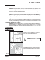
1
ADVIA® 60 Hematology System Operator’s Guide Closed Tube Model 075D0002-01 Rev. A, 2003-09 © 2003 Bayer HealthCare LLC No part of this manual or the products it describes may be reproduced by any means or in any form without prior consent in writing from Bayer HealthCare LLC. ADVIA is a trademark of Bayer HealthCare LLC. Vacutainer is a trademark of Bectin Dickson and Company. Manufactured in France for Bayer HealthCare LLC. The information in this manual was correct at the time of printing. However, Bayer HealthCare LLC continues to improve products and reserves the right to change specifications, equipment, and maintenance procedures at any time without notice. If the system is used in a manner differently than specified by Bayer HealthCare LLC, the protection provided by the equipment may be impaired. See warning and hazard statements. User Manual Closed Tube Model Daily Startup and Shutdown Procedures Start-up procedure 1. Press the ON/OFF switch located on the ADVIA® 60 Hematology System rear panel. Wait for approximately three (3) minutes. 2. Verify that the printer is connected and turned to ON. 3. Wait for the end of the STARTUP CYCLE or press the STARTUP key and validate. 4. The background values must be less than: WBC 0.3 x 10³ WBC/mm3 RBC 0.02 x 10⁶ RBC/mm3 HGB 0.0 g/dL PLT 10 x 103 PLT/mm3 If the background values are not within these limits, the ADVIA 60 Hematology System will automatically perform another startup cycle. If the STARTUP fails, refer to Section 9 for troubleshooting procedures. 5. Run a blood sample to prime the system: a. Enter the patient identification or run # (according to the identification mode chosen) using the ID/SEQ key. b. Place the sample tube into the tube holder. c. Close the door of the tube holder in its sampling position to start the analysis if this starting mode has been setup, or press the START key after closing the door. The tube holder carriage moves up in the piercing position, and the sample aspiration begins. The green/red LED will blink during sampling. d. The LED stops blinking and the tube holder carriage moves down when the sampling is completed. 6. Perform a QC procedure using ADVIA 60 TESTpoint™ Hematology Controls (Prod. No. B03-4200-54, B034201-54, and B03-4202-54). 7. Perform a calibration procedure only if necessary. 8. Run patient samples : a. Enter patient identification: • Press the ID/SEQ key. • Enter the identification # (up to 13 characters) or Run # (ranging from 1 to 10000). • Press the ENTER key. b. Place the sample tube into the tube holder. c. Close the door of the tube holder in its sampling position to start the analysis if this starting mode has been setup, or press the START key after closing the door. The tube holder carriage moves up in the piercing position, and the sample aspiration begins. The green/red LED will blink during sampling. d. The LED stops blinking and the tube holder carriage moves down when the sampling is completed. Shutdown procedure 1. Perform a general cleaning/rinse cycle of the ADVIA 60 Hematology System using the STAND BY key on the front panel. This cycle lasts for approximately one (1) minute. The instrument will cycle into the STAND BY mode. 2. Press the ON/OFF switch to OFF. NOTE: When the instrument is left in STAND BY mode, a STARTUP CYCLE must be performed prior to any analysis cycle © 2003 Bayer HealthCare LLC. All rights reserved. Table of Contents CHAPTER 1. INTRODUCTION ................................................................................... 1-1 1.1. INTENDED USE ................................................................................................................................ 1.2. PRESENTATION ............................................................................................................................... 1.3. OPEN TUBE AND CLOSED TUBE MODELS .................................................................................... 1.4. NOTES ............................................................................................................................................. 1-1 1-2 1-2 1-2 CHAPTER 2. INSTALLATION ..................................................................................... 2-1 2.1. INSPECTION .................................................................................................................................... 2.2. UNPACKING ..................................................................................................................................... 2.3. PACKAGE CONTENTS ..................................................................................................................... 2.4. WORKING CONDITIONS ................................................................................................................. 2.4.1. Environment ............................................................................................................................... 2.4.2. Location ..................................................................................................................................... 2.4.3. Grounding .................................................................................................................................. 2.4.4. Humidity and temperature conditions ..................................................................................... 2.5. VISUAL CHECKS ............................................................................................................................. 2.5.1. Mechanical check ...................................................................................................................... 2.5.2. Connection check ...................................................................................................................... 2.6. REAGENT PACK CONNECTIONS .................................................................................................... 2.7. REAGENT AND WASTE HANDLING PRECAUTIONS ...................................................................... 2.8. ELECTRICAL CONNECTIONS ......................................................................................................... 2.9. GENERAL POINTS ........................................................................................................................... 2.10. PRINTER (optional) ....................................................................................................................... 2.10.1. Connection ............................................................................................................................... 2.11. REAGENT PRIMING ....................................................................................................................... 2-1 2-1 2-2 2-3 2-3 2-3 2-3 2-3 2-3 2-3 2-4 2-5 2-6 2-6 2-6 2-6 2-7 2-7 CHAPTER 3. SPECIFICATIONS ................................................................................. 3-1 3.1. PERFORMANCE SPECIFICATIONS ................................................................................................. 3.1.1. Expected Values ........................................................................................................................ 3.2. REAGENT SPECIFICATIONS ........................................................................................................... 3.2.1.Diluent : sysDIL ........................................................................................................................... 3.2.2. Lyse : sysLYSE ............................................................................................................................ 3.2.3. Detergent : sysKLEN .................................................................................................................. 3.2.4. sysCLEAR (bleach solution) ...................................................................................................... 3.2.5. TIMEPAC ..................................................................................................................................... 3.3. LIMITS ............................................................................................................................................. 3.3.1. Cleaning ..................................................................................................................................... 3.3.2. Caps of the sampling tubes ...................................................................................................... 3.3.3. Blood specimens ....................................................................................................................... 3.3.4. Known interfering substances .................................................................................................. 3-1 3-4 3-6 3-6 3-6 3-7 3-7 3-7 3-7 3-7 3-8 3-8 3-8 CHAPTER 4. TECHNOLOGY...................................................................................... 4-1 4.1. MEASUREMENT PRINCIPLES ......................................................................................................... 4.1.1. RBC / WBC / PLT detection principles ..................................................................................... 4.1.2. Hemoglobin measurement principle ....................................................................................... 4.1.3. Hematocrit measurement principle ......................................................................................... 4.2. CELL DISTRIBUTION STUDY .......................................................................................................... 4.2.1. White blood cell distribution .................................................................................................... 4.2.1.1. Analysis principle ................................................................................................................. 4.2.1.2. Diluent and lysing action ..................................................................................................... 4.2.1.3. Volumetric study ................................................................................................................... 4.2.1.4. Results ................................................................................................................................... 4.2.2. Distribution of Red Blood Cells ................................................................................................. 4.2.3. Platelet distribution ................................................................................................................... i 4-1 4-1 4-3 4-3 4-3 4-3 4-3 4-4 4-4 4-4 4-5 4-5 4.3. STUDY OF GENERAL FLAGS ......................................................................................................... 4.3.1. Platelet flags .............................................................................................................................. 4.3.2. HGB flag ..................................................................................................................................... 4.3.3. WBC Flags .................................................................................................................................. 4.3.4. Comments on the flagging capabilities ................................................................................... 4-6 4-7 4-7 4-7 4-8 CHAPTER 5. DESCRIPTION ...................................................................................... 5-1 5.1. INSTRUMENT ................................................................................................................................... 5.1.1. Front panel and command keys ............................................................................................... A - Using the keyboard ...................................................................................................................... B - Command keys ............................................................................................................................ 5.1.2. Rear panel / Main fuses ............................................................................................................ 5.1.3. Left side internal view .............................................................................................................. 5.1.4. Front internal view .................................................................................................................... 5-1 5-1 5-1 5-2 5-4 5-4 5-5 CHAPTER 6. STARTUP AND SAMPLE RUN ............................................................ 6-1 6.1. STARTUP CHECKS .......................................................................................................................... 6.1.1. INSTRUMENT STARTUP ............................................................................................................... 6.1.2. Reagent pack ............................................................................................................................. 6.2. SAMPLE COLLECTION AND MIXING ............................................................................................. 6.2.1. Sample collection ..................................................................................................................... 6.2.2. Mixing ......................................................................................................................................... 6.3. DAILY QUALITY CONTROL/CALIBRATION VERIFICATION ............................................................ 6.3.2. US mode identification with barcode reader .......................................................................... 6.3.1. US mode identification without barcode reader .................................................................... 6.3.3. Standard identification mode ................................................................................................... 6.3.4. Sample tube holder selection .................................................................................................. 6.3.5. Analysis ...................................................................................................................................... 6.4. RUNNING SAMPLES ....................................................................................................................... 6.4.1. Sample identification ................................................................................................................ 6.4.2. Automatic cleaning ................................................................................................................... 6.4.3. End of the day rinsing ............................................................................................................... 6.5. RESULTS ......................................................................................................................................... 6.5.1. Displayed results ........................................................................................................................ 6.5.2. Result printout ............................................................................................................................ 6-1 6-1 6-2 6-2 6-2 6-2 6-3 6-4 6-4 6-5 6-5 6-6 6-7 6-7 6-7 6-7 6-8 6-8 6-9 CHAPTER 7. CALIBRATION AND QUALITY CONTROL ........................................... 7-1 7.1. INTRODUCTION ............................................................................................................................... 7-1 7.2. SMART CARD OPTION .................................................................................................................... 7-2 7.3. AUTOCALIBRATION ........................................................................................................................ 7-2 7.3.1. Select operator .......................................................................................................................... 7-2 7.3.2. Change lot number .................................................................................................................... 7-3 7.3.3. Change expiration date ............................................................................................................ 7-4 7.3.4. Change target values ................................................................................................................ 7-4 7.3.5. Change number of calibrator samples .................................................................................... 7-5 7.3.6. Run calibration .......................................................................................................................... 7-6 7.4. CALIBRATION COEFFICIENTS ........................................................................................................ 7-9 7.4.1. Changing calibration coefficients ............................................................................................ 7-9 7.4.2. Print coefficients .......................................................................................................................7-10 7.4.3. Coefficient limits .......................................................................................................................7-10 7.5. QUALITY CONTROL PROGRAM .................................................................................................... 7-11 7.5.1. Introduction ............................................................................................................................... 7-11 7.5.2. QC - Automatic ..........................................................................................................................7-12 7.5.2.1. Insert QC smart card ............................................................................................................7-12 7.5.2.2. Select operator ....................................................................................................................7-13 7.5.2.3. Select commercial control level ........................................................................................7-13 7.5.2.4. Run commercial control ......................................................................................................7-14 7.5.2.5. Accepting/rejecting results .................................................................................................7-14 7.5.2.6. Exiting QC automatic ..........................................................................................................7-15 7.5.3. Analysis .....................................................................................................................................7-16 ii 7.5.4. QC print targets ......................................................................................................................... 7-16 7.5.5. QC statistics ...............................................................................................................................7-17 7.5.5.1. Select statistics ....................................................................................................................7-17 7.5.5.2. Select level ..........................................................................................................................7-17 7.5.6. QC Graphs .................................................................................................................................7-18 7.5.6.1. Select graphs .......................................................................................................................7-18 7.5.6.2. Select level ..........................................................................................................................7-18 CHAPTER 8. INSTRUMENT CONFIGURATION ........................................................ 8-1 8.1. RESULTS OPTIONS ........................................................................................................................ 8-1 8.1.1. Reprint results ............................................................................................................................ 8-2 8.1.2. Printout ....................................................................................................................................... 8-2 8.1.3. Units ............................................................................................................................................ 8-2 8.1.4. Printer selection ........................................................................................................................ 8-3 8.1.5. Temperature printout ................................................................................................................ 8-3 8.1.6. Print limits .................................................................................................................................. 8-4 8.1.7. Print differential results ............................................................................................................. 8-4 8.2. CHANGE LABORATORY LIMITS ..................................................................................................... 8-4 8.2.1. Result low limits ........................................................................................................................ 8-4 8.2.2. Result high limits ....................................................................................................................... 8-5 8.2.3. Print limits and flag values ....................................................................................................... 8-5 8.2.4. Flag limits ................................................................................................................................... 8-6 8.3. SPECIAL FUNCTIONS ..................................................................................................................... 8-7 8.3.1. Change operator ........................................................................................................................ 8-7 8.3.2. Change password ...................................................................................................................... 8-8 8.3.3. Startup cycle .............................................................................................................................. 8-8 8.3.4. Autocleaning frequency ............................................................................................................ 8-9 8.3.5. Internal setup printout ............................................................................................................... 8-9 8.3.6. Cycle end audible signal .........................................................................................................8-10 8.3.7. Identification mode ..................................................................................................................8-10 8.3.8. Start mode ................................................................................................................................ 8-11 8.4. DATE AND TIME .............................................................................................................................. 8-11 8.4.1. Change time ..............................................................................................................................8-12 8.4.2. Date format ............................................................................................................................... 8-12 8.4.3. Change date ..............................................................................................................................8-12 8.5. HOST OPTIONS ..............................................................................................................................8-13 8.5.1. Host communication .................................................................................................................8-13 8.5.2. Baud rate ...................................................................................................................................8-13 8.5.3. Transmission .............................................................................................................................8-13 8.6. BARCODE SETUP ...........................................................................................................................8-14 8.7. Patient Memory Card ....................................................................................................................8-14 8.7.1. Introduction ............................................................................................................................... 8-14 8.7.2. Memo operation mode ............................................................................................................. 8-15 8.7.2.1. Memo on/off ......................................................................................................................... 8-15 8.7.2.2. Transmission mode ..............................................................................................................8-15 8.7.3. Running the samples ................................................................................................................ 8-16 8.7.4. Print list ..................................................................................................................................... 8-17 8.7.5. Trans. one .................................................................................................................................. 8-17 8.7.6. Trans. all ....................................................................................................................................8-18 8.7.7. Trans. from to ............................................................................................................................ 8-18 8.7.8. Clear smart card .......................................................................................................................8-18 CHAPTER 9. MAINTENANCE AND TROUBLESHOOTING ...................................... 9-1 9.1. MAINTENANCE AND SERVICE ........................................................................................................ 9.1.1. Overview .................................................................................................................................... 9.1.2. Daily maintenance .................................................................................................................... 9.1.2.1. Startup and standby cycles .................................................................................................. 9.1.2.2. Automatic cleaning .............................................................................................................. 9.1.2.3. Instrument general cleaning ............................................................................................... 9.1.3. Service functions ....................................................................................................................... iii 9-1 9-1 9-1 9-1 9-1 9-1 9-2 9.1.3.1. Backflush ............................................................................................................................... 9-2 9.1.3.2. Drain chambers .................................................................................................................... 9-2 9.1.3.3. Prime reagents ..................................................................................................................... 9-2 9.1.3.4. Concentrated cleaning ......................................................................................................... 9-3 9.1.3.5. Mechanical checks ............................................................................................................... 9-4 9.1.3.6. Cycle functions ...................................................................................................................... 9-6 9.1.3.7. Technician functions ............................................................................................................ 9-6 9.1.3.8. Automatic cleaning .............................................................................................................. 9-6 9.1.4 Disposal of System Waste and Supplies .................................................................................. 9-6 9.2. TROUBLESHOOTING ...................................................................................................................... 9-7 9.2.1. Overview .................................................................................................................................... 9-7 9.2.2. Identification procedure ........................................................................................................... 9-7 9.2.2.1. System operations ................................................................................................................ 9-7 9.2.2.2. Reagents ............................................................................................................................... 9-7 9.2.2.3. Precision ............................................................................................................................... 9-7 9.2.2.4. Calibration ............................................................................................................................ 9-8 9.2.3. Troubleshooting parameters ..................................................................................................... 9-8 9.2.3.1. WBC and HGB ....................................................................................................................... 9-8 9.2.3.2. WBC ......................................................................................................................................9-10 9.2.3.3. HGB .......................................................................................................................................9-10 9.2.3.4. RBC, HCT and PLT ............................................................................................................... 9-11 9.2.3.5. RBC .......................................................................................................................................9-12 9.2.3.6. HCT .......................................................................................................................................9-13 9.2.3.7. PLT ........................................................................................................................................9-13 9.2.4. Troubleshooting system operations ........................................................................................9-13 9.2.4.1. Power ...................................................................................................................................9-13 9.2.4.2. Display ..................................................................................................................................9-13 9.2.4.3. Motors ...................................................................................................................................9-14 9.3. ERROR MESSAGES .......................................................................................................................9-15 9.4. PURPOSE OF THE VALVES ...........................................................................................................9-18 9.5. MENU OVERVIEW ..........................................................................................................................9-19 9.6. PNEUMATIC DIAGRAM ...................................................................................................................9-20 CHAPTER 10. SERVICE AND SUPPLIES ............................................................... 10-1 10.1 Supplies .........................................................................................................................................10-1 10.2 For Service ....................................................................................................................................10-2 10.2.1 Bayer Authorized Representative ..........................................................................................10-2 10.2.2 Bayer Offices Worldwide ........................................................................................................10-2 APPENDIX A. SYSTEM AND REAGENT ROLLS ...................................................... A-1 A.1 System and Reagent Symbols ....................................................................................................... A-1 APPENDIX B. BIOHAZARD PROTECTION .............................................................. B-1 B.1 Protecting Yourself from Biohazards ............................................................................................. B-1 B.2 References ....................................................................................................................................... B-2 APPENDIX C. BIBLIOGRAPHY ................................................................................. C-1 C.1 Bibliography .................................................................................................................................... C-1 Index .................................................................................................................... Index-1 iv CHAPTER 1. INTRODUCTION 1.1. INTENDED USE The ADVIA 60-CT (Closed Tube) system is a fully automated (Microprocessor controlled) hematology analyzer used for the in vitro diagnostic testing of whole blood specimens. ADVIA 60-CT16 : - WBC, LYM%, LYM#, MON%, MON#, GRA%, GRA#, - RBC, HGB, HCT, MCV, MCH, MCHC, RDW, PLT, MPV, and 3 distribution curves: WBC, RBC, PLT. ADVIA 60-CT18 : - WBC, LYM%, LYM#, MON%, MON#, GRA%, GRA#, - RBC, HGB, HCT, MCV, MCH, MCHC, RDW, - PLT, MPV, PDW**, PCT**, and 3 distribution curves: WBC, RBC, PLT. from 10 µL of whole blood (taken from EDTA). WBC : RBC : HGB : HCT : MCV : MCH : MCHC : RDW : PLT : MPV : LYM% : LYM# : MON% : MON# : GRA% : GRA# : PDW : PCT : White blood cell count Red blood cell count Hemoglobin Hematocrit Mean cell volume Mean corpuscular hemoglobin Mean corpuscular hemoglobin concentration Red cell distribution width Platelet count Mean platelet volume Lymphocyte percent Lymphocyte number Monocyte percent* Monocyte number* Granulocyte percent Granulocyte number Platelet distribution width ** Plateletcrit ** * The MON count (both # and %) is a composite count that includes monocytes, eosinophils and basophils. ** PCT and PDW are not available in the UNITED STATES The rate of determinations is 55 samples per hour in the optimum configuration. The system is totally automated including the cap piercing of the sample tube, with an internal dilution system and a graphic printer (optional) for recording all test results including flags and graphic printouts. 075D0002-01 Page 1/1 1. INTRODUCTION 1.2. PRESENTATION The instrument, which is small in size, has 9 main parts: 1 - The electrical supply. 2 - The electronic board. 3 - The dilution pneumatics. 4 - A control panel including a keyboard and a LCD screen. 5 - A cap piercing mechanism. 6 - A reagent compartment. 7 - A printer (optional) that prints out the results and the plotting of the distribution curves. 8 - A smart card reader (optional) for quality control, result records and calibration direct entries. 9 - A barcode reader (optional) for a direct entry of the alphanumerical identifications. All the controls are grouped together on one panel, at the front of the system. Diag.1.1 1.3. OPEN TUBE AND CLOSED TUBE MODELS The ADVIA 60 is also available in two mechanical models. The ADVIA 60-OT is an Open Tube model whereby the operator needs to remove the stopper from the blood tube before introducing the sample via the sampling probe. The ADVIA 60-CT is a Closed Tube model permitting sampling of the blood specimen without removing the stopper from the blood collection tube. 1.4. NOTES Bayer HealthCare retains the right to make changes to the instrument and this document. Your local technical support provider or distributor will provide the latest revision of this document. No part of this document may be copied or reproduced in any form or by any means without prior written consent of Bayer HealthCare. Page 1/2 075D0002-01 CHAPTER 2. INSTALLATION 2.1. INSPECTION A thorough inspection is performed prior to the release of an ADVIA 60-CT Hematology System. It is important to verify receipt of all parts. Notify any descrepancies with the carrier. As instructed, the installation procedures must be followed in the order listed below. 2.2. UNPACKING The instrument is enveloped in a special, protective foam before being placed in a cardboard box. Cut the four angles of the box to unpack the system. Remove the cardboard box containing the ADVIA 60-CT installation kit from its location. Diag.2.1 - The ADVIA 60-CT "pack" installation kit : 075D0002-01 XEA 335 A Page 2/1 2. INSTALLATION 2.3. PACKAGE CONTENTS - The ADVIA 60-CT boxes contain the following parts : - ADVIA 60-CT - Printer (optional) - User's daily startup and shutdown procedure - User's manual - ADVIA 60-CT power cable (European) : or - ADVIA 60-CT power cable (US) : DAC 011 A DAC 012 A - The ADVIA 60-CT "pack" installation kit (XEA 335 A) includes : DESIGNATION PART NUMBER QTY GBC 052 AS 1 O ring 6 mm x 1.5 mm FAA 036 A 2 O ring 0.74 mm x 1.02 mm FAA 054 A 2 ADVIA 60 common installation kit XEA 312 A 1 Sampling needle ADVIA 60-CT Table 2.1 - The ADVIA 60-CT common installation kit (XEA 312 A) includes : DESIGNATION Table 2.2 PART NUMBER QTY Fuse 1A, 220 V 5 x 20 mm DAR 040 A 2 Fitting 1.6mm EAB 032 A 1 ADVIA 60 cover FBH 015 A 1 ADVIA 60 cover key FAJ 004 A 1 Allen key 1.5 mm MAB 003 A 1 Allen key 2.0 mm MAB 001 A 1 Allen key 2.5 mm MAB 069 A 1 Bent wrench 2.5 mm MAB 002 A 1 TORX key MAB 090 A 1 Tygon tube 1.52 mm EAE 007 A 2 Tygon tube 2.29 mm EAE 009 A 2 O ring 30.8 x 3.6 mm FAA 017 A 1 O ring 15 x 1.5 mm FAA 029 A 1 Grease KM 1011 XEA 019 A 1 Page 2/2 075D0002-01 2. INSTALLATION 2.4. WORKING CONDITIONS 2.4.1. Environment ADVIA 60-CT should be operated in an indoor location only. Operation at an altitude over 2000 meters (6562 feet) is not recommended. The instrument is designed to be safe for transient voltages according to INSTALLATION CATEGORY II and POLLUTION DEGREE 2. Please ask your local technical support provider or distributor for any information about operating location when it does not comply with the specifications. 2.4.2. Location ADVIA 60-CT should be placed on a clean and level table or work station. Please note that ADVIA 60-CT, printer and reagents weigh approximately 30 kilograms (66 lbs). Avoid exposure to sunlight. Proper ventilation requires that a space of at least 20 cm (8 inches) must be left behind the apparatus. 2.4.3. Grounding Proper grounding is required. Check that the wall ground (earth) plug is correctly connected to the laboratory grounding electricity installation. If there is no ground then use a ground stake. Current electricity norms must be applied. 2.4.4. Humidity and temperature conditions ADVIA 60-CT can function between 18 to 32°C (65 to 90°F). Maximum relative humidity is 80% for temperatures up to 31°C decreasing linearly to 50% relative humidity at 40°C. If it is kept at a temperature less than 10°C (50°F), the instrument should be allowed to sit for an hour at the correct room temperature before use. 2.5. VISUAL CHECKS 2.5.1. Mechanical check Using the key from the installation kit, unloosen the locker. Open the pneumatic protection door. WARNING: Do not open or close the instrument front door when the door of the piercing mechanism is open. Diag.2.2 Unscrew the 5 cover fixation screws and remove the cover: pull it backward and lift it up to the rear of the instrument. Diag.2.3 075D0002-01 Page 2/3 2. INSTALLATION Push the black plastic carriage locking clip as far as possible to the left and place the sample needle carriage as far forward as possible to the right-hand side. Check that the aspiration needle is not bent and make sure it is in its upper position. Diag.2.4 1 - Carriage locking clip 2 - Sample needle carriage 3 - Needle Check the position of the chambers. Each chamber should be in its proper position with its clips and the electrode block is attached firmly to the RBC chamber. 1 - Clip 2 - RBC chamber Diag.2.5 Unscrew the 2 screws of the WBC/HGB chamber protection cover slightly. Remove the cover and check that the chamber is fixed properly in its clips and the electrode block is attached firmly to the chamber. Re-install the HGB/WBC chamber cover. 1 - Chamber protection cover Diag.2.6 2.5.2. Connection check Check that the connectors on the printed circuit board are securely in place. Re-install the instrument cover. Diag.2.7 Page 2/4 075D0002-01 2. INSTALLATION Remove the fuse holder from its location on the rear panel pressing on the holder lock and check the fuse characteristics : they should be 1 Amperes, 220 Volts Slow-Blow. Diag.2.8 2.6. REAGENT PACK CONNECTIONS The ADVIA 60 Hematology System TIMEPAC® Reagents includes the entire set of reagents in a "Pack" and is able to receive the waste liquids. Three soft pockets contain the 3 reagents sysDIL®, sysKLEN® and sysLYSE® and are closed by means of the valve connectors located at the bottom of the pack. The fourth pocket is empty and is intended for receiving waste liquids. To order additional reagents, contact your local technical support provider or distributor. Remove the reagent output protections, as well as the waste input protection. Diag. 2.9 Install the pack directly into the compartment of the instrument. Push the pack down in order to plug correctly the pack on the male connectors. Diag. 2.10 The free male connector must be plugged on the pack upper valve in order to receive the waste liquids. CAUTION: To prevent leaks in the reagent pack avoid unplugging reagent pack more than once. 075D0002-01 Page 2/5 2. INSTALLATION 2.7. REAGENT AND WASTE HANDLING PRECAUTIONS Reagents have to be stored at room temperature (18°C to 25°C). Lyse reagent contains cyanides and has to be handled according to the local or national regulations. Always follow the recommended precautions. Collect all the waste generated during testing to facilitate compliance with the local environmental regulations. BIOHAZARD: Wear personal protective equipment. Use universal precautions. Refer to Appendix B for recommended precautions when working with biohazardous materials. 2.8. ELECTRICAL CONNECTIONS The ADVIA 60-CT is connected to the laboratory electrical supply using the power cable included in the installation kit. Connect the power cable to the plug located on the rear left-hand side of the device. Two 1A fuses are located, under the power plug. The instrument can be operated at any other voltage (from 100 to 240V) or frequency (from 50 to 60Hz) without modification. 1 - ON/OFF SWITCH 2 - FUSE HOLDER 3 - MAIN SUPPLY PLUG 4 - RS 232 OUTPUT 5 - PRINTER OUTPUT Diag.2.11 If the instrument has to be connected to a laboratory computer, use the plug (4) RS 232. 2.9. GENERAL POINTS The ADVIA 60-CT responds to the UL 3101 norm. Refer to the Declaration of Conformity Statement for details. System performance is guaranteed by Bayer HealthCare under the following conditions only: - services and repairs are provided by your local technical support provider or distributor - the electrical supply of the laboratory follows the national or international regulations - the system is operated under the following instructions. 2.10. PRINTER (optional) It is mandatory to connect a printer which fulfills the following conditions : - the printer is recommended by your local technical support provider or distributor, - the printer is approved by the CE norms (EEC only). - the printer is certified CSA. Page 2/6 075D0002-01 2. INSTALLATION 2.10.1. Connection The printer is connected to ADVIA 60-CT with the cable delivered with the instrument. Lock the connector in place by tightening the 2 screws on each end of the connector to the ADVIA 60-CT. Attach the other end of the cable to the printer and lock the printer connector in place by the means of the 2 clips located on the connector itself. Diag.2.12 On the ADVIA 60-CT, it is necessary to select the printer format RESERVED 1 of the "PRINTER" menu (function 4) accessible through the "OPTIONS" menu (function 5 of the main menu) then "RESULTS" (function 1). NOTE: 2.11. REAGENT PRIMING When the ADVIA 60-CT is first installed, it contains no reagents. All the reagents have to be primed now. Turn ON instrument by pressing the ON/OFF switch located on the rear panel. When the instrument turns on, the display shows: PLEASE WAIT FOR 3 MIN ESCAPE : ESC This time is required at the startup for the instrument initialization and stabilization, specifically for the HGB diode to reach its operationnal temperature. After three minutes, the LED of the front panel turns from red to green and the display shows the following: STARTUP NOT INITIATED PRESS A KEY TO CONTINUE... This message appears when the instrument is setup with the manual startup cycle to prevent any analysis cycle before running a startup cycle. Press any key, the main menu is displayed: MAIN MENU HH : MM 075D0002-01 1 RESULTS 2 QC Page 2/7 2. INSTALLATION From the MAIN MENU, move the cursor to the function 4 SERVICE and press ENTER. The service menu is displayed : SERVICE HH : MM Move the cursor to 3 > 2 DRAIN CHAMBER 3 PRIME REAGENTS PRIME REAGENTS and press the ENTER key. Select the function 1 CHANGE PACK and follow the instructions given by the LCD in order to install the pack. REAGENT PACK HH : MM > 1 CHANGE PACK 2 CBC LEFT < 150> Once the new PACK is installed a priming cycle will be automatically carried out and the following menu displayed. PRIME ********** NOTE: WAIT FOR 2 MIN 3 S Before analyzing samples, visually inspect reagent lines and pumps for air bubbles. Repeat priming if air bubbles are still present. Call the local technical support provider or distributor if priming does not eliminate air bubbles. From the REAGENT PACK menu, the function 2 CBC LEFT displays the number of analysis cycles left to run with the same pack. It is also possible to run a priming cycle at any time using the selection 3 PRIME of the REAGENT PACK menu. CAUTION: To prevent leaks in the reagent pack avoid unplugging reagent pack more than once. Page 2/8 075D0002-01 CHAPTER 3. SPECIFICATIONS 3.1. PERFORMANCE SPECIFICATIONS ADVIA 60-CT performs automated blood counts, and requires no manual operations for aspirating blood, dilution, measuring, calculations, print-out, and computer transfer. The parameters given according to the internal setup : ADVIA 60-CT16: - WBC, LYM%, LYM#, MON%, MON#, GRA%, GRA#, - RBC, HGB, HCT, MCV, MCH, MCHC, RDW, PLT, MPV, and 3 distribution curves: WBC, RBC, PLT. ADVIA 60-CT18: - WBC, LYM%, LYM#, MON%, MON#, GRA%, GRA#, - RBC, HGB, HCT, MCV, MCH, MCHC, RDW, - PLT, MPV, PDW*, PCT*, and 3 distribution curves: WBC, RBC, PLT. * : PCT and PDW are not available in the United States. - Dimensions: - Height : approximately 440 mm (16.5 inches) - Width : approximately 360 mm (14.2 inches) - Depth : approximately 330 mm (12.6 inches) Diag.3.1 - Weight : * approximately 14 Kgs (31 lbs). - Power supply : * 100 Vac to 240 Vac + 10%, 50 Hz to 60 Hz - LCD screen : * 2 lines of 40 characters, backlighted - Power Consumption : * Maximum : 150 VA (-30%, + 10%) * In use : 110 VA (-30%, + 10%) * Stand-by mode : 35 VA (-30%, +10%) - Conditions for use : * ADVIA 60-CT can function between 18 to 32°C (65 to 90°F). Maximum relative humidity 80% for temperatures up to 31°C decreasing linearly to 50% relative humidity at 40°C. - Smart Card option : * Reader : GCI 400 - ADVIA 60 SETpointTM Calibrator memory card : - GFM 2K - Patient memory card : - MCOS 24K (capacity : 60 CBCs) - ADVIA 60 TESTpointTM Control memory card : - MCOS 24K (16 parameters) - MCOS 16K (8 parameters) 075D0002-01 Page 3/1 3. SPECIFICATIONS - Storage capacity : Number of QC available # of QC Aspirations/day QC8P card QC16P card 1 80 80 2 44 60 3 31 41 Table 3.1 - Hemoglobin : * HGB/WBC chamber, LED 550 nm. - Size of apertures : * WBC = 80 µm * RBC/PLT = 50 µm - Final dilutions : * WBC = approximately 1/250 * RBC / PLT = approximately 1/15000 - Throughput : * 55 samples / hour approximately - Capacity of internal memory : * Last sample only * 60 samples with the memory Smart Card - Anticoagulent: EDTA is recommended - Volume of whole blood sample : * 10 µL - Reagent consumption : * Software version V 1.6 CYCLE Table 3.2 sysDIL sysLYSE sysKLEN sysCLEAR Analysis cycle 17.5 mL 0.60 mL 0.85 mL x Prime all reagents 40.0 mL 11.6 mL 5.2 mL x Prime diluent 27.0 mL x x x Prime lyse x 11.6 mL x x Prime cleaner x x 6.3 mL x Startup cycle 21.0 mL 0.6 mL 1.4 mL x Standby cycle x x 13.6 mL x Auto clean cycle 16.4 mL 0.6 mL 15.2 mL x Concentrated cleaning 16.4 mL 0.6 mL 1.5 mL 6.0 mL HGB blank cycle 6.0 mL 1.3 mL x x Backflush cycle x x x x Page 3/2 075D0002-01 3. SPECIFICATIONS - Reagent pack * Total capacity : 4.2 liters - Measuring principles: * WBC.RBC.PLT = Impedance change * Hematocrit = Numeric integration * HGB = Cyanmethemoglobin method (550 nm) - Barcode reader option: * EAN 8, EAN 13, C 39, C 128, ITF (2/5), CODABAR, STF, C 93 with or without checksum. - Reproducibility: (based on 20 consecutive samplings from one fresh, normal, whole blood sample) PARAMETERS % CV - WBC : < 2.5% - RBC : < 2% - HGB : < 1.5% - HCT : < 2% at 45% - MCV : < 1% at 90 µm3 - PLT : < 5% at 300 x 103/mm3 - LYM : < 5% at 40% - MON : < 10% at 10% - GRA : < 5% at 50% Table 3.3 TEST LEVEL at 103/mm3 at 5 x 10⁶/mm3 at 15 g/dL - Linearity: Linearity was tested using commercially available low range and full range linearity test kits. The kits were analyzed and data was computed according to the manufacturer's instructions. Each kit included six levels and one level was used as the reference value. Each level was run four times. The results of this study are as follows : PARAMETERS Table 3.4 NOTE: 075D0002-01 LINEARITY RANGE LIMITS WBC (x 103 cells/mm3) 0 to 100 +/- 0.5 or +/- 5 % (whichever is greater) RBC (x 106 cells/mm3) 0 to 8.0 +/- 0.3 or +/- 3 % (whichever is greater) HGB (g/dL) 0 to 26 +/- 0.3 or +/- 3 % (whichever is greater) HCT (%) 0 to 80 +/- 2 or +/- 3% (whichever is greater) PLT (x 103 cells/mm3) for HGB concentrations > 2 g/dL PLT (x 103 cells/mm3) for HGB concentrations < 2 g/dL 0 to 2200 +/- 10 or +/- 10 % (whichever is greater) 0 to 4000 +/- 10 or +/- 10 % (whichever is greater) Platelet linearity depends on hemoglobin concentration. Page 3/3 3. SPECIFICATIONS - Carryover : Carry-over was tested by analyzing samples with high concentrations of WBC's, RBC's, HGB and PLT's. Each sample was run in triplicate, followed by three background cycles. The % carryover is calculated using the following formula : Carryover = WBC RBC HGB PLT 63.0 7.58 23.4 988 %Carryover (actual) 0.3 0.00 0.0 0.0 %Carryover (claim) <0.5% <0.5% <0.5% <0.5% Level Table 3.5 Background 1 - Background 3 --------------------------------------------- X 100 Sample 3 - Background 3 3.1.1. Expected Values Normal ranges were established at a study performed in Tarrytown (NY, USA). The results were derived from the central 95% of the values in the distribution of 50 apparently healthy individuals. MALE (N=25) FEMALE (N=25) 4.5 - 10.8 4.3 - 10.4 LYM (%) 20 - 47 19 - 48 MON (%) 3 -9 3 -9 GRA (%) 46 - 72 48 - 71 RBC (x 10 6 /mm3 ) 4.7 - 6.1 4.2 - 5.4 13.8 - 17.0 11.3 - 15.5 HCT (%) 42 - 50 36 - 46 MCV (µm 3) 80 - 94 81 - 99 MCH (pg) 27 - 32 27 - 32 MCHC (g/dL) 31 - 34 31 - 34 RDW (%) 13 - 16 13 - 15 PLT (x 10 3 /mm3 ) 185 - 402 132 - 440 MPV (µm3 ) 7.1 - 9.5 7.8 - 9.3 WBC (x 10 3 /mm 3) HGB (g/dL) Table 3.6 PCT and PDW have not been established as indications in the United States for this product. Their use should be restricted to research or investigational use only. Expected values vary with sample population and/or geographic location. It is recommended that each laboratory establish its own normal ranges based on the local population. Page 3/4 075D0002-01 3. SPECIFICATIONS - Accuracy : Approximately 200 patient specimens were analyzed on the ADVIA 60-CT and the ADVIA® 120 Hematology Systems at three different locations. The following table summarizes the data: SITE 1 PARAMETER Table 3.7 075D0002-01 SITE 2 SITE 3 N R N R N R WBC (x 10 3 /mm 3 ) 205 0.996 180 0.992 208 0.986 RBC (x 10 6 /mm3 ) 205 0.993 180 0.983 208 0.992 HGB (g/dL) 205 0.996 180 0.986 208 0.994 HCT (%) 205 0.985 180 0.972 208 0.984 MCV (µm 3) 205 0.947 180 0.940 208 0.902 MCH (pg) 205 0.974 180 0.945 208 0.916 MCHC (g/dL) 205 0.360 180 0.314 208 0.330 RDW (%) 205 0.656 180 0.400 208 0.607 PLT (x 10 3 /mm 3 ) 205 0.990 180 0.972 208 0.985 MPV (µm3 ) 205 0.877 N/A N/A 208 0.801 LYM (%) 204 0.980 177 0.994 200 0.983 MON (%) 204 0.809 177 0.940 200 0.819 GRA (%) 204 0.980 177 0.982 200 0.979 Page 3/5 3. SPECIFICATIONS 3.2. REAGENT SPECIFICATIONS In order for the instrument to operate correctly, high-quality reagents must be used. Bayer HealthCare provides all the necessary reagents. To order additional reagents, contact your local technical support provider or distributor. Do not use beyond the expiration date and store the reagents at a temperature between 18 to 25°C. The reagents are ready to use and require no preparation. 3.2.1.Diluent : sysDIL (1 x 3.2 L) For in vitro diagnostic use as an isotonic solution for the determination and differentiation of blood cells. Composition : · Stabilized buffer solution · Sodium hydroxide 0.04 % · Sodium azide 0.09 % CAUTION: Sodium azide can react with copper and lead plumbing to form explosive metal azides. On disposal, if disposal into a drain is in compliance with federal, state, and local requirements, flush reagents with a large volume of water to prevent the buildup of azides. Physio-chemical properties : Boiling point : About 100°C, pH : neutral. 3.2.2. Lyse : sysLYSE (1 x 200 mL) For in vitro diagnostic use as an eythrocyte-lysing agent for leukocyte counting, differentiation, and hemoglobin determination on the ADVIA 60 Hematology system. Composition : · Potassium cyanide, 0.03%. Physio-chemical properties : Boiling point : approximately 100°C, pH : basic. Page 3/6 075D0002-01 3. SPECIFICATIONS 3.2.3. Detergent : sysKLEN Function : For in vitro diagnostic use in hte cleaning of the Advia 60 Hematology system. Composition: Organic buffer, < 0.2%. Physio-chemical properties: Boiling point : around 100°C, pH = 9.7 ± 0.2 at 20°C. 3.2.4. sysCLEAR (bleach solution) sysCLEAR is used in the concentrated cleaning procedure. Diluted bleach solution can also be used. Follow the recommended precautions for safe use. See the Material Safety Data Sheet for first aid. REF: 02488831 Part number: B01-4198-01 (4 x 500 mL) For in vitro diagnostic use as a cleaning and bleaching solution for the ADVIA 60 Hematology system. Composition: · Sodium Hypochlorite (4%) · Sodium Hydroxide (0.26%) Physio-chemical properties: Boiling point : 100°C, pH : 12.9 ± 0.5 at 20°C. 3.2.5. TIMEPAC See the reagent specifications above. REF: 07622536 Part number: B01- 4199-54 TIMEPAC (4 x 145 aspirations) The TIMEPAC reagent pack contains the following reagents: - sysDIL - sysLYSE - sysKLEN 3.3. LIMITS As with any hematological analysis, users must be alert to the possible effect on results of unknown interferences from medications or endogenous substances. All patient results must be evaluated by the laboratory and the physician in light of the total clinical status of the patient. 3.3.1. Cleaning In section 9, specific start-up, shutdown, and maintenance procedures are listed. The maintenance procedures identified are mandatory for the proper use and operation of the ADVIA 60-CT. FAILURE TO EXECUTE ANY OF THESE RECOMMENDED PROCEDURES MAY RESULT IN DECREASED RELIABILITY OF THE SYSTEM. 075D0002-01 Page 3/7 3. SPECIFICATIONS 3.3.2. Caps of the sampling tubes Some caps of the sampling tubes are more adapted to "cap piercing" sampling systems. Plastic caps cannot be used. Rubber caps can be of different quality materials. Use the best quality materials in order to avoid any rubber particles entering the sample tube when the cap is pierced. It is also recommended to use caps specially designed to avoid any blood retention in the upper part of the cap. Diag.3.3 1 Type 1 - Wrong 2 Type 2 - Good Avoid using Type 1 caps as they may block the atmosphere connection because of the blood trapped inside the cap. This type of cap also can drop some blood when opening the tube, and introduce pressure when closing the tube. Type 2 caps are recommended as they are specially designed to prevent these problems. NOTE: It is recommended not to pierce more than three times through the caps. 3.3.3. Blood specimens Verification of any abnormal test result (including flagged results or results outside of the normal range) should be performed using reference methods or other standard laboratory procedures for the conclusive verification of results.The sections below list known limitations of automated blood cell counters which use the principle of impedance. BIOHAZARD: Wear personal protective equipment. Use universal precautions. Refer to Appendix B for recommended precautions when working with biohazardous materials. 3.3.4. Known interfering substances WBC White Blood Cells (Leukocytes): WBC results that exceed the linearity limits of the system will require dilution of the blood sample. Re-assaying the diluted sample will help to obtain the correct assay value. NOTE: Dilute the sample using autologous plasma or Saline. NRBC - Immature nucleated red blood cells will be counted in the WBC (White Blood Cell) parameter. If the number of nucleated red blood cells is sufficient to activate an L1 alarm, such interference will be detected. However, the manual differential white blood cell count - performed on the stained blood film - will reveal the presence of NRBC's. Page 3/8 075D0002-01 3. SPECIFICATIONS Following the manual differential white blood cell count, the WBC assay value must be corrected for the presence of nucleated red blood cells. The formula utilized for correcting the WBC parameter, when nucleated red blood cells are present, is: CORRECT WBC: counted WBC X 100 ------------------------------------100 + (# of NRBC/100 WBC) Unlysed Red Cells - In particularly rare instances, the erythrocytes in the blood sample may not completely lyse. These non-lysed red blood cells may be detected on the WBC histogram with an L1 alarm or as an elevated baseline on the side (leading edge) of the lymphocytes population. Non-lysed erythrocytes will cause a falsely elevated WBC count. Multiple myeloma - The precipitation of proteins in multiple myeloma patients may give elevated WBC counts. Hemolysis - Hemolyzed specimens contain red cell stroma which may elevate white cell counts. Leukemia - A spurious low WBC count may result in this disease state because of the possible increased fragility of the leukocytes leading to some destruction of these cells during counting. These white cell fragments will also interfere with the white cell partial differential parameters; LYM% + #, MON% + #, GRAN% + #. A spurious low WBC count may also be seen in patients with lymphocytic leukemias due to the presence of abnormally small lymphocytes which may not be counted by the instrument. Chemotherapy - Cytotoxic and immunosuppressive drugs may increase the fragility of the leukocytes which may cause low WBC counts. Cryoglobulins - Increased levels of cryoglobulin that may be associated with myeloma, carcinoma, leukemia, macroglobulinemia, lymphoproliferative disorders, metastic tumors, autoimmune disorders, infections, idiopathic disease, aneurism, pregnancy, thromboembolic phenomena, diabetes, etc can elevate the WBC, RBC or PLT counts and the HGB value. The specimen can be warmed up to 37°C and reanalyzed immediately or a manual WBC, RBC, or PLT count can be performed. RBC Red Blood Cells (Erythrocytes): The red blood cell dilution contains all the formed elements in the blood : erythrocytes, leukocytes, and platelets. During the counting of the erythrocytes (red blood cells), platelets are not counted since their size falls below the minimum threshold. Leukocytes (White blood cells), on the other hand, are included in the RBC count. However, since the normal ratio between red blood cells and white blood cells is so extreme, the influence of the WBC on the RBC is negligible. High WBCs - In rare cases where the WBC is extremely high, the RBC count may be corrected, especially if the RBC count is extremely low. Agglutinated red blood cells - May cause a falsely decreased RBC count. Blood samples containing the agglutinated red blood cells may be identified by observing abnormal MCH and MCHC values, as well as by examination of the stained blood film. Cold agglutinins - IgM immunoglobulins which are elevated in cold agglutinin disease may lower RBC and PLT counts and increase MCV. 075D0002-01 Page 3/9 3. SPECIFICATIONS HGB (Hemoglobin): Turbidity of the blood sample - Due to any number of physiologic and/or therapeutic factors may produce falsely elevated HGB results. To obtain accurate hemoglobin results when increased turbidity of the blood sample occurs, determine the cause of the turbidity and follow the appropriate method below : 1. Elevated WBC : An extremely elevated WBC will cause excessive light scatter. In these cases use reference (manual) methods.The diluted sample should be centrifuged, and the supernatant fluid measured on a spectrophotometer. 2. Elevated lipids : Elevated lipids in the blood sample will give the plasma a "milky" appearance. This condition can occur with hyperlipidemia, hyperproteinemia (as in gammapathies) and hyperbilirubinemia. Accurate hemoglobin determinations can be achieved by using reference (manual) methods and a plasma blank. Increased turbidity may also be seen in case where the red blood cells are resistant to lysing. This condition will cause a falsely elevated Hgb result, but may be detected by observing the abnormal MCH, MCHC values, and the increased baseline on the leading edge of the WBC histogram. Erroneous hemoglobin results will cause the results of the MCH and MCHC to be erroneous as well. Fetal bloods - The mixing of fetal and maternal bloods may produce a falsely elevated HGB value. HCT (Hematocrit): Red blood cells agglutination - May produce erroneous HCT and MCV values. Red blood cells agglutination may be detected by observing the abnormal MCH and MCHC values, as well as by examination of the stained blood film In such cases, manual methods may be required to obtain an accurate HCT value. MCV (Mean Corpuscular Volume): Red blood cell agglutination - May produce an erroneous MCV value. Red blood cell agglutination may be detected by observing the abnormal MCH and MCHC values, as well as by examination of the stained blood film. In such cases, manual methods may be required to obtain an accurate MCV value. Excessive numbers of large platelets and/or the presence of an excessively high WBC count - May interfere with the accurate determination of the MCV value. In such cases, careful examination of the stained blood film may reveal the error. MCH (Mean Corpuscular Hemoglobin): The MCH is a function of th HGB value and the RBC count. The limitations listed for the HGB and RBC will have an effect on the MCH and may cause erroneous values. MCHC (Mean Corpuscular Hemoglobin Concentration): The MCHC is a function of the HGB and HCT values. The limitations listed for the HGB and HCT will have an effect on the MCHC and may cause erroneous values. Page 3/10 075D0002-01 3. SPECIFICATIONS RDW (Red Blood Cell Distribution Width): The red blood cell distribution width is a function of the RBC count. The red blood cell dilution contains all of the formed elements in the blood : erythrocytes, leukocytes and platelets. During the counting of the erythrocytes (Red Blood Cells) platelets are not included in RBC count since their size falls below the minimum threshold. However, leukocytes (White Blood Cells) are counted and included in the RBC count. Since the normal ratio between RBC and WBC is so extreme, the influence of the WBC is extremely low and the RBC count may need to be corrected, especially if the RBC count is extremely low. Agglutinated RBC - May cause a falsely decreased RBC count and erroneous RDWs. Blood samples containing the agglutinated RBC may be detected by observing abnormal MCH and MCHC values, as well as by examination of the stained blood film. Nutritional deficiency or blood transfusion - May cause elevated RDW results due to iron, vitamin B12 or folate conditions. High RDWs may also be present from bi-modal RBC distribution in blood transfusion. PLT (Platelets): Very small erythrocytes (microcytes) , erythrocytes fragments (schizocytes), and WBC fragments - May interfere with the proper counting of platelets and cause elevated PLT counts. Agglutinated erythrocytes - May trap platelets, causing an erroneously low platelet count. The presence of agglutinated erythrocytes may be detected by observation of abnormal MCH and MCHC values and by careful examination of the stained blood film. Giant platelets in excessive numbers - May cause an erroneously low platelet count since these large platelets may exceed the upper threshold for the platelet parameter and are not counted. Chemotherapy - Cytotoxic and immunosuppressive drugs may increase the fragility of these cells which may cause low PLT counts. Reference (manual) methods may be necessary to obtain an accurate platelet count. Hemolysis - Hemolyzed specimens contain red cell stroma which may elevate platelet counts. A.C.D. blood - Blood anticoagulated with Acid Citrate Dextrose may contain platelet aggregates which could depress the platelet count. RBC inclusions - Erythrocyte inclusions, such as Howell-Jolly bodies, Heinz bodies, siderotic and basophilic granules, etc may produce a spuriously increased platelet count. Platelet agglutination - Clumped platelets due to poor collection techniques or platelet satellitosis caused by EDTA activation of immunoglobulins may cause a decreased platelet count and/or an elevated WBC count. The specimen should be recollected in sodium citrate anticoagulant and reanalyzed for only the platelet count. The final PLT result must be corrected for the sodium citrate dilution effect. MPV (Mean Platelet Volume): Giant platelets that exceed the upper threshold of the Platelet parameter - May not be counted as platelets. Consequently, these larger platelets will not be included in the instrument's calculation of Mean Platelet Volume. Very small erythrocytes (microcytes), erythrocytic fragments (Schizocytes), and white blood cell fragments - May interfere with the proper counting and sizing of Platelets. 075D0002-01 Page 3/11 3. SPECIFICATIONS Agglutinated erythrocytes - May trap Platelets, causing an erroneous MPV result. The presence of agglutinated erythrocytes may be detected by observation of the abnormal MCH and MCHC values and by careful examination of the stained blood film. Chemotherapy - May also affect the sizing of PLTs. CAUTION: Blood samples collected in EDTA will not maintain a stable Mean Platelet Volume. Platelets collected in EDTA swell with time and temperature. LYM# (Lymphocyte count absolute value): The lymphocyte count is derived from the WBC count. The presence of nucleated red blood cells (NRBC), certain parasites, and erythrocytes that are resistant to lysis may interfere with an accurate LYM count. Limitations listed for the WBC count pertain to the LYM # count as well. LYM% (Lymphocyte percentage): The lymphocyte percent is a function of the WBC count and the number of lymphocytes. The presence of nucleated RBC (NRBC), certain parasites, and erythrocytes that are resistant to lysis may interfere with an accurate LYM% count. Limitations listed for the WBC count pertain to the LYM% as well. MON# (Mononcyte cell count absolute)*: The monocyte cell count absolute is derived from the WBC count.The presence of large lymphocytes, atypical lymphocytes, blasts, and excessive number of basophils may interfere with an accurate number of monocytes MON% (Monocyte percentage)*: The monocyte percentage is a function of the WBC count and the number of monocytes. The presence of large lymphocytes, atypical lymphocytes, blasts, and excessive number of basophils may interfere with an accurate MON% count. * : The MON count (both # and %) is a composite count that includes monocytes, eosinophils and basophils. GRA# (Granulocyte cell count absolute): The granulocyte cell count is derived from the WBC cell count. The excessive presence of eosinophils, metamyelocytes, myelocytes, promyelocytes, blasts, and plasma cells may interfere with an accurate granulocyte count. GRA% (Granulocyte percentage): The granulocyte percentage is a function of the WBC count and the number of the granulocytes. The excessive presence of eosinophils, metamyelocytes, myelocytes, promyelocytes, blasts, and plasma cells may interfere with an accurate GRA% count. Page 3/12 075D0002-01 CHAPTER 4. TECHNOLOGY 4.1. MEASUREMENT PRINCIPLES 4.1.1. RBC / WBC / PLT detection principles The counting principle used for red blood cells (RBC), white blood cells (WBC), and platelets (PLT) is based on the variation in impedance generated by the passage of cells through a calibrated microaperture. 1 - The sample is diluted in an electrolytic diluent (current conductor). The conductivity of the diluent differs considerably from the non conductivity of the blood cells. 2 - The dilution is pulled through the calibrated microaperture. Two electrodes are placed on each side of the aperture. Electric current passes through the electrodes continuously. 3 - When the cell passes through the aperture, electric resistance (or impedance) between the two electrodes increases proportionately with the cell volume. Diag. 4.1 1 2 3 4 5 6 7 8 075D0002-01 Volts Pulse Time Electrodes Analyzing Electronic Circuit I = Constant Vacuum = Constant Solution to be Analyzed Page 4/1 4. TECHNOLOGY We can derive from Ohm’s law : V = Voltage I = Current V = RI R = Resistance Since I is constant, R increases with each cell passage (through the aperture), thus V increases proportionately to the cell volume. 4 - The generated impulses have a very low voltage, which the amplification circuit increases, so that the electronic system can analyze them and eliminate the background noise. 5 - Two measuring chambers and detection circuits separately carry out the analysis of white blood cells, and that of platelets and red blood cells. 1 2 3 Volts Threshold Time Diag. 4.2 6 - The dilutions used for these different measurements are the following: * WBC = 10 µL of whole blood mixed with 2.50 mL of diluent, the final dilution is at 1/250, then 0.60 mL of lyse are added before the count, resulting in a final dilution of 1/300. * RBC / PLT = 30 µL of the dilution at 1/250 are mixed with 2.50 mL of the diluent, resulting in a dilution of 1/21,000. 7 - Each type of cell (WBC, RBC, PLT) is analyzed by the microprocessor which also handles the cell distribution (histograms). 8 - To count the platelets, the ADVIA 60-CT uses high performing electronics, which avoids the use of complex hydraulic systems for the elimination of faulty impulses generated at the rear side of the aperture. When red blood cells reenter the analysis zone, this creates impulses with a height comparable to platelet impulses, but with a different shape. The instrument uses a very sophisticated impulse sorting system. This system rejects any impulse which does not have the typical platelet shape. This sorting system maintains a very reliable aperture and a traditional hydraulic system. 9 - ADVIA 60-CT provides distribution curves, by analysis on 256 counting channels for the WBC, RBC and Platelets. Page 4/2 075D0002-01 4. TECHNOLOGY 4.1.2. Hemoglobin measurement principle 1 - During the STARTUP cycle an HGB blank test sequence including two blank measures is run. If the difference between these two measures is too large a third measurement is performed. HGB reference blank measurement will occur if the operator: - has required a CALIBRATION operation. - has required a QC operation. - has left the instrument more than 10 minutes after the STARTUP. - has left the instrument more than 60 minutes after ANALYSIS. - has not carried out the STARTUP cycle after switching on the instrument. NOTE: 2 - On every cycle an Hgb blank is carried out on diluent and compared to the previous HGB blank analysis. 3 - 0.60 mL of lyse agent is added to the 2.5 mL of 1/250 dilution. 4 - The hemoglobin freed by the lyse of the red blood cells combines with potassium cyanide to form chromogenous cyanmethemoglobin compound. 5 - The compound is then measured by spectrophotometry, through the WBC cuvette at 550 nm. 6 - The result is given with the units set up in Section 8 "Instrument Configuration". 4.1.3. Hematocrit measurement principle 1 - The height of the impulse generated by the passage of a cell through the microaperture is directly proportional to the volume of the analyzed cell. 2 - The hematocrit is measured as a function of the numeric integration of the MCV. 3 - The result is given with the units set up in Section 8 "Instrument Configuration". 4.2. CELL DISTRIBUTION STUDY ADVIA 60-CT carries out volumetric distributions (histograms) for WBC and RBC on 256 analysis channels, and 128 channels for PLT with the following measuring range: * WBC = approximately 30 to 460 fL. * RBC = approximately 25 to 300 fL. * Plt = approximately 2 to 33 fL. 4.2.1. White blood cell distribution The WBC volumetric distribution study reveals the following three leukocyte subpopulations : Lymphocytes, Monocytes, Granulocytes. 4.2.1.1. Analysis principle The principle is based on the volumetric study of leukocytes after use of a (patented) diluent and a lysing reagent. NOTE: 075D0002-01 The lysing action varies according to the temperature of the dilution (ambient temperature). In order to correct these fluctuations within the operating temperature range (18°C to 32°C, 65°F to 90°F), a temperature sensor is placed on the diluent circuit and the position of the separation thresholds varies according to the temperature. Page 4/3 4. TECHNOLOGY 4.2.1.2. Diluent and lysing action The diluent preserves and prepares the cell membrane for the differential reaction. The lyse has a differential mode of action on cytoplasmic membranes. The lymphocyte cytoplasmic membranes allow the release of water soluble cytoplasm and shrink the membrane around the nucleous, the monocytes undergo an intermediate reaction, while the granulocytes have a limited reaction due to a molecule and their cytoplasmic structure which protects them from the shrinking action of the lyse. 4.2.1.3. Volumetric study After the differential lysing action, ADVIA 60-CT analyzes the heights of each impulse, coming from the aperture and ranked in the 256 counting channels. A curve is obtained with number of cells on the y-axis and cell volume on the x-axis. The cells will be broken down accordingly: * the lymphocytes between 30 - 100 fL . * the monocytes between 100 - 150 fL . * the granulocytes between 150 fL and a maximum (unlimited volumetrically). Pathologic cells will, of course, place themselves in different zones in the distribution curve. Mobile and fixed flags will alert the lab operator of the presence of such pathologic elements. NOTE: The WBC distribution curve disappears when the WBC result is rejected. Diag. 4.3 1 2 3 4 5 Leukocyte Flags LYM MON GRA Number of Cells 4.2.1.4. Results The lymphocytes, monocytes, and granulocytes are expressed in percents (%) and absolute numbers (#) : LYM%, LYM#, MON%, MON#, GRA%, GRA#. Page 4/4 075D0002-01 4. TECHNOLOGY 4.2.2. Distribution of Red Blood Cells The study of the distribution of RBC detects erythrocyte anomalies linked to anisocytosis. A Red Cell Distribution Width (RDW) will enable you to follow the evolution of the width of the curve in relation to the cell number and average volume. K SD —— MCV RDW = With K SD = = MCV = System constant Determined standard deviation according to statistical studies on cell distribution. Mean Corpuscular Volume of erythrocytes 4.2.3. Platelet distribution The platelet distribution study counts platelets, detects platelet anomalies and alerts the lab operator in the event of a non-platelet cell population (schistocytes, microcytes, etc...). 1 - Platelets are counted, between a low threshold placed at 2 fL and a variable high threshold. The variable high threshold varies according to the microcyte population, which is present in the platelet analysis zone. 1 2 3 Diag. 4.4 Small Cells (SCL) Schistocytes (SCH) Microcytes (MIC) 2 - Measuring the MPV : The MPV (Mean Platelet Volume) is directly derived from the analysis of the platelet distribution curve. The MPV is expressed in µm3 or fL. 3 - Calculating PCT : PLT (103 /mm3) x MPV (µm3) PCT% = —————————————— 10000 4 - Measuring the PDW (Platelet Distribution Width): This count is derived from the platelet curve. PDW = Width of the curve between 15% of the number of platelets starting from 2 fL (S1) and 15% of the number of platelets beginning with the variable top threshold (S2). Diag. 4.5 075D0002-01 1 PDW Page 4/5 4. TECHNOLOGY 4.3. STUDY OF GENERAL FLAGS With the print-out of results the following general flags may occur : * following either WBC, RBC, HCT or PLT indicates that the system, analyzed the sample three times but that all three counts differed and were outside the systems precision limits. The result should be verified by repeating the sample or checked according to one of the laboratory reference methods. $ following a parameter : indicates that the instrument performed 3 counts, 2 of the 3 counts are within : - 7% for WBC - 5% for RBC - 15% for PLT If the maximum of the first two raw counts is lower than : - 3000 for WBC, the limit becomes 9% - 16000 for RBC, the limit becomes 8% - 400 for PLT, the limit becomes 20% Result for the concerned parameter can be accepted. --- D If results exceed the linearity limits shown below, a <D> will be printed after the result. If the results greatly exceed the limits, a result will not be shown on the LCD or the printout. The flag DIL will be shown on the LCD, and ---D or ---0 will be printed and transmitted to the LIS. Repeat the sample using a 1:2 dilution and repeat each time the flag reappears. Limits to the linearity ranges : NOTE: - WBC - RBC - PLT - PLT - HGB - HCT > > > > > > 100 x 103/mm3 8.0 x 10⁶/mm3 2200 x 103/mm3 (HGB > 2 g/dL) 4000 x 103/mm3 (HGB < 2 g/dL) 26 g/dL 80% Dilute the sample using autologous plasma or Saline. H : located next to a result of a parameter shows that the value is above the upper limit set up by the operator. L : located next to a result of a parameter shows that the value is below the lower limit set up by the operator. NOTE: Temperature : Operating reagent temperature should be within the recommended limits (18°C to 32°C, 65°F to 90°F). The operating temperature can be printed out on the result form. When these limits are exceeded, the values obtained cannot be guaranteed with certainty and the result transmitted on the RS shows an error code. Page 4/6 075D0002-01 4. TECHNOLOGY 4.3.1. Platelet flags MIC following the Platelet result indicates the excessive presence of microcytes in the Platelet measurement zone. Verify the result using a Platelet Rich Plasma (PRP) or a manual count. This flag can be adjusted by the user. SCH following the Platelet result indicates the presence of schistocytes or Platelet aggregates in the Platelet measurement zone. Review/scan slide before reporting result. This flag can be adjusted by the user. SCL following the Platelet result indicates the presence of small cells in the 2 and 3 fL zone. A second sample cycle should be carried out and the results verified. If this flag should persist, perform an automatic cleaning cycle and resample. If the flag persists verify using a Platelet Rich Plasma (PRP) of the sample and make a manual slide count for the Platelets. This flag can be adjusted by the user. 4.3.2. HGB flag ! : located next to the HGB result shows that the HGB blank carried out during the analysis and the previous analysis blank differed and were outside the system's precision limits. Nevertheless the instrument provides a result according to the previous HGB blank. This result can be reported. If this suspicious flag occurs more than three consecutive times run the checkup procedure. 4.3.3. WBC Flags The ADVIA 60-CT has a system of WBC differential flags alerting the operator to the possible presence of pathological cells, abnormal volume distribution histograms or abnormal elevated populations such as in the case of the eosinophils and basophils. 1 - Flag L1 : This flag shows an abnormal number of cells in comparison with lymphocytes (in the 30 - 60 fL zone). The pathologic elements which may be found in this area include platelet agregates, nucleated red blood cells or atypical lymphocytes. The flag corresponds to the number of cells counted in the first five channels out of the total number of lymphocytes. The lab operator can adjust the limit which triggers this flag. 075D0002-01 Page 4/7 4. TECHNOLOGY 2 - Flag M2 : Located in the 130 to 160 fL zone, this flag informs the lab operator of the presence of lymphoblasts, myelocytes, abnormal lymphocytes, or basophilia (too many basophils). This flag can be adjusted by the operator. It corresponds to the number of cells counted by the detection zone out of the total number of granulocytes. The limit which triggers this flag is adjusted by the lab operator. 3 - Flag G1 : Situated in the 160 to 220 fL zone, this detects the presence of eosinophils, myelocytes, and sometimes, neutrophilia. This flag can be adjusted by the operator. It corresponds to the number of cells counted in the detection area over the total number of granulocytes. The limit which triggers this flag is adjusted by the lab operator. 4 - Flag G2 : This flag makes it possible to follow an abnormal granulocyte peak displacement, shows anomalies in the membrane of the granulocytes, and also possible lyse or hydraulic anomalies. This flag is also triggered, if the blood is too old (after 6 - 8 hours). The flag is also triggered if the granulocyte volume is less than 250 fL . 5 - Flag G3 : This flag is situated in the zone which is greater than 400 fL. It detects the presence of metamyelocytes. The flag is set off when the percentage in number, compared to the number of granulocytes is higher than the level set. The lab operator can adjust the limit which triggers this flag. 4.3.4. Comments on the flagging capabilities All anomalies and/or abnormal distributions signaled by the ADVIA 60-CT should be verified manually for the presence of pathological elements. As a result of the differential resistance of cytoplasmic membranes in the different cell types, pathological elements can be found in a number of different zones. This also applies to the presence of normal or non-pathological cells that have been subject to chemotherapy or some other form of treatment in alarm zones. This will result in a false alarm. Page 4/8 075D0002-01 CHAPTER 5. DESCRIPTION 5.1. INSTRUMENT 1 2 3 4 Cover Door to pneumatic part Piercing carriage door Front panel Diag.5.1 1 - ADVIA 60-CT Cover The instrument cover is fixed by the means of 5 screws. Before any attempt to remove the cover, open the pneumatic access door on the front of the system. WARNING: Do not open or close the instrument front door when the piercing carriage door is open. Do not open the piercing carriage door when the aspiration needle is in its lower position. 2 - Pneumatic access door. This door gives an access to the pneumatic parts. It also allows the operator to check the hydraulic cycle operation. It is recommended to keep the door locked during the measuring cycles as it is equipped with an electrical interference shield. 3 - Piercing carriage door. This door gives access to the sample tube holder which moves up during the sampling cycle in order to pierce the tube cap. Then the aspiration needle moves down into the sample tube inside the piercing needle and aspirates the 10 µL whole blood sample. Its internal and external rinsing is patented and is fully automatic. 4 - Control panel. This panel allows the operator to communicate with the instrument : - To access the different cycles - To identify the patients - To setup the instrument, etc... 5.1.1. Front panel and command keys A - Using the keyboard All functionalities of the instrument have been dispatched in "menus". Each menu has a certain number of functions. These menus are all accessible through the MAIN MENU displayed at the instrument startup. Navigation in the menus is done using the ">" cursor command keys UP and DOWN . Entry in a function is done by moving the cursor in front of the function and pressing the ENTER key. NOTE: 075D0002-01 When menus and functions are known, it is possible to enter directly the menu or function number to access it (and press ENTER when a function is requested). Page 5/1 5. DESCRIPTION Diag.5.2 1 2 3 4 5 STARTUP key STAND BY key Identification Key (ID/SEQ) Start analysis cycle key (START) Numerical keyboard 6 7 8 9 10 DELETE key ENTER key ESCAPE key Display scroll keys Cycle light indicators B - Command keys 1 - STARTUP key: When this key is pressed, a startup cycle including a cleaning and rinsing procedure is carried out. Detergent left in the chambers is rinsed with diluent and the system is ready for the analysis cycles. This cycle has a duration of approximately 130 seconds (this cycle can be run 2 or 3 times if blank values are not within the acceptable limits). 2 - STAND BY key: this key is used for shutdown at the end of the working day. When this key is pressed, it is mandatory to carry out a startup cycle before any analysis cycle. This cycle has a duration of approximately 65 seconds. 3 - Identification Key (ID/SEQ): This key is used to enter the patient identification (13 characters maximum, letters or numbers), and the run number. NOTE: The patient identification can be entered also using the barcode reader when the instrument is set up in the "US" identification mode. 4 - Start Analysis Cycle key (START): This key starts the analysis when the manual mode for starting the analysis has been set up. Page 5/2 075D0002-01 5. DESCRIPTION 5 - Numerical keyboard : keys from 0 to 9 allow the operator to enter the following figures : - Date - Calibration values - Laboratory limits - Patient number for analysis - Leukocytes differential flag values. These 2 keys pressed simultaneously will allow an automatic adjustment of the LCD. 6 - DELETE Key: When it is pressed, this key deletes the information entered on the LCD display. 7 - ENTER Key: This key is pressed to validate the informations entered on the LCD display. 8 - ESCAPE Key (ESC): When it is pressed, this key allows the operator to exit a function and to come back to the previous menu. This key can be used to stop an hydraulic cyle (see NOTE below). It also opens the tube holder compartment door. 9 - UP/DOWN Keys: These keys allow the user to scroll up or down in the instrument menus to access the different functions and to choose alphabetic letters in the patient identification. 10 - When the START key is activated, the indicator light flashes during the sampling time. When the indicator stops flashing, the operator is allowed to remove the tube from the sampling position. This cycle has a duration of 65 seconds approximately. When the ESC key is pressed during a hydraulic cycle, it is necessary to run a STARTUP cycle to rinse the instrument before any analysis. NOTE: C - Screen details SERVICE HH : MM > 2 DRAIN CHAMBER 3 PRIME REAGENTS The position of the cursor is given by the ">" as shown above. The the screen indicate that more menus are available up and down. 075D0002-01 and on the right side of Page 5/3 5. DESCRIPTION 5.1.2. Rear panel / Main fuses 1 2 3 4 5 6 Rear panel RS 232 computer connection Printer output ON/OFF switch F1,F2 Main fuses Main power socket 1 2 3 4 Vacuum/pressure syringe Liquid electrovalve block 1 Dilution block Liquid electrovalve block 2 Diag.5.3 Diag.5.4 5.1.3. Left side internal view Diag.5.5 Page 5/4 075D0002-01 5. DESCRIPTION 5.1.4. Front internal view 1 2 3 4 5 6 7 8 9 10 11 LCD display Keyboard Cycle light indicators Sampling needle carriage Start cycle key RBC chamber WBC chamber Spectrophotometer Piercing mechanism Tube holder switches Piercing carriage door switch Diag.5.6 075D0002-01 Page 5/5 5. DESCRIPTION Page 5/6 075D0002-01 CHAPTER 6. STARTUP AND SAMPLE RUN 6.1. STARTUP CHECKS 6.1.1. INSTRUMENT STARTUP Turn ON instrument by pressing the ON/OFF switch located on the rear panel. The display shows the following : PLEASE WAIT FOR 3 MIN ESCAPE : ESC Then the front panel LED turns from red to green to indicate that the initialization phase is completed. Wait for the end of the automatic startup cycle or press the STARTUP key, the instrument will perform a startup cycle and perform a blank cycle for a background count (an analysis cycle on reagent without any blood sample). If the background count does not exceed the following : - WBC :0.3x 10³/mm3 (0.3 on the display) - RBC : 0.02 x 10⁶/mm3 (0.02 on the display) - PLT : 10 x 103/mm3 (10 on the display) the Startup is completed and the message "STARTUP PASSED" is printed out with results (US mode). If the results are above these limits, the instrument will automatically perform another background cycle. If the problem persists after 3 cycles, a message "STARTUP FAILED, CHECK REAGENTS" is displayed, run the checkup procedure. During the startup cycle an HGB blank test is carried out. If the HGB blank is not within acceptable values after 3 cycles, a flag "STARTUP FAILED, CHECK REAGENTS" and "HGB REFERENCE FAILED" is printed out with startup results.Run the checkup procedure. If the startup fails the message "STARTUP FAILED" is printed out with all the results until a new startup is carried out. During the startup, if the HGB blank test is unacceptable, the background count is not carried out and a new attempt is automatically performed (3 maximum). NOTE: - When the instrument has not been used for 4 hours, a startup cycle must be run before running sample analyses. NOTE: 075D0002-01 If analyses are performed when the startup cycle has not been carried out, the message "STARTUP NOT INITIATED" Is displayed and printed out. Page 6/1 6. STARTUP AND SAMPLE RUN 6.1.2. Reagent pack A low reagent level is indicated by the number of CBCs left (from 5 to 0) when the operator tries to run an analysis cycle : WARNING : PACK LOW LEVEL : 5 ESC TO EXIT START TO CONFIRM If this occurs operator has the choice either to run the cycle pressing the START key or to replace the Reagent pack with a new one. 6.2. SAMPLE COLLECTION AND MIXING 6.2.1. Sample collection Sample collection should be done on venous blood by the means of vacuum or atmospheric sample collection tubes. It is possible to collect capillary blood into a microtainer with a minimum volume of 100 µL (for example in a pediatric lab.) and analyze this on the ADVIA 60-CT. EDTA is the recommended anticoagulent for analysis on the ADVIA 60-CT. Samples should be processed as soon as possible, and within 6-8 hours of collection. BIOHAZARD: Wear personal protective equipment. Use universal precautions. Refer to Appendix B for recommended precautions when working with biohazardous materials. CAUTION: The sample collection tube should be filled with the exact quantity of blood indicated on the tube itself. Any incorrectly measured blood sample collection will show a possible variation in the results. Bayer HealthCare has a list of recommended sample collection tubes at your disposal. 6.2.2. Mixing NOTE: It is critical to assure that all the blood samples are thoroughly and gently mixed (with a gentle up and down and rolling motion), prior to each sample aspiration. Page 6/2 075D0002-01 6. STARTUP AND SAMPLE RUN 6.3. DAILY QUALITY CONTROL/CALIBRATION VERIFICATION Refer to your laboratory quality assurance program to ensure quality throughout the entire testing process. It is recommended that the system be controlled using Bayer TESTpoint Hematology Controls Low, Normal, and High (Prod. Nos. B03-4200-54, B03-4201-54, and B03-4202-54, respectively). Refer to the package insert supplied with the control for instructions for use. Controls are intended to be integrated into a clinical laboratory’s own quality control program and procedures. The following is a suggested protocol. The actual frequency of control in a laboratory will be based upon many factors such as workflow, system experience, government regulation, etc. These controls should be assayed: 1 At the beginning of each shift. 2 Whenever a new reagent container (same or different lot) is used. 3 Following the performance of any system maintenance or cleaning. A satisfactory level of performance is achieved when the control values obtained for each level are within the acceptable range for the system as published in the package insert provided with the Bayer TESTpoint Hematology Controls. Refer to Section 7 "Calibration and Quality Control" for instructions for use. Before analyzing samples it is recommended that the operator analyze three levels of control material (low, normal and high) to verify that the system is within acceptable limits. Two Identification modes are available : 1 - US mode : that requires the patient (or control) identification on each analysis. This mode allows the use of the barcode reader. 2 - Standard mode : increments a run number on each analysis. BIOHAZARD: Wear personal protective equipment. Use universal precautions. Refer to Appendix B for recommended precautions when working with biohazardous materials. 075D0002-01 Page 6/3 6. STARTUP AND SAMPLE RUN 6.3.1. US mode identification without barcode reader Press the ID/SEQ key of the front panel to enter the control ID. The identification menu is displayed : PAT. ID. ? : CURRENT: EXIT : ESC SAVE : ENTER The identification can be entered using 13 characters, letters or numbers. Letters can be entered using the UP and DOWN keys of the front panel. Press ENTER for each letter to step to the next one. Press the ENTER key when the identification is entered (or ESC key to save the displayed one) : a message "PLEASE CLOSE HOLDER DOOR" is displayed. NOTE: The identification displayed in "actual" stays in memory until a cycle is run. 6.3.2. US mode identification with barcode reader When the blood control tube is equipped with a barcode label, press the ID/SEQ key of the front panel to enter the control ID. The identification menu is displayed : PAT. ID. ? : CURRENT: EXIT : ESC SAVE : ENTER Read the control identification barcode label using the barcode reader. When the reading is completed, a beep occurs and the control lot identification is displayed on the screen. Press the ENTER key when the identification is entered (or ESC key to save the displayed one) : a message "PLEASE CLOSE HOLDER DOOR" is displayed. Install the sample tube in the tube holder and close the sample door to start the analysis if this mode has been selected or close the sample door and press the START key. Diag. 6.1 Page 6/4 075D0002-01 6. STARTUP AND SAMPLE RUN 6.3.3. Standard identification mode Press the ID/SEQ key to enter the control Run #. The following menu is displayed: RUN #? : NEXT : EXIT : ESC SAVE : ENTER Enter the run number (from 1 to 9999) using the numeric keys of the front panel. Press the ENTER key to record the number (or the ESC key to save the displayed one) : A message "PLEASE CLOSE HOLDER DOOR" is displayed. 6.3.4. Sample tube holder selection The sample tube holder has 4 positions according to the sample tube characteristics. The required position is selected when it is facing the inside of the sampling compartment. Turn the sample tube holder in either side to place the required position until the 'click' is heard. The tube holder is associated with 3 switches which are able to detect the sampling position, the presence of the tube holder and a wrong position of the tube holder. The 4 positions can be used for the following sample tubes (list is not exhaustive): Diag. 6.2 075D0002-01 Position 1 : Vacutainers Position 2 : Mini Vacutainers Position 3 : Control and calibration vial position Position 4 : Micro sample collection devices. Page 6/5 6. STARTUP AND SAMPLE RUN 6.3.5. Analysis Install the sample tube in the tube holder and close the sample door to start the analysis if this mode has been selected or close the sample door and press the START key. Diag. 6.3 NOTE: If the instrument has not been used for 1 hour, when the first analysis is requested, the instrument will start an HGB reference cycle, the message "PLEASE WAIT" is displayed. At the end of this cycle the first analysis will be automatically performed when the sample door mode is selected or press the START key again to start the analysis. BIOHAZARD: Wear personal protective equipment. Use universal precautions. Refer to Appendix B for recommended precautions when working with biohazardous materials. NOTE: The current Identification stays in memory until the analysis cycle has begun and can be displayed on the screen by pressing the ID/SEQ key. The analysis cycle lasts 65 seconds. At the end of the result printout, the LED turns to green and the instrument is ready for the next analysis. If any of the control results are outside the acceptable ranges perform the following : a - Rerun the control b - Clean the system and rerun the control c - Open a new vial d - Recalibrate the system Page 6/6 075D0002-01 6. STARTUP AND SAMPLE RUN 6.4. RUNNING SAMPLES 6.4.1. Sample identification After completing the start up procedure, run a non pathological blood from the previous day, followed by the quality control analyses and then the sample analyses. 1 - Enter either the patient identification (US mode) or the patient run # (Standard mode). 2 - Install the sample tube in the tube holder and close the sample door to start the analysis if this mode has been selected or close the sample door and press the START key. 6.4.2. Automatic cleaning When the instrument has run 50 samples from the date changing, an automatic cleaning procedure is carried out. This cycle lasts 10 minutes approximately while the autocleaning menu is displayed: AUTO CLEANING *************************** PLEASE WAIT 2MNS 13S The automatic cleaning frequency can be adjusted by the user from 1 to 50 as described in the instrument setup. NOTE: 6.4.3. End of the day rinsing It is necessary to run a standby/shutdown cycle at the end of the day. Press the STAND BY key, the instrument performs a complete cleaning with detergent, and puts the system into the stand by mode. The instrument can be switched off if the working day is completed or left in this standby mode overnight or until the next analysis. NOTE: 075D0002-01 When the instrument is left in standby mode, it is necessary to carry out a STARTUP cycle before any sample analysis. Page 6/7 6. STARTUP AND SAMPLE RUN 6.5. RESULTS When the analysis cycle is completed, results are displayed and printed out according to the setup of the instrument: 6.5.1. Displayed results The first group of parameters is displayed: WBC 7.5 RBC 5.22 HGB 15.6 HCT MCV 46.5 89 MCH MCHC 29.9 33.5 PLT 233 The second group of parameters can be accessed when moving the cursor to the top: MPV RDW %LYM %MO %GRA #LYM #MO #GRA 8.8 13.0 39.3 7.3 53.4 2.21 0.41 3.00 * Identification 1. US mode The patient identification can be reviewed when moving the cursor to the bottom: MM/DD/YY HH:MM PAT. ID : 2. Standard mode The patient run number can be reviewed when moving the cursor to the bottom: MM/DD/YY HH:MM RUN# * Flags: The PLT and LMG flags can be reviewed moving the cursor to the bottom PLT FLAGS : LMG FLAGS : The last sample run can be displayed again at any time when moving the cursor from NOTE: the main menu to the function 1 Page 6/8 RESULTS and pressing ENTER. 075D0002-01 6. STARTUP AND SAMPLE RUN 6.5.2. Result printout 1. US mode Diag. 6.4 On the result printout can be found: 1 - The sample identification that was entered by the operator. 2 - The sequence number. 3 - The STARTUP status. 4 - The PLT flags. 5 - The CBC results. 6 - The WBC flags. 7 - The Diff results. 8 - The histogram representations. 9 - The date sample was run. 10 - The time sample was run. The sequence number is updated to 1 everyday and increases by 1 on each cycle. The sequence number cannot be modified by the operator. 075D0002-01 Page 6/9 6. STARTUP AND SAMPLE RUN 2. Standard mode Diag. 6.5 On the result printout can be found: 1 - The sample run # that was entered by the operator. 2 - The sequence number. 3 - The PLT flags. 4 - The CBC results. 5 - The WBC flags. 6 - The Diff results. 7 - The histogram representations. 8 - The date sample was run. 9 - The time sample was run. The sequence number is updated to 1 everyday and increases by 1 on each cycle. The sequence number cannot be modified by the operator. NOTE: The STARTUP status is printed out only when it has failed (message : "STARTUP FAILED"). Page 6/10 075D0002-01 CHAPTER 7. CALIBRATION AND QUALITY CONTROL 7.1. INTRODUCTION Calibration should be performed upon installation of the ADVIA 60-CT Hematology system. Subsequent recalibration is required if there is a significant shift in control values after replacement of instrument parts, a change in reagent lot number, or whenever indicated by quality control data. Calibration can be achieved in two different ways. 1 - Bayer SETpoint Calibrator (B03-4203-51) is used to calibrate the ADVIA 60-CT Hematology System. Refer to the package insert supplied with the calibrator for instructions for use and assay values specific to the ADVIA 60 Hematology system used. 2 - Calibration coefficients are known and can be entered directly. NOTE: Calibration must be performed on a clean and reproducible instrument, blank values must be within the acceptable limits. BIOHAZARD: Wear personal protective equipment. Use universal precautions. Refer to Appendix B for recommended precautions when working with biohazardous materials. From the MAIN MENU, move the cursor to the function 3 CALIBRATION and press ENTER. The CALIBRATION menu is displayed : CALIBRATION HH:MM 075D0002-01 1 AUTO CALIBRATION 2 COEFFICIENTS Page 7/1 7. CALIBRATION AND QUALITY CONTROL 7.2. SMART CARD OPTION When the instrument is equipped with a smart card reader proceed as follows. Install the card in the smart card reader. Make sure that the cursor is positioned on function 1 AUTOCALIBRATION and press ENTER. Diag. 7.1 As the lot number, the expiration date and the target values are recorded into the memory card, only 2 operations remain : - The operator selection, - The selection of the number of the calibrator samples. Once these two steps are done, run calibration. NOTE: The card can be removed as soon as the lot number has been validated. 7.3. AUTOCALIBRATION From the calibration menu, move the cursor to function 1 AUTOCALIBRATION and press ENTER. The CALIBRATION menu will unfold step by step through the operator selection, the calibrator lot, identification, the target values and the number of samples to be analyzed before starting the calibration. 7.3.1. Select operator Move the cursor to one of the 4 required operator identification and press ENTER. A star (*) is displayed next to the chosen identification and the menu turns to the calibrator identification. SELECT OP. HH:MM Page 7/2 1 OP_1 > *2 OP_2 075D0002-01 7. CALIBRATION AND QUALITY CONTROL 7.3.2. Change lot number CHANGE LOT # ? CURRENT : SETPOINT NO : ESC YES : ENTER The current lot number is displayed. If the lot number of the calibration blood has to be changed, press ENTER. The following menu is displayed: LOT # : CURRENT : SETPOINT EXIT : ESC SAVE : ENTER Without barcode reader: enter the new lot number (10 characters maximum). Numbers can be entered directly using the numerical keyboard, letters can be entered using the DOWN and UP keys. For each letter, press the ENTER key to validate it and enter the next one, validate a second time to record the lot number. The menu turns to the expiration date. If the lot number has not changed, press the ESC key, the menu turns directly to the expiration date. With barcode reader: When the calibration blood tube is equipped with a barcode label, read the calibration blood identification barcode label using the barcode reader. When the reading is completed, a beep occurs and the control lot identification is displayed on the screen. Press ENTER to validate. The menu turns to the expiration date. Diag. 7.2 075D0002-01 Page 7/3 7. CALIBRATION AND QUALITY CONTROL 7.3.3. Change expiration date The current expiration date is displayed. CHANGE EXP. DATE ? (DD.MM.YY) CURRENT : NO : ESC YES : ENTER If it is correct, press the escape key, the menu turns to the target values. If the expiration date needs to be changed, press ENTER, the following menu is displayed : EXP. DATE : (DD.MM.YY) CURRENT : EXIT : ESC SAVE : ENTER Change the expiration date according to the format requested on the display and press ENTER. The menu turns to the target value modifications. 7.3.4. Change target values The following menu with the current WBC target value is displayed : CHANGE TARGET WBC ? CURRENT : NO : ESC YES : ENTER If the WBC target value is correct, press ESC, the menu turns to RBC target value. If the value has to be changed, press ENTER, the following menu is displayed: TARGET WBC : CURRENT : EXIT : ESC SAVE : ENTER Enter the new WBC target value if required and press ENTER or press ESC directly. The menu turns to RBC target value. Repeat the same procedure for RBC, HGB, HCT, PLT, and MPV. When this last value has been modified (or ESC pressed), the number of calibrator samples to be analyzed is displayed. Page 7/4 075D0002-01 7. CALIBRATION AND QUALITY CONTROL 7.3.5. Change number of calibrator samples CHANGE # SAMPLE ? CURRENT : NO : ESC YES : ENTER ADVIA 60-CT calibration may be performed using 3 to 11 sample aspirations. The autocalculation module performs statistics on these results in order to obtain the best calibration coefficients. NOTE: In order to obtain the best calibration possible, it is recommended to run the calibration blood a minimum of 5 times. NOTE: The first result is not taken into account in the statistical calculations used for the calibration. The first sample is used as a "primer". Press ENTER to change the required number of calibrator samples (or press ESC). The following menu is displayed : # SAMPLE CURRENT : EXIT : ESC SAVE : ENTER Enter the new number of calibrator samples and press ENTER if required or press ESC. The run calibration menu is displayed. 075D0002-01 Page 7/5 7. CALIBRATION AND QUALITY CONTROL 7.3.6. Run calibration Prepare the calibrator according to the specific instructions (temperature, mixing, etc...). RUN CAL. ? NO : ESC YES : ENTER Press the ENTER key. The start calibration menu is displayed : START CALIBRATION ESC TO EXIT CALIBRATION # 1/X ENTER TO ASPIRATE Instrument is requesting the first run of the calibrator. Install the calibrator tube in the correct holder position. Close the sample door to start the analysis if this mode has been selected or close the sample door and press the cycle button located on the front panel. When the analysis cycle ends, the first result menu is displayed: WBC RBC HGB HCT PLT MPV PRESS ENTER X.X X.XX XX.X XX XXX X.X TO CONTINUE Check that the results are within the limits given in the calibrator instructions and press ENTER. The first validation menu is displayed: VALID. CALIBRATION ESC TO DISCARD # 1/X ENTER TO VALID If the results are not within the acceptable limits, use the ESC key to reject the results and to restart the first run. NOTE: Results having flags ($ , * or ! for HGB) are automatically rejected. If the results are correct, press the ENTER key. The menu of the second calibration run is displayed: START CALIBRATION ESC TO EXIT CALIBRATION # 2/X ENTER TO ASPIRATE Run the second calibrator sample and follow the same procedure until the number of samples set up is obtained. When the last result has been validated, the instrument calculates the statistical calibration factors for each parameter : Mean, target, coefficient of variation, percentage difference between the target value and the mean (% CHG), new calibration coefficient as well as a recall of the previous calibration coefficient and a pass/fail status appear on the result printout. Page 7/6 075D0002-01 7. CALIBRATION AND QUALITY CONTROL A - Calibration passed : If the statistical figures are within the acceptable limits: - Coefficient of variation is within the limits given in Table 7.1 and, - The percentage difference between the target and the mean value is less than 20. The calibration passed and the results are printed out. The calculated coefficients become the new calibration coefficients and the calibration is completed. Table 7.1 PARAMETERS WBC RBC HGB HCT PLT MPV CV 2.5 2.0 1.5 2.0 5.0 3.0 The first run of the calibrator is not taken into account in the statistical calculations (a "P" is printed out next to the run number). NOTE: CALIBRATION DATE : 04/02/98 OPERATOR : JMG LOT Nr : SETpoint Diag. 7.3 TIME : 09:21 RUN 1 P 2 3 4 5 6 WBC 6.3 6.3 6.4 6.5 6.6 6.5 RBC 5.30 5.35 5.40 5.31 5.28 5.30 HGB 16.0 16.1 16.2 16.1 16.1 16.0 HCT 47.2 47.1 47 46.8 47.5 47.1 PLT 230 224 230 240 228 235 MEAN TARGET CV % CHG OLD CAL CURRENT 6.46 6.5 1.76 0.61 0.98 0.97 5.32 5.34 0.89 0.37 0.90 0.88 16.1 16.1 0.43 0 1.13 1.13 47.1 47 0.54 0.21 0.98 1.03 231.4 221 2.69 4.70 0.91 0.95 STATUS OK OK OK OK OK The following message is displayed: CALIBRATION ENDED WITH NEW COEFF. PRESS A KEY TO CONTINUE ... Press any key to return to the MAIN MENU. 075D0002-01 Page 7/7 7. CALIBRATION AND QUALITY CONTROL B - Calibration failed: If the statistical figures are not within the acceptable limits, the following menu is displayed: CALIBRATION FAILED !!! PRESS A KEY TO CONTINUE ... - Coefficient of variation is not within the limits given in Table 7.1 or, - The percentage difference between the target and the mean value is greater than 20. The calibration failed and the results are printed out. The calculated coefficients are rejected, the instrument keeps in memory the previous calibration coefficients and returns to the calibration menu. NOTE: If the calibration has failed on one or more parameters no parameter is calibrated. NOTE: If the calibration fails, the user can restart the calibration, or enter directly the calibration coefficients through the coefficient function of the calibration menu, using the password. CALIBRATION DATE : 04/02/98 CALIBRATION FAILED OPERATOR : JMG LOT Nr : SETpoint RUN 1 P 2 3 4 5 6 WBC 6.3 6.3 6.4 6.5 8.2 6.5 RBC 5.30 5.35 5.40 5.31 5.28 5.30 HGB 16.0 16.1 16.2 16.1 16.1 16.0 HCT 47.2 47.1 47 46.8 47.5 47.1 PLT 230 224 230 240 228 235 MEAN TARGET CV % CHG REJ. COEFF CURRENT 6.78 6.5 11.7 4.30 1.16 0.97 5.32 5.34 0.89 0.37 0.90 0.88 16.1 16.1 0.43 0 1.13 1.13 47.1 47 0.54 0.21 0.98 1.03 231.4 221 2.69 4.70 0.90 0.95 FAILED OK OK OK OK STATUS Diag. 7.4 TIME : 09:21 If the printer is not used, when the calibration fails, the following menu is displayed: SAVED COEFF. WBC 0.97 REJECT. COEFF. 1.16 RBC 0.88 0.90 HGB 0.95 0.90 Rejected and saved coefficients are displayed using the DOWN and UP keys in order to check and to record the faulty parameter(s). Press the ESC key to return to the main calibration menu. Page 7/8 075D0002-01 7. CALIBRATION AND QUALITY CONTROL 7.4. CALIBRATION COEFFICIENTS Calibration can be achieved directly by changing the calibration coefficients when they are known. Move the cursor to function 2 COEFFICIENTS and press ENTER. The following menu is displayed: COEFFICIENTS HH:MM 1 CALIB. COEFF. 2 PRINT COEFF. 7.4.1. Changing calibration coefficients Move the cursor to function 1 CALIB.COEFF. and press ENTER. The following menu is displayed PASSWORD ? : HH:MM A specific password is requested to enter the function. Enter the password "123" or the userdefined password and press ENTER. The following menu is displayed: CALIB. COEFF. HH:MM 1 WBC < 0.97 > 2 RBC < 0.98 > Enter the new coefficients for WBC and RBC, then move the cursor down to the HGB, HCT, PLT, MPV, RDW and PDW positions and enter the required new coefficients. CALIB. COEFF. HH:MM 3 HGB < 1.13 > 4 HCT < 1.03 > CALIB. COEFF. HH:MM 5 PLT < 0.95 > 6 MPV < 0.92 > CALIB. COEFF. HH:MM 7 RDW < 1.00 > 8 PDW < 1.00 > When the required coefficients have been changed, press ENTER to record the setup. RDW and PDW can be calibrated by means of calibration coefficients. These coefficients are incremented to 1.00 by default. The RDW and PDW are calculated according to the below formulas : * RDW result = RDW coeff x RDW measured * PDW result = PDW coeff x PDW measured - Press ENTER to modify one of both coefficients. Type in a new value and press ENTER to validate. NOTE: 075D0002-01 PDW is not available in the United States. Page 7/9 7. CALIBRATION AND QUALITY CONTROL 7.4.2. Print coefficients From the COEFFICIENTS menu, it is possible to print the coefficient values. Move the cursor to function PRINT COEFF. and press ENTER. The printout of the coefficient values starts automatically. COEFFICIENTS DATE : 01/11/1997 CURRENT RDW COEFF PDW COEFF WBC : 0.97 TIME : HH : MM RBC HGB HCT PLT MPV 0.88 1.13 1.03 0.95 0.92 : 1.00 : 1.00 Diag. 7.5 NOTE: PDW is not available in the United States. 7.4.3. Coefficient limits Check that the calibration coefficients corresponds to the following ranges : LIMITS Minimum Mean Maximum Diag. 7.6 WBC 0.89 1.09 1.29 RBC 0.73 0.89 1.05 HGB 0.83 1.11 1.39 HCT 0.87 1.08 1.29 PLT 0.99 1.20 1.41 MPV RDW 0.75 0.75 0.94 1.00 1.13 1.25 PDW 0.75 1.00 1.25 If not, call your local technical support provider or distributor. Page 7/10 075D0002-01 7. CALIBRATION AND QUALITY CONTROL 7.5. QUALITY CONTROL PROGRAM 7.5.1. Introduction The ADVIA 60-CT quality control program contains five different functions : 1 - AUTOMATIC - The function of this submenu is to allow the operator to analyze the commercial control bloods and store the results on the memory smart card. 2 - ANALYSIS - The function of this submenu is to allow the operator to analyze a commercial control with fixed WBC thresholds whatever is the ambient temperature. 3 - PRINT TARGETS - The function of this submenu is to allow the operator to print the targets values of the commercial control bloods. 4 - STATISTICS - The function of this submenu is to allow the operator to print the cumulative statistics of the commercial control files. 5 - GRAPHS - The function of this submenu is to allow the operator to print the Levey-Jennings graphs of the commercial control files. From the MAIN MENU, move the cursor to the function 2 QC and press ENTER. The QC menu is displayed: QC HH:MM 075D0002-01 1 AUTOMATIC 2 ANALYSIS Page 7/11 7. CALIBRATION AND QUALITY CONTROL 7.5.2. QC - Automatic From the QC menu, move the cursor to function 1 AUTOMATIC and press ENTER. The AUTOMATIC menu will unfold step by step through the smart card insertion, the operator selection, the commercial control identification and lot, etc ... 7.5.2.1. Insert QC smart card First, the analyzer checks if a CARD READER is present, if a card reader is not present, the QC PROGRAM will be aborted and the following message will appear: ERROR : NO SMART CARD READER PRESS A KEY TO CONTINUE ... After a key is pressed, the analyzer comes back to the QC menu because it is impossible to run automatic QC without a smart card reader. Then, the analyzer checks if a QC smart card is present. If the smart card has not been inserted, the following message appears: ERROR : NO SMART CARD... INSERT NEW CARD NO : ESC YES : ENTER Insert the QC smart card into the slot in the upper left corner of the analyzer and press ENTER to VALIDATE. If you have no QC smart card, press ESC to come back to the QC menu because it is impossible to run automatic QC without a QC smart card. If the smart card is present the previous message does not appear and automatically the lot # and the expiration date will be read and are processed and the following message appears: LOT# TESTPOINT EXP. DATE 06.03.98 NEW QC NO : ESC YES :ENTER The current lot number is displayed. Verify the lot number of the commercial control. The message "NEW QC" is to explain that this smart card is used for the first time. With every BLOOD CONTROL LOT a smart card is available and to avoid confusion, the number of runs stored on the QC smart card or the message NEW QC is displayed. If the analyzer index and the smart card index (index = the number of QC stored on the smart card) are different, a warning message is also displayed. Page 7/12 075D0002-01 7. CALIBRATION AND QUALITY CONTROL List of the different messages : NEW QC : The smart card has not been used, it is the beginning of a new QC. XX QC RUN : xx QC are stored on this smart card and the index in the smart card and in the analyzer are identical. QC DIFF.: The QC index in the analyzer and the index on the QC smart card are different, certainly it comes from the confusion between 2 QC smart cards. If the user presses the ENTER key, the change is validated and index analyzer equal index QC smart card. If the user presses the ESC key, the analyzer requests a new smart card, it reads the new smart card information and displays the information. SMART CARD FULL : From 31 to 80 QC sample runs (varies according to type of smart card) have been stored, it is impossible to continue running the QC with this smart card, you must change your smart card. Press the ENTER key to validate the smart card, or if it is not the proper smart card, press the ESC key. If the user presses the ESC key, the analyzer requests a new smart card, reads the new smart card information and displays the information. When the smart card is validated, the program initializes the first day values to zero (as a protection against wrong results). This operation is automatic and a waiting message is displayed during this time (about 2 seconds for 8 parameters and 4 seconds for 16 parameters). If the smart card is removed during the QC operation, the instrument goes back to the main menu. NOTE: 7.5.2.2. Select operator Move the cursor to one of the 4 required operator identification and press ENTER. A star (*) is displayed next to the chosen identification and the menu turns to the commercial control identification. SELECT OP HH:MM > * 1 OP.1 2 OP.2 If the operator name is changed during the use of the QC card, it is memorized on the card. The QC card allows up to 5 operator name changes. NOTE: 7.5.2.3. Select commercial control level SELECT LEVEL HH:MM > * 1 LOW BLOOD 2 HIGH BLOOD Using the upper or lower arrows select the commercial control level that will be analyzed. Press ENTER . A message "LOADING LEVEL PLEASE WAIT" appears during a half of a second, the information on the smart card is read during this time. After reading the smart card, the following message appears: TESTPOINT ESC TO EXIT LOW START QC PRESS START TO ASPIRATE The current lot number is displayed. Verify the lot number of the commercial control blood. 075D0002-01 Page 7/13 7. CALIBRATION AND QUALITY CONTROL 7.5.2.4. Run commercial control Prepare the commercial control according to the specific instructions (temperature, mixing, etc ... ). Install the calibrator tube in the correct holder position. Close the sample door to start the analysis if this mode has been selected or close the sample door and press the cycle button located on the front panel. When the analysis cycle ends, the first QC results menu is displayed : WBC XX.X H RBC X.XX HGB XX.X L XX.X HCT The complete listing of results can be rewieved when moving the cursor up or down. Press the ESC key to exit this menu. Diag. 7.7 7.5.2.5. Accepting/rejecting results The results are compared to the control assay ranges stored on the memory card. If any result is out of range, an H (High) or L (Low) will be shown on the display and the printout. If a third count must be processed, a dollar ($) or a star (*) will be shown on the display and the run is rejected. If the HGB blank is not within acceptable limits a (!) is displayed and the run is rejected. You must rerun the control. The following message appears when the ENTER key is pressed: RUN REJECTED PRESS A KEY TO CONTINUE... If the results are correct (no star, dollar or ! will appear), the following menu appears: VALID LOW Page 7/14 NO : ESC YES : ENTER 075D0002-01 7. CALIBRATION AND QUALITY CONTROL 1 - ACCEPT: If the results are accepted they will be stored on the QC smart card and the display will return to the SELECT LEVEL. SELECT LEVEL HH:MM > * 1 LOW BLOOD 2 HIGH BLOOD It is possible to exit the QC or run another control level. The control level stored on the smart card disappears in the listing of the control levels remaining (it is impossible to store 2 identical control levels) to avoid confusion. After the second run (in order to save time), the analyzer loads automatically the last control blood level without going to the SELECT LEVEL (no choice is requested). 2 - REJECT AND RERUN: If any results are not within the acceptable limits, it is possible to reject the results and to repeat the control analysis. 7.5.2.6. Exiting QC automatic EXIT BEFORE END OF QC: If at any time the operator wants to exit this QC AUTOMATIC MENU, the ESC key can be pressed and the following warning message appears: EXIT QC ? NO : ESC YES : ENTER After 1, 2 or 3 correct results are stored, it is possible to exit QC and save it. The analyzer checks the number of correct results before exiting the QC PROGRAM. Then the following message appears: VALID QC ? NO : ESC YES : ENTER Then the following message appears: QC STORED PRESS A KEY TO CONTINUE... 1 - VALID QC: If the QC is validated, the index of the QC smart card is increased as well as the internal index of the analyzer. 2 - INVALID QC: If the QC is not validated, the following message appears: QC NOT VALID PRESS A KEY TO CONTINUE ... All the previous data stored this day are erased to avoid confusion in the processing of the printing results. This operation is automatic and a waiting message is displayed during this time (about 2 seconds for 8 parameters and 4 seconds for 16 parameters ). 075D0002-01 Page 7/15 7. CALIBRATION AND QUALITY CONTROL 7.5.3. Analysis This function 2 ANALYSIS allows the operator to run a control as a normal analysis cycle but with specific LMG thresholds for QC blood (independant from the temperature). From the QC main menu, move the cursor to the function 2 LOT # : CURRENT : MX223N and press ENTER. EXIT : ESC SAVE : ENTER Enter a lot identification when required and press ENTER. An HGB blank reference measurement is done prior to the analysis. When this cycle is completed, the following message is displayed: ANALYSIS PRESS THE SAMPLING BAR... Install one of the 3 levels of control blood tubes in the correct holder position. Close the sample door to start the analysis if this mode has been selected or close the sample door and press the cycle button located on the front panel. The analysis is carried out. Results are displayed and printed out. Diag. 7.8 7.5.4. QC print targets The commercial control blood target values can be printed at any time. Normally these values are printed on the assay sheets of your control. From the QC MENU, move the cursor to the function 3 PRINT TARGETS and press ENTER. The QC targets are printed out. QC HH:MM Page 7/16 2 ANALYSIS 3 PRINT TARGETS 075D0002-01 7. CALIBRATION AND QUALITY CONTROL 7.5.5. QC statistics The commercial control files can be printed for permanent records at any time. It is recommended to print the files at the end of each month. Each file printout includes the following information: File Name (blood level), Lot number of control, expiration date of control, date and time of print request, date and time of the run, operator and the data points for each stored quality control run, the reference means and upper and lower limits, the actual mean results of the quality controls runs, the 2 standard deviation value, and the percent coefficient of variation. 7.5.5.1. Select statistics From the QC MENU, move the cursor to the function 4 QC HH:MM STATISTICS and press ENTER. 4 STATISTICS 5 GRAPHS The QC card is read. 7.5.5.2. Select level SELECT LEVEL HH:MM > * 1 ALL 2 LOW BLOOD SELECT LEVEL HH:MM 3 NORMAL BLOOD 4 HIGH BLOOD Using the UP or DOWN arrows select the commercial control level to print or select ALL to print all three levels automatically. Press ENTER and the following message appears: PRINTING QC RESULTS PLEASE WAIT... All statistics will be printed. 075D0002-01 Page 7/17 7. CALIBRATION AND QUALITY CONTROL QC LOW LOT # : XXX EXP DATE : 12/31/97 No DATE TIME 2 12/01/97 10:12 3 ---------- --------------- --------------- ------ TIME : 12:10PM DATE : 12/14/97 OP JOHN ---------------------- WBC 7.8 H ------------------- RBC 4.22 ---------------- HGB 11.6 L ---------------- HCT 33.0 ---------------- MCV 81 ---------- MCH 28.7 ------------- MCHC 35.6 ------------- : : : WBC 7.4 6.8 7.9 RBC ---------------- HGB ---------------- HCT ---------------- MCV ---------- MCH ------------- MCHC ------------- PLT ---------- : : : WBC 7.8 0.3 2.0 RBC ---------------- HGB ---------------- HCT ---------------- MCV ---------- MCH ------------- MCHC ------------- PLT ---------- PLT 257 ---------- ETC .... REFERENCE : MEAN LOW HIGH ACTUAL : MN 2SD CV ETC ...... Diag. 7.9 7.5.6. QC Graphs The ADVIA 60-CT plots Levey-Jennings charts for each parameter of the quality control files stored. The Levey-Jennings chart will plot one point for every control data point stored. Beneath each chart, the Reference Mean, two SD value and the actual mean, two SD value and % CV are provided. 7.5.6.1. Select graphs From the QC MENU, move the cursor to function 5 QC HH:MM GRAPHS and press the ENTER key. 4 STATISTICS > 5 GRAPHS The QC smart card is read. 7.5.6.2. Select level SELECT LEVEL HH:MM SELECT LEVEL HH:MM Page 7/18 > * 1 ALL 2 LOW BLOOD 3 NORMAL BLOOD 4 HIGH BLOOD 075D0002-01 7. CALIBRATION AND QUALITY CONTROL Using the UP or DOWN arrows select the commercial control level to print one set of graphs or all graphs automatically. Press ENTER and the following message appears: PRINTING QC RESULTS PLEASE WAIT... All graphs will be printed as shown in the example below. NOTE: QC graphs will be printed out even when values are equal to zero. Diag. 7.10 1 - High target value 2 - Mean 3 - Low target value 075D0002-01 Page 7/19 7. CALIBRATION AND QUALITY CONTROL Page 7/20 075D0002-01 CHAPTER 8. INSTRUMENT CONFIGURATION The ADVIA 60-CT has several operator options : - Specific laboratory limits. - Date and time formats. - Result format. - RS 232 options. - Special functions. These options can be configurated according to the operator needs through the SETUP function of the MAIN MENU. From the MAIN MENU, move the cursor to the function 5 and press ENTER. The SETUP menu is displayed: SETUP HH:MM > 1 RESULTS 2 CHG LAB LIMITS 8.1. RESULTS OPTIONS The RESULTS menu allows the operator to access the following options : - To reprint the results of the last sample in memory. - To select or not the histogram printouts. - To select the unit type. - To select the printer type. - To select the temperature printout. - To select or not the limit printouts. - To select or not the Differential result printout. From the setup menu, move the cursor to the function 1 RESULTS and press ENTER, the RESULTS menu is displayed: RESULTS HH:MM 075D0002-01 > 1 REPRINT LAST RESULT 2 PRINTOUT < WITH > Page 8/1 8. INSTRUMENT CONFIGURATION 8.1.1. Reprint results To reprint the results of the last sample in memory, move the cursor to function 1 of the RESULTS menu and press ENTER. The results of the last sample is automatically reprinted with the date and time, the associated identification, sample run and sequence numbers, the possible flags and the histograms if their printout is selected. 8.1.2. Printout The distribution curves (histograms) for WBC, RBC, and PLT can be printed out when this option is selected. From the RESULTS menu, move the cursor to function 2 and press ENTER. The HISTOGRAM menu is displayed: PRINTOUT HH:MM > * 1 WITH HISTOGRAMS 2 WITHOUT HISTOGRAMS Move the cursor to the required function and press ENTER : results will be printed and reprinted with or without the histograms according to the selection The third function "HISTO WITHOUT RBC" allows to print out the results with the PLT and WBC histograms : This function avoids the lapse of time taken to print out all the histograms at the end of an analysis cycle (recommended with the CITIZEN printer). 8.1.3. Units The operator has the choice between 4 different unit systems accessible when moving the cursor from the RESULTS menu to the function 3 UNITS and pressing ENTER. The UNITS menu is displayed: UNITS HH:MM > * 1 STANDARD 2 SI The 4 different systems are: WBC RBC PLT HCT HGB MCV MCH MCHC MPV Table 8.1 STANDARD SI INTER 1 INTER 2 103/mm3 106/mm3 103/mm3 % g/dL µm3 pg g/dL µm3 109/L 1012/L 109/L L/L mmol/L fL fmol mmol/L fL 103/mm3 106/mm3 103/mm3 % g/dL fL pg g/dL fL 109/L 1012/L 109/L L/L g/L fL pg g/L fL Move the cursor in front of the required unit system and press ENTER. Page 8/2 075D0002-01 8. INSTRUMENT CONFIGURATION 8.1.4. Printer selection Four different printers and a no printout option can be selected from the RESULTS menu. Move the cursor to the function 4 PRINTER and press ENTER. The PRINTER menu is displayed: PRINTER HH:MM > * 1 RESERVED 1 2 RESERVED 2 Move the cursor to the required printer : - 1 RESERVED 1 : - 2 RESERVED 2 : - 3 RESERVED 3 : - 4 STANDARD : - 5 NONE EPSON (80 columns) printer STAR printer SEIKO (thermal) printer CITIZEN (dot matrix) printer or if no printout is required, move the cursor to the function 5 NONE and press ENTER. In this last case, results will have to be recorded by the operator from the display at the end of each analysis. 8.1.5. Temperature printout The temperature of the diluent during the analysis has to remain in between the specified limits (18 - 32°C, 65 - 90°F). Results obtained for temperature outside these limits cannot be certified. When the temperature printout is requested, move the cursor to the function 5 PRINT TEMP and press ENTER. The TEMPERATURE menu is displayed: PRT. TEMP. HH:MM > * 1 YES 2 NO Move the cursor to the required option and validate. The temperature measured on the diluent circuit will be printed out on each result (sample and QC results). 075D0002-01 Page 8/3 8. INSTRUMENT CONFIGURATION 8.1.6. Print limits The laboratory limits can be printed out underneath each result when this option is selected. From the RESULTS menu, move the cursor to function 6 and press ENTER. The PRINT LIMITS menu is displayed: PRT LIMITS HH:MM > * 1 YES 2 NO The * indicates the current state. Move the cursor to YES to print the results with the limits and press ENTER. Move the cursor to NO to print the results without the limits and press ENTER. 8.1.7. Print differential results The operator has the choice to print out or not the differential results. From the RESULTS menu, move the cursor to function 7 PRT LMG HH:MM and press ENTER. The PRT LMG menu is displayed: > * 1 YES 2 NO The current state of the printout is indicated by the *. Move the cursor to YES to print out the differential and press ENTER. Move the cursor to NO to print out the results without the differential and press ENTER. 8.2. CHANGE LABORATORY LIMITS Laboratory limits can be set by the operator according to its own specifications. Results that exceed the laboratory limits are identified with a flag : H for results above the upper limit, L for results below the lower limit. From the SETUP menu, move the cursor to the function 2 CHG LAB LIMITS and press ENTER. The LAB LIMITS menu is displayed: CHG LAB LIMITS HH:MM > 1 LOW LIMITS 2 HIGH LIMITS 8.2.1. Result low limits Move the cursor to function 1 LOW LIMITS and press ENTER. The LOW LIMITS menu is displayed: LOW LIMITS HH:MM Page 8/4 > 1 WBC LOW < XX > 2 RBC LOW < XX > 075D0002-01 8. INSTRUMENT CONFIGURATION Move the cursor next to the required parameter and press ENTER, the following menu (RBC for example) is displayed: RBC LOW ?: CURRENT : EXIT : ESC SAVE : ENTER Enter the required low value and press ENTER or press the ESC key to keep the current value. Move the cursor to the next required parameter and follow the same procedure. Press the ESC key when all modifications have been carried out. 8.2.2. Result high limits From the CHG LAB LIMITS menu, move the cursor to function 2 HIGH LIMITS and press ENTER. HIGH LIMITS HH:MM > 1 WBC HIGH < XX > 2 RBC HIGH < XX > Follow the same procedure for the result low limits. 8.2.3. Print limits and flag values Move the cursor to function 3 PRINT LIMITS and press ENTER. The high and low limits as well as the flag values are printed out: Diag. 8.1 NOTE: 075D0002-01 PCT and PDW are not available in the United States. Page 8/5 8. INSTRUMENT CONFIGURATION 8.2.4. Flag limits A - Platelet flag adjustment From the menu CHG LAB LIMITS, move the cursor to the function 4 FLAGS and press ENTER. The flag menu is displayed: FLAGS LIMITS HH:MM > 1 SCL 2 SCH <8.0> <8.0> Move the cursor in front of the flag to be adjusted and press ENTER. The following menu (SCL for exemple) is displayed : SCL : ? ACTUEL : 8.00 EXIT : ESC SAVE : ENTER Enter the flag required value and press ENTER or press ESC to keep the current value. Move the cursor in front of the next flag and repeat the same procedure. Press the ESC key when all required values have been adjusted to return to the previous menu. NOTE: The lower the flag value, the higher the triggering sensitivity. The factory adjustment values are: SCL : 8.00 SCH : 8.00 MIC : 8.00 B - WBC morphological flags These flags have to be adjusted by the user according to the representative population of the samples to be analyzed. Specialized hospital laboratories may not have the same detection requirements as outpatient laboratories. Move the cursor in front of the required flag and press ENTER. Enter the new flag value and press ENTER. Press the ESC key when all required values have been adjusted to return to the previous menu. The factory adjustment values are: L1 : M2 : G1 : G3 : Page 8/6 8.00 8.00 15.00 8.00 075D0002-01 8. INSTRUMENT CONFIGURATION 8.3. SPECIAL FUNCTIONS These special functions accessible through a password allow the user to : 1 - Identify 4 users 2 - Change the password 3 - Choose the startup mode 4 - Adjust the cleaning frequency 5 - Print the internal setup of the instrument 6 - Set ON/OFF the cycle end audible signal 7 - Choose the identification mode 8 - Choose the start analysis mode From the SETUP menu, move the cursor to function 3 SPECIAL and press ENTER. The message : <PASSWORD ?> is displayed. Enter the password <123> or the user-defined password and press ENTER. If the password is correct, the SPECIAL menu is displayed: SPECIAL HH:MM > 1 CHANGE OP 2 CHG PASS. <123> 8.3.1. Change operator Some of the instrument functions (calibration, QC) requires the operator identification. Four different identifications can be entered and modified at any time by the user. From the SPECIAL menu, move the cursor to the function 1 CHANGE OP HH:MM and press ENTER. The CHANGE OP menu is displayed: > * 1 OP1 2 OP2 Move the cursor in front of the required operator identification to be changed and press ENTER. The following menu is displayed(example for operator 1): OP1 ? : CURRENT : EXIT : ESC SAVE : ENTER Enter the new operator identification (4 characters maximum). Alphabetic identification can be entered using the UP and DOWN keys. Press the ENTER key to memorize the identification or ESC to keep the current figure. Repeat the same procedure for the 3 other identifications if necessary. 075D0002-01 Page 8/7 8. INSTRUMENT CONFIGURATION 8.3.2. Change password The use of a password is mandatory to access some important functions such as: - Changing the calibration factors. - Accessing the technician functions. - Changing the password. The original password is <123>. If the change is requested, move the cursor from the SPECIAL menu to function 2 and press ENTER. The PASSWORD menu is displayed: CHG PASS ? : CURRENT : EXIT : ESC SAVE : ENTER The current password is displayed. Enter the new password if necessary, 3 numerical characters maximum using the numeric keyboard or ESC to keep the current one. 8.3.3. Startup cycle The STARTUP cycle is used every day at the instrument startup to rinse out any detergent in the system. The STARTUP cycle includes a background count which must be determined before any samples are analyzed. This is necessary to ensure that there are no extraneous interferences that may be detected as background noise and affect the cell count. If the results of the background count are not within those specified, the analyzer performs a second STARTUP cycle automatically. This STARTUP cycle can be run automatically at each instrument startup, or manually accessed using the STARTUP key. To configure the instrument according to the operator's needs, from the SPECIAL menu, move the cursor to the function 3 STARTUP and press ENTER. The STARTUP menu is displayed: STARTUP HH:MM 1 AUTO 2 MANUAL Move the cursor to the required setup and press ENTER. When the MANUAL setup is selected, the following message will be displayed at the instrument startup: STARTUP NOT INITIATED PRESS A KEY TO CONTINUE... Press a key to access the MAIN menu then the STARTUP key if an analysis is required. NOTE: A message "STARTUP NOT INITIATED" will be printed out with the analysis results when the STARTUP cycle is not carried out after the startup of the instrument. Page 8/8 075D0002-01 8. INSTRUMENT CONFIGURATION 8.3.4. Autocleaning frequency An automatic cleaning involving a cleaner solution is normally carried out every 50 samples. The user has the possibilty to setup the automatic cleaning frequency according to the number of samples usually run in his laboratory. From the SPECIAL menu, move the cursor to the function 4 CLEAN. FREQ. and press ENTER. The CLEAN. FREQ.menu is displayed: CLEAN FREQ. ? : CURRENT : EXIT : ESC SAVE : ENTER Enter the required frequency and press ENTER. The new automatic cleaning frequency is recorded. 8.3.5. Internal setup printout The setup of all user options can be printed out using this function. Move the cursor to function 5 PRINT CONFIG. and press ENTER. The instrument internal setup (limits and configuration) is printed out. Diag. 8.2 075D0002-01 Page 8/9 8. INSTRUMENT CONFIGURATION 8.3.6. Cycle end audible signal A cycle end audible signal (BEEP) can be set up with the SPECIAL menu. Move the cursor to the function 6 BUZZER and press ENTER. The audible signal menu is displayed: BUZZER HH:MM > * 1 ON 2 OFF Move the cursor on the required option and press ENTER. The new setup is recorded. 8.3.7. Identification mode Two identification modes are available: - US mode: the operator must type in the identification of each patient using letters or numbers. The identification will be printed out with the results. The STARTUP results are printed out too. or - Standard mode: the operator can enter a run number before running an analysis series. This run number will be incremented on each cycle and printed out with the results. Move the cursor to the function 7 ID MODE HH:MM ID MODE and press ENTER. The following menu is displayed: > 1 STANDARD * 2 US Move the cursor to the required option and press ENTER. The new "ID MODE" is recorded. NOTE: The US Identification mode allows the use of the barcode reader for an alphanumerical identification. Page 8/10 075D0002-01 8. INSTRUMENT CONFIGURATION 8.3.8. Start mode The START MODE function allows the operator to choose between 2 different analysis starting modes: - In the automatic mode, the analysis can start directly when the operator closes the sample door, - In the manual mode, the analysis can start after closing the sample door, when the operator presses the START cycle key. To setup the instrument according to the operator's needs, from the SPECIAL menu, move the cursor to the function 8 START MODE and press ENTER. The START MODE menu is displayed: START MODE HH:MM NOTE: > * 1 AUTO 2 MANUAL Pressing the ESC key will open the door of the tube holder compartment. 8.4. DATE AND TIME Date and time can be changed according to the country specifications. From the SETUP menu, move the cursor to the function 4 DATE TIME and press ENTER. The DATE TIME menu is displayed: DATE TIME HH:MM 075D0002-01 > 1 CHG TIME 2 DATE FORMAT Page 8/11 8. INSTRUMENT CONFIGURATION 8.4.1. Change time Move the cursor to the function 1 and press ENTER. The CHANGE TIME menu is displayed: NEW TIME (HH.MM) ? : CURRENT : EXIT : ESC SAVE : ENTER Enter the required time in the format HH.MM and press ENTER. The new time is recorded. 8.4.2. Date format From the DATE TIME menu, move the cursor to the function 2 DATE FORMAT (the current setting is displayed) and press ENTER. The DATE FORMAT menu is displayed: DATE FMAT HH:MM 4 different date formats can be used : > * 1 MM.DD.YY 2 DD.MM.YY MM.DD.YY DD.MM.YY YY.MM.DD YY.DD.MM Move the cursor in front of the required selection and press ENTER. The new date format is recorded. 8.4.3. Change date From the DATE TIME menu, move the cursor to the function 3 CHANGE DATE and press ENTER. The CHG DATE menu is displayed: NEW DATE (MM.DD.YY) ? : CURRENT : EXIT : ESC SAVE : ENTER Enter the new date according to the format recalled in the menu and press ENTER. The new date is recorded. Page 8/12 075D0002-01 8. INSTRUMENT CONFIGURATION 8.5. HOST OPTIONS The ADVIA 60-CT is capable of transmitting data to an external laboratory computer via the RS 232 interface. If you have an external computer to be connected to the analyzer, plug one end of the computer cable (provided by your computer vendor) into the external computer. Plug the other end of the computer cable into the cable receptacle at the rear of the instrument. The ADVIA 60-CT has to be setup according to the external computer specifications. The following functions have to be used by your laboratory computer specialist only. The default setup of the serial port are as follow : 1 - Byte number : 8 2 - Parity : none 3 - Stop byte : 1 4 - Xon / Xoff : none NOTE: From the SETUP menu, move the cursor to the function 5 HOST OPTIONS and press ENTER. The HOST OPTIONS menu is displayed: HOST OPTIONS HH:MM > 1 HOST COMM. 2 BAUD RATE Move the cursor in front of the required option and press ENTER to access the different settings. Move the cursor in front of the required setting and press ENTER to record the new setup. The different options and their settings are as follow: 8.5.1. Host communication 1 - FORMAT 1 2 - FORMAT 2 3 - STANDARD 4 - TR OFF (transmission OFF) 8.5.2. Baud rate 1 - 300 2 - 1200 3 - 2400 4 - 4800 5 - 9600 8.5.3. Transmission From the HOST OPTIONS menu, move the cursor to the function 3 TRANSMISSION and press ENTER. The last results are transmitted to the external laboratory computer via the RS 232. 075D0002-01 Page 8/13 8. INSTRUMENT CONFIGURATION 8.6. BARCODE SETUP The barcode reader can be set up according to the barcode label specifications (checksum). From the SETUP menu, move the cursor to the function 6 BARCODE and press ENTER. The BARCODE menu is displayed: CHECKSUM HH:MM > 1 YES 2 NO Move the cursor to the required option and validate. NOTE: Make sure that the "US" identification mode has been set up for the use of the barcode reader. 8.7. Patient Memory Card 8.7.1. Introduction This menu is available only if the ADVIA 60-CT is equipped with a smart card reader. The MEMORY program contains the seven following functions: 1 - MEMO : allows the operator to enable/disable the memory function. 2 - TRANSMISSION : allows the operator to select the transmission mode : printer or host. 3 - PRINT LIST : the purpose of this submenu is to print or to send to the host the list of the patient identification stored on the memory smart card. 4 - TRANS. ONE : this submenu allows the operator to print or to send to the host one result only. 5 - TRANS. ALL : this submenu allows the operator to print out or to send to the host all the results recorded on the memory smart card. 6 - TRANS. FROM TO : print out or send to the host results from XXX to YYY. 7 - CLEAR CARD : erase all the results recorded on the memory smart card. From the SETUP menu move the cursor to the function 7 MEMO. CARD and press ENTER. The memory menu is displayed: MEMO. CARD HH:MM Page 8/14 > 1 MEMO < ON > 2 TRANSMISSION 075D0002-01 8. INSTRUMENT CONFIGURATION 8.7.2. Memo operation mode 8.7.2.1. Memo on/off Insert the Patient Memory Card into the slot in the upper left corner of the analyzer. From the MEMOCARD menu, move the cursor to function 1 MEMO and press ENTER. The following menu is displayed: MEMO HH:MM > * 1 ON 2 OFF This function allows the user to enable or disable the result backup on the Patient Memory Card. The * indicates the current state of the memory function. To activate the MEMO function, move the cursor to 1 - ON and press ENTER. To disable move the cursor to 2 - OFF and press ENTER. NOTE: As soon as the MEMO is ON, the identification mode turns to "US" mode. It is mandatory to enter the identification before running the analysis. 8.7.2.2. Transmission mode The operator can chose the transmission mode of the results recorded on the Patient Memory Card : printer or host computer. From the MEMOCARD menu, move the cursor to function 2 MEMO and press ENTER. The following menu is displayed: TRANSMISSION HH:MM > * 1 PRINTER 2 HOST Move the cursor to the required mode and validate. The * indicates the current state of the memory function. 075D0002-01 Page 8/15 8. INSTRUMENT CONFIGURATION 8.7.3. Running the samples Place the sample tube in the sample holder and close the door to start the analysis cycle. The following menu is displayed: PAT ID ? : CURRENT NO SAVE : ESC SAVE : ENTER Enter the patient identification and press ENTER. Then, the analyzer checks if a Patient Memory Card is present. - If the Patient Memory Card has not been inserted, the following message appears: ERROR : NO SMART CARD... INSERT A NEW CARD NO : ESC YES : ENTER Insert the Patient Memory Card and press ENTER. - If the smart card is not a Patient Memory Card, the following message warns the operator: ERROR : BAD SMART CARD... INSERT A NEW CARD NO : ESC YES : ENTER Replace the smart card with a Patient Memory Card and press ENTER. - If the smart card has been introduced in a wrong way ERROR : BAD CARD INSERTION INSERT A NEW CARD NO : ESC YES : ENTER Remove the smart card and insert it with the arrow facing toward the system, and on the upper side and press ENTER. When the smart card is full: ERROR : MEMORY CARD FULL INSERT A NEW CARD NO : ESC YES : ENTER Insert a new one and press ENTER. When the analysis is completed the results are stored on the smart card, as well as the patient identification. NOTE: The graphic results are not recorded on the smart card. Sixty results can be stored on the smart card. When this one is full, the operator must either replace the smart card with a new one or clear the results from the old smart card. The results are stored under a backup number which corresponds to the index of the results recorded on the smart card. NOTE: Results cannot be recorded if the MEMO is OFF. Page 8/16 075D0002-01 8. INSTRUMENT CONFIGURATION 8.7.4. Print list The PRINT LIST option allows the operator to print out all the patient identifications recorded on the Patient Memory Card, the time and the date of the analysis as well as the backup number (column MEMO). From the MEMOCARD (Patient Card) menu, move the cursor to 3 PRINT LIST and press ENTER. MEMO CARD HH:MM > 3 PRINT LIST 4 TRANS ONE Example : Diag. 8.3 MEMO DATE TIME NAME 1 2 3 4 5 10/23/1998 10/23/1998 10/23/1998 10/26/1998 10/26/1998 16:39 17:18 17:26 17:33 17:36 123 951 1235 784 895 8.7.5. Trans. one Reprinting or sending to the host one result involves the backup number. This backup number is displayed with results underneath the patient identification. MM/DD/YY HH:MM PAT. ID : MEMO : From the MEMOCARD (Patient Card) menu, move the cursor to 4 TRANS. ONE and press ENTER. the following menu is displayed : MEMO ? : CURRENT NO SAVE : ESC SAVE : ENTER Enter the backup number of the results to be printed out and press ENTER. The results are printed out with the backup number (on the right, below the time). NOTE: 075D0002-01 The total number of results stored on the card is displayed in CURRENT [ ]. Page 8/17 8. INSTRUMENT CONFIGURATION 8.7.6. Trans. all This function allows the operator to reprint or to send to the host all the results stored on the smart card. Check the quantity of paper before running a REPRINT ALL. From the MEMOCARD (Patient Card) menu, move the cursor to 5 TRANS. ALL and press ENTER. The results are printed out or sent to the host in the usual format without graphs. If the printout has to be interrupted press ESC until it has been detected by the program (the program checks the keys only at the end of a result printout). 8.7.7. Trans. from to This function allows the operator to printout or to send to the host only one part of the results recorded on the smart card. From the MEMOCARD (Patient Card) menu, move the cursor to 6 TRANS. FROM TO and press ENTER. TRANS. FROM TO Move the cursor to 1 > 1 - BEGIN 2 - END < < > > BEGIN and press ENTER : the BEGIN submenu is displayed: BEGIN ? : CURRENT : 1 NO SAVE : ESC SAVE : ENTER Type in the backup number of the first result to be transmitted and press ENTER. Move the cursor to 2 END and press ENTER : the END submenu is displayed: END ? : CURRENT : 3 NO SAVE : ESC SAVE : ENTER Type in the backup number of the last result to be transmitted and press ENTER. Move the cursor to 3 SEND RESULTS and press ENTER. The results are printed out in the usual format without the graphs. 8.7.8. Clear smart card A single result cannot be erased by itself. This function allows the operator to erase all the results recorded on the smart card. From the MEMOCARD (Patient Card) menu, move the cursor to 7 CLEAR CARD and press ENTER. A message is displayed to alert the operator before erasing any data. Press the ENTER key to erase any data or press the ESC key to cancell this function. The results are erased and the backup number is updated to 0. The smart card can be reused. Page 8/18 075D0002-01 CHAPTER 9. MAINTENANCE AND TROUBLESHOOTING 9.1. MAINTENANCE AND SERVICE 9.1.1. Overview One of the principle factors contributing to accurate and reliable results is a well-maintained instrument. Routine maintenance is required to keep your instrument functioning properly. The ADVIA 60-CT is designed to keep this maintenance automated and to a minimum. This section describes the daily and periodic maintenance procedures. BIOHAZARD: Wear personal protective equipment. Use universal precautions. Refer to Appendix B for recommended precautions when working with biohazardous materials. 9.1.2. Daily maintenance These cleaning procedures are required daily to maintain optimum performance of your ADVIA 60-CT. CAUTION: Failure to perform any of these recommended cleaning procedures may result in decreased reliability of your system. 9.1.2.1. Startup and standby cycles At the beginning of each day, a startup cycle must be performed. This can be performed automatically without the operator's involvement or manually by pressing the STARTUP key. At the end of each day, press the STAND BY key. The instrument goes into "STANDBY" at the end of the cycle. This cycle takes 1 minute. Leave the instrument in this mode overnight (instrument has to be switched off in this mode) with sysKLEN in the chambers. 9.1.2.2. Automatic cleaning A cleaning cycle is activated automatically after the number of samples programmed by the operator. The cycle frequency can be adjusted to the laboratory workload. This one can be run directly from the service menu. 9.1.2.3. Instrument general cleaning In general, intrument has to be cleaned with a wet piece of cloth. Use water and a drop of liquid soap if necessary to clean the outside of the instrument. Never use solvant or abrasive materials. Wipe off any trace of blood as soon as possible. Disconnect instrument from the main electrical supply before any cleaning intervention and make sure the instrument is clean and dry before reconnection. 075D0002-01 Page 9/1 9. MAINTENANCE AND TROUBLESHOOTING 9.1.3. Service functions Several service functions are available for the user to clean and check his instrument. These functions are accessible from the Main menu. Move the cursor to the Function 4 SERVICE and and press ENTER. The SERVICE menu is displayed: SERVICE HH:MM > 1 BACKFLUSH 2 DRAIN CHAMBERS 9.1.3.1. Backflush This cycle allows the user to clean the instrument aperture in case of blockages. Move the cursor to the function 1 BACKFLUSH and press ENTER. The backflush cycle is carried out and lasts approximately 22 seconds. Check that the liquids are aspirated from the WBC and RBC chambers through the apertures. Check that the liquids are also pushed back into the chambers. If it is not the case, the aperture may be blocked. Perform a concentrated cleaning procedure. 9.1.3.2. Drain chambers This cycle allows the user to check that the chambers are drained properly and to maintain some parts of the hydraulic manifold as it flushes the liquid out of the instrument. This cycle lasts approximately 25 seconds. Move the cursor to the function 2 DRAIN CHAMBERS and press ENTER. 9.1.3.3. Prime reagents This cycle allows the user to prime the reagents when replacing one or all reagent containers (or the pack). Move the cursor to the function 3 PRIME REAGENTS and press ENTER. Run the required priming cycle. Select the function 1 : CHANGE PACK and follow the instructions given by the LCD in order to install the pack. Once the new PACK installed a priming cycle will be automatically carried out. Visually inspect reagent lines and pump. Check that they are clear of air bubbles. Page 9/2 075D0002-01 9. MAINTENANCE AND TROUBLESHOOTING 9.1.3.4. Concentrated cleaning This procedure provides a strong cleaning of the RBC and WBC apertures. This cycle lasts approximately 3 minutes and involves the operator intervention to fill the chambers with sysCLEAR or diluted bleach solution. The diluted bleach solution is obtained by a 1:5 dilution of a commercial bleach solution containing from 10 to 15% of sodium hypochlorite. NOTE: WARNING: Commercial strength bleach is 10 to 15% sodium hypochlorite. When handling bleach, which can be used as a cleaning and antiviral agent, wear protective clothing, gloves, and safety glasses. It is harmful if swallowed and may cause eye or skin irritation. Use bleach that is free of heavy metals. To prepare a 1:5 dilution of a commercial bleach solution, dilute one part of bleach with five parts of clean distilled water, or clean deionized water. The prepared solution is stable for one week when stored at room temperature. Move the cursor to the function 4 CONCENTRATED CLEANING and press ENTER. The following menu is displayed: PLEASE OPEN COVER DOOR PRESS A KEY TO CONTINUE ... Open the instrument cover door and press any key. The following menu is displayed: POUR 3ML OF SYSCLEAR IN WBC CHAMBER PRESS A KEY TO CONTINUE ... Using a 5 mL syringe, pour 3 mL of sysCLEAR into the WBC chamber and press any key. The next menu is displayed : POUR 3ML OF SYSCLEAR IN RBC CHAMBER PRESS A KEY TO CONTINUE ... Pour 3 mL of sysCLEAR inside the RBC chamber and press any key. The next menu is displayed: PLEASE CLOSE COVER DOOR PRESS A KEY TO CONTINUE ... Close the instrument cover door and press any key. The following menu is displayed: CONCENTRATED CLEANING WAIT 2MNS 27S ************ Stars are displayed at the beginning of the cycle. Every 10 seconds a star is cleared off. The procedure involves different cycles, backflush, aspiration, rinsing, which allow a perfect cleaning of the apertures. After this procedure is completed, run a startup cycle, then sample analysis can begin. 075D0002-01 Page 9/3 9. MAINTENANCE AND TROUBLESHOOTING 9.1.3.5. Mechanical checks Move the cursor to the function 5 MECHANIC and press ENTER. The MECHANIC menu is displayed : MECHANIC HH:MM > 1 CHECK SENSORS 2 NEEDLE U/P A - Sensor operations : This function allow the user to check the correct detection of the motor home positions. Move the cursor to the function 1 CHECK SENSORS and press ENTER. The CHECK SENSORS menu is displayed : NEEDLE SENSOR : CARRIAGE SENSOR : 0 0 - NEEDLE SENSOR : open the instrument front door and push upward the sampling needle support to the top. Check the correct detection on the display : 0 turns to 1 followed by 10 stars. - CARRIAGE SENSOR : with the sampling needle in its upper position, move the sampling carriage on the right hand side position. Check the correct detection on the display : 0 turns to 1 followed by 10 stars. NEEDLE SENSOR : CARRIAGE SENSOR : 1 ********** 1 ********** Press any key to exit the function. B - Needle up and down operation : Move the cursor to the function 2 NEEDLE U/D and press ENTER. Closely observe the needle translation, the movement has to be smooth and regular. C - Carriage left/right operation : Move the cursor to the function 3 CARRIAGE L/R and press ENTER. Closely observe the needle translation, the movement has to be smooth and regular. D - Liquid syringe : Move the cursor to the function 4 LIQUID SYRINGE and press ENTER. Closely observe the syringe translation, the movement has to be smooth and regular. E - Pressure/vacuum syringe : Move the cursor to the function 5 PRESSURE SYRINGE and press ENTER. Closely observe the syringe translation, the movement has to be smooth and regular. F - Valves : Move the cursor to the function 6 VALVES and press ENTER. Closely observe the valve operations, the movement has to be straight and regular. NOTE: On the ADVIA 60-CT, the valve #3 is not installed. Page 9/4 075D0002-01 9. MAINTENANCE AND TROUBLESHOOTING G - Change contrast : The display contrast can be adjusted for a better readability. Move the cursor to the function 7 CHG CONTRAST and press ENTER. The CHG CONTRAST menu is displayed: CONTRAST HH:MM + - : : PRESS ENTER TO VALID Press the UP key of the numeric keyboard to increase the contrast, or press the DOWN key to reduce the contrast. When the best contrast is obtained, press ENTER to validate the setup. The contrast adjustment menu can be accessed from any other menu by pressing simultaneously the DEL and "." keys. NOTE: H - Park : Raises the vacuum/waste syringe piston in the upper position and the dilution block syringes in the lower position. I - Piercing : Allows the operator to check the needle height adjustment acccording to the sample tube holder position. Close the sample compartment door. Move the cursor to the function 9 PIERCING and press ENTER : A piercing test is carried out and the needle low position measured. The following menu is displayed: NEEDLE X CURRENT XXX STANDARD XXX If the needle height has to be readjusted, call your local technical support provider or distributor. 075D0002-01 Page 9/5 9. MAINTENANCE AND TROUBLESHOOTING 9.1.3.6. Cycle functions These functions allow the user to check the cycle numbers run on the instrument. From the SERVICE menu, move the cursor to the function 6 CYCLE and press ENTER. The CYCLE menu is displayed : CYCLES HH:MM > 1 STARTUP <4097> 2 STANDBY <6234> The following cycles can be reviewed : STARTUP, STANDBY, and CBCs. 9.1.3.7. Technician functions This functions allows the field service engineer to check the instrument on a technical basis. These functions can be accessed only with the use of a specific password. 9.1.3.8. Automatic cleaning Move the cursor to function 8 and press ENTER. AUTO CLEANING PLEASE WAIT 3MNS 00S *************************** The cycle frequency can be adjusted to the laboratory workload. 9.1.4 Disposal of System Waste and Supplies Laws and regulations enacted to protect the environment and to encourage resource conservation require the disposal of hazardous and biohazardous wastes in a specified manner. Some of the wastes from the ADVIA 60-CT Hematology System can be classified as hazardous or biohazardous wastes. It is essential that the laboratory take appropriate steps to determine the laws and regulations applicable to their disposal and to effect compliance. If it is necessary to sample instrument wastes and effluent in order to evaluate compliance with applicable regulations, the laboratory should contact a local licensed biohazardous waste disposal firm for assistance. The principal wastes associated with the use of the ADVIA 60-CT Hematology System are the TIMEPAC reagent containers, and the sysCLEAR reagent. Test tubes with human specimens and control materials should also be handled and disposed of in accordance with the prevailing regulations and guidelines of agencies with jurisdiction over the laboratory. Refer to the product label and to Material Safety Data Sheets for details concerning any special precautions related to the handling of ADVIA 60-CT TIMEPAC containers. Material Safety Data Sheets are available from Bayer HealthCare. Page 9/6 075D0002-01 9. MAINTENANCE AND TROUBLESHOOTING 9.2. TROUBLESHOOTING 9.2.1. Overview Your ADVIA 60-CT may occasionally require troubleshooting if : - System operations are faulty. - The background count is unacceptably high. - Your control values are out-of-ranges, or patient results are suspicious (e.g., consistently high RBC counts or inability to verify results by manual methods). - Precision is poor. - Calibration is drifting. To locate parts of the analyzer mentioned in the following discussions, see the diagrams in section 5. NOTE: 9.2.2. Identification procedure The first step in any troubleshooting session is to identify the source of the system malfunction: system operations, reagents, precision, or calibration. These steps should be carried out in sequence as described below : 9.2.2.1. System operations Press the START key and observe the instrument operation as described in section 4. If the ADVIA 60-CT appears to be operating properly, continue with the identification procedure. If operations are faulty, identify the source of the malfunction and initiate appropriate troubleshooting procedures. 9.2.2.2. Reagents If your background count is unacceptable, your control values are out-of-range, or your patient results are suspicious, reagent deterioration or contamination can be suspected. Replace your reagents and perform the concentrated cleaning procedure. Obtain a background count, and if appropriate, reassay controls or patient samples. If the background count is acceptable, but control values are still out-of-range (or patient results are still suspicious), continue with identification procedure. If replacing reagents and performing concentrated cleaning does not correct the background count, call your local technical support provider or distributor. 9.2.2.3. Precision Analyze a fresh patient sample 5 to 10 times and calculate the coefficient of variation (%CV). NOTE: 075D0002-01 The %CV is calculated by dividing the standard deviation of the measurements by the mean and multiplying this result by 100. Page 9/7 9. MAINTENANCE AND TROUBLESHOOTING 2 SD = where : X = mean Σ ( X - Xi ) n-1 SD = standard deviation X i = individual value The following CVs should be obtained : n = # of observations Parameters %CV WBC RBC HGB HCT PLT <2.5% <2.0% <1.7% <2.0% <5.0% Proceed with the identification procedure if precision is acceptable. If the precision of any parameter is not within these specifications, identify the out-of-range parameter(s) and initiate appropriate troubleshooting procedures. 9.2.2.4. Calibration If the system appears to be operating properly, fresh uncontaminated reagents are being used, and the precision is within the specifications, the ADVIA 60-CT may need calibration. Calibrate your instrument as described in section 7. 9.2.3. Troubleshooting parameters The procedures described below should be performed whenever the precision of a parameter is not within the specifications noted above, or a parameter result is incorrect or suspicious. 9.2.3.1. WBC and HGB Perform the following if both your WBC and HGB results are incorrect or suspicious. Press the START key and closely observe the specific operations of the analyzer listed below in the order specified. Identify the malfunction(s) and initiate appropriate troubleshooting procedure(s). Sample probe : During the initial dilution cycle, is the sample probe between the edge of the WBC chamber and the center of the chamber, close to the bottom? Page 9/8 075D0002-01 9. MAINTENANCE AND TROUBLESHOOTING If it is, continue. If sample probe is not in its correct position in the mixing chamber, call your local technical support provider or distributor. Diag. 9.1 Diluent dispenser : Do you see bubbles in the diluent dispenser? Is the plunger of the diluent dispenser moving up and down smoothly during sample analysis? If you do not see bubbles and the diluent dispenser is operating properly, continue. If you see bubbles, or if the diluent dispenser is not operating properly, refer to Mechanical Checks for appropriate actions. Diag. 9.2 Transfer of lyse to WBC chamber : Is the lyse pump plunger moving up and down smoothly? Can you see air bubbles? If the lyse pump is operating properly, and no bubbles are seen (thus system operations all appear to be acceptable), call your local technical support provider or distributor. If bubbles are seen, change the reagent pack and prime the reagent lines. Diag. 9.3 Call your local technical support provider or distributor if this does not correct the WBC and HGB results. 075D0002-01 Page 9/9 9. MAINTENANCE AND TROUBLESHOOTING 9.2.3.2. WBC Check the following if only the WBC count is incorrect or suspicious. Concentrated cleaning procedure : Was the concentrated cleaning procedure performed earlier as part of the identification procedure? If not, perform the concentrated cleaning procedure. Calibration : Was the system calibrated earlier as part of the identification procedure. If it was not, and the WBC count is still erroneous after the concentrated cleaning procedure was performed, calibrate the instrument as described in section 7. Continue troubleshooting if: - This does not correct the WBC results. - The instrument has already been calibrated as part of the identification procedure. - Earlier attempts to calibrate the WBC during the identification procedure failed. Analyze a sample and observe the operation of the liquid valve <6>. Valve <6> : Is liquid valve <6> opening and closing during analysis cycle? If the liquid valve <6> is not opening and closing, replace the valve. If this does not correct the WBC count, call your local technical support provider or distributor. If the valve is operating properly, call your local technical support provider or distributor. 9.2.3.3. HGB Check the following if only the HGB results are incorrect or suspicious. Concentrated cleaning : Was the concentrated cleaning performed? If it was not, perform the concentrated cleaning. Continue troubleshooting if this does not correct the HGB result. Calibration : Was the system calibrated earlier as part of the identification procedure? If HGB was not calibrated as part of the identification procedure, calibrate it as described in section 7. If all attempts to calibrate this parameter have failed, press the START key and closely observe the specific operations of the analyzer listed below in the order specified. Identify the malfunction(s) and initiate appropriate troubleshooting procedure(s). If upon close inspection all operations still appear to be acceptable, call your local technical support provider or distributor. Lyse pump : Is the lyse pump plunger moving up and down smoothly? Can you see air bubbles? If the lyse pump is operating properly and bubbles are not observed, continue. If lyse pump is not operating properly or bubbles are observed, see Mechanical Checks for troubleshooting procedure. Page 9/10 075D0002-01 9. MAINTENANCE AND TROUBLESHOOTING HGB LED : Is the HGB LED illuminated when the system power is on? If it is and therefore all system operations are acceptable, call your local technical support provider or distributor. If the HGB LED is not illuminated when the system power is on, call your local technical support provider or distributor. Diag. 9.4 9.2.3.4. RBC, HCT and PLT Check the following if RBC, HCT, and PLT results all appear to be erroneous. Concentrated cleaning procedure : Was the concentrated cleaning performed earlier as part of the identification procedure? If not, perform the concentrated cleaning procedure. Then press the START key and closely observe the specific operations of the analyzer listed below in the order specified. Identify the malfunction(s) and initiate appropriate troubleshooting procedure(s). If upon close inspection all operations still appear to be acceptable, call your local technical support provider or distributor. Sampling syringe : Is the sample syringe moving up and down? If the 10 µL syringe is operating properly during sample analysis, continue. If the sampling syringe is not operating properly during sample analysis, call your local technical support provider or distributor. Diluent dispenser : Do you see any bubbles in the diluent dispenser? Is the plunger of the diluent dispenser moving up and down smoothly during sample analysis? If no air bubbles are seen, and the diluent dispenser plunger is operating properly, continue. If bubbles are seen, or the plunger of the diluent dispenser is not operating properly, see Mechanical Checks for actions. First dilution : Is there a stream of air bubbles in the WBC chamber during the initial dilution cycle? Is the sample probe between the edge of the chamber and the center of the chamber, close to the bottom? If there is a stream of bubbles, and the sample probe is in its proper position in the WBC chamber (between the edge of the chamber and the center of the chamber, close to the bottom) continue by observing the RBC chamber (see RBC chamber below). If there is no stream of bubbles in the WBC chamber during the initial dilution cycle, check the operation of the liquid valve <12>. If the liquid valve <12> is not opening and closing, replace the valve. If this does not correct the problem, call your local technical support provider or distributor. If the valve is operating properly, call your local technical support provider or distributor. If the sample probe is not in its proper position in the WBC chamber (between the edge of the chamber and the center of the chamber, close to the bottom) call your local technical support provider or distributor. 075D0002-01 Page 9/11 9. MAINTENANCE AND TROUBLESHOOTING RBC chamber : Is the sample probe between the edge of the chamber and the center of the chamber, close to the bottom? If the sample probe is in its proper position in the RBC chamber (between the edge of the chamber and the center of the chamber, close to the bottom), and therefore system operations appear to be acceptable, call your local technical support provider or distributor. If the sample probe is not in its proper position in the RBC chamber (between the edge of the chamber and the center of the chamber, close to the bottom) call your local technical support provider or distributor. Diag. 9.5 9.2.3.5. RBC Perform the following actions if only the RBC parameter is erroneous or suspicious. Analyze a normal control and observe the specific operation of the analyzer listed below in the order specified. Identify the malfunction(s) and initiate appropriate troubleshooting procedure(s). If upon close inspection all operations still appear to be acceptable, call your local technical support provider or distributor. Valve <6> : Is liquid valve <6> opening and closing during analysis cycle? If the liquid valve <6> is not operating properly, replace the valve. If the valve is still not operating properly, call your local technical support provider or distributor. If the valve is operating properly, continue the procedure. Sampling syringe : Is the sample syringe moving up and down ? If the 10 µL syringe is not operating properly during sample analysis, see Mechanical Checks for troubleshooting actions. If the sampling syringe is operating properly during sample analysis, continue the procedure. Diluent dispenser : Do you see any bubbles in the diluent dispenser? Is the plunger of the diluent dispenser moving up and down smoothly during sample analysis? If no air bubbles are seen, and the diluent dispenser plunger is operating properly, continue. If bubbles are seen, or the plunger of the diluent dispenser is not operating properly, see Mechanical Checks for troubleshooting actions. Valves <8> and <11> : Are liquid valves <8> and <11> opening and closing during analysis cycle? If the liquid valves <8> and <11> are not opening and closing, replace the valves. If this does not correct the RBC count, call your local technical support provider or distributor. If the valves are operating properly, call your local technical support provider or distributor. Page 9/12 075D0002-01 9. MAINTENANCE AND TROUBLESHOOTING 9.2.3.6. HCT Check the following if only the HCT result is incorrect or suspicious. Calibration : Was the system calibrated earlier as part of the identification procedure? If HCT was not calibrated as part of the identification procedure, calibrate it as described in section 7. Call your local technical support provider or distributor if this does not correct the HCT result. 9.2.3.7. PLT Perform the following actions if only the PLT parameter is erroneous or suspicious. Review patient results. Sample flags : Were there frequent $ flags next to the PLT results? If there were, replace your reagents and perform the concentrated cleaning procedure if it was not already done as part of the identification procedure. If you do not observe $ flags on patient samples, continue. Calibration : Was the system calibrated earlier as part of the identification procedure? If PLT was not calibrated as part of the identification procedure, calibrate it as described in section 7. Call your local technical support provider or distributor if this does not correct the PLT result. Sample probe : During the initial dilution cycle, is the sample probe between the edge of the mixing chamber and the center of the chamber, close to the bottom? If it is, continue. If sample probe is not in its correct position in the mixing chamber, call your local technical support provider or distributor. 9.2.4. Troubleshooting system operations The procedures described on the next pages should be performed whenever system operations are faulty. Identify the source of the malfunction and initiate the appropriate troubleshooting procedure. A "SERVICE" menu is available in the ADVIA 60-CT program to help the user in the troubleshooting of the hydraulic transfers and mechanical operations. This SERVICE menu can be accessed when the following checks have been passed. 9.2.4.1. Power Check the following if you are unable to analyze a sample because of a lack of power to the ADVIA 60-CT. Is the power switch on? If it is, continue. Is the power cord plugged into the wall outlet? If it is, continue. Is there current in the wall outlet? If there is, continue. If there is no current, call your maintenance department. Are the fuses still functioning? If they are, and you are to identify the source of the power failure, call your local technical support provider or distributor. If the fuses are defective or blown, replace them. 9.2.4.2. Display When the instrument is switched on, if an audible alarm occurs and the display remains dark, check the following : Are both connectors properly fitted? - Check the flat cable connection on the display. - Check the flat cable connection on the electronic board. If they are, and you have therefore been unable to identify the source of the display failure (and no audible alarm occured), press simultaneously on the "." and DEL keys to access the display adjustment menu. Follow the procedure described in Mechanical Checks. If the display remains dark, call your local technical support provider or distributor. 075D0002-01 Page 9/13 9. MAINTENANCE AND TROUBLESHOOTING Diag. 9.6 9.2.4.3. Motors After having switched on the instrument or whenever a problem occurs, the display may show some error messages concerning a failure in one of the motor initialization. These messages appear for a few seconds on the display and are as follow : - "ERROR : NEEDLE MOTOR" - "TRANSFER MOTOR ERROR" - "ERROR : LIQUID SYRINGE MOTOR" - "ERROR : PRESSURE SYRINGE MOTOR" - "ERROR : PIERCING MOTOR" - "ERROR : CARRIAGE MOTOR" Are the motor connectors properly fitted on the electronic board? If they are, and you have therefore been unable to identify the source of the motor failure, call your local technical support provider or distributor. Diag. 9.7 1 - Pressure syringe motor 2 - Liquid syringe motor 3 - Transfer motor 4 - Needle motor 5 - Fan motor 6 - Piercing motor When the STARTUP checks have been passed, it is now possible to access the SERVICE menu in order to check the operation of the hydraulics and the mechanical parts. Page 9/14 075D0002-01 9. MAINTENANCE AND TROUBLESHOOTING 9.3. ERROR MESSAGES Some error messages can be displayed and require the operator intervention. Follow the instructions given for each message and refer to the specified section. If the problem cannot be solved, call your local technical support provider or distributor. A - "ERROR : PRESSURE SYRINGE MOTOR": This message can be displayed at the instrument startup. It shows a malfunction on the pressure syringe motor. Check that the motor is properly connected on the electronic board. If it is, call your local technical support provider or distributor. B - "ERROR : LIQUID SYRINGE MOTOR": This message can be displayed at the instrument startup. It shows a malfunction on the liquid syringe motor. Check that the motor is properly connected on the electronic board. If it is, call your local technical support provider or distributor. C - "ERROR : TRANSFER MOTOR": This message can be displayed at the instrument startup. It shows a malfunction on the transfer motor. Check that the motor is properly connected on the electronic board. If it is, call your local technical support provider or distributor. D - "ERROR : NEEDLE MOTOR" : This message can be displayed at the instrument startup. It shows a malfunction on the needle motor. Check that the motor is properly connected on the electronic board. If it is, call your local technical support provider or distributor. E - "ERROR : PIERCING MOTOR": This message can be displayed at the instrument startup. It shows a malfunction on the piercing motor. Check that the motor is properly connected on the electronic board. If it is, call your local technical support provider or distributor. F - "PRINT IN PROGRESS": This message appears when the operator attempts to print some data during a result printout. G - "ERROR : OUT OF PAPER": This message is displayed when the printer runs out of paper. - Press ESC to clear off the message and to complete the cycle, the last result can be reprinted after installing a new roll of paper or, - install a new roll of paper and press ENTER. the result will be automatically printed out. H - "ERROR : PRINTER OFF LINE": This message is displayed when the printer is off line. Check the status of the online lamp (or select lamp, according to the type of the printer in use). Press ENTER when the line is on to print automatically the result. I - "ERROR : NO PRINTER" : This message is displayed when the printer is off or not connected. Reconnect the printer or set up the instrument without printer. J - "ERROR : PRINTER NOT SELECTED" : This message is displayed when a printout is requested and no printer has been selected. Select a printer. 075D0002-01 Page 9/15 9. MAINTENANCE AND TROUBLESHOOTING K - "ERROR : BAD DATE ! TRY AGAIN" : This message is displayed in the date change function when the new date entered is incompatible with the date format previously entered. Re-enter a correct date or change the date format. L - "ERROR : BAD TIME ! TRY AGAIN" : This message is displayed in the change time function when the new time has not the required format : HH.MM. Re-enter the correct time. M - "CYCLE ABORTED BY USER" : This message is displayed whenever the ESC key is pressed during an hydraulic cycle. A confirmation message "CYCLE ABORTED?" is displayed. If the abortion is confirmed by pressing the ENTER key, an initialization cycle is carried out to reinitialize the motors in their home position. NOTE: After aborting a hydraulic cycle, it is necessary to run a STARTUP cycle to rinse the instrument before any further sample analysis. N - "BAD VALUE...MINI : XXX, MAXI : XXX" : this message is displayed in the following occasions : - When the sample run number is above 9999. - When the target values entered in the AUTOCALIBRATION are out of the limits. - When the number of sample selected in the AUTOCALIBRATION is out of the limits (3 to 11). - When the calibration factors entered in the CALIBRATION function are out of the factor limits. - When the laboratory limit values entered in the CHANGE LAB. LIMITS are out of the limits. - When the flag values entered in the FLAG ADJUST function are out of the limits. - When the automatic cleaning frequency value entered in the AUTO CLEANING function is out of the limits. NOTE: Maximum and minimum values available are displayed with the corresponding error message. Correct the values according to the specified indications. O - "STARTUP NOT INITIATED" : This message is displayed when the instrument is turned on, and the startup cycle is not automatically carried out. This STARTUP cycle is mandatory after each STANDBY cycle in order to flush the instrument from the cleaning reagent. A blank cycle on reagents is performed to check the cleanliness of the instrument. When this message is displayed and a sample analysis is requested, press the STARTUP cycle key. P - "STARTUP FAILED, CHECK REAGENTS" : This message is displayed when the instrument gives out of range blank values after 3 consecutive startup cycles. Check the expiration dates, replace the reagents if necessary or perform a concentrated cleaning. Page 9/16 075D0002-01 9. MAINTENANCE AND TROUBLESHOOTING Q - "HGB REFERENCE FAILED": this message is displayed when the instrument gives out of range HGB blank values after 3 consecutive HGB reference blank cycles. Check the expiration dates, replace the reagents if necessary or perform a concentrated cleaning. R - "4 HOURS ELAPSED BETWEEN LAST OPERATION": this message is displayed when the instrument has not been used for at least 4 hours. In order to prevent any drift in the results, a STARTUP has to be performed before a sample is run. Press the ENTER key in order to run the STARTUP. Run a STANDBY cycle if the instrument has to be stopped. S - "ERROR : TUBE HOLDER POSITION" : This message is displayed when the tube holder is not in its proper position. Turn it slightly to the right or to the left until a click is heard. T : "ERROR : NO SAMPLE TUBE HOLDER" : This message is displayed when an analysis cycle is requested but no sample tube holder is installed. U : "PLEASE, CLOSE SAMPLE TUBE HOLDER DOOR" : This message is displayed when the instrument is set up with the manual start mode and the START key is pressed with the tube holder door open. V - "TUBE HOLDER DOOR ERROR, PLEASE OPEN THE DOOR MANUALLY" : This message is displayed together with an audible alarm when the tube holder door is blocked. Open the instrument cover and by means of a flat screwdriver or a finger nail, push slightly on the solenoid washer to open the door. Diag. 9.8 W - "SENSOR ERROR OR DILUENT EMPTY" :This message is displayed when the instrument detects a problem on the drainage operation.The waste detection cell may be faulty, or the diluent may have run out. X - "MAX. OP. SAVED" : The operator attempted to change a sixth operator's name on the current QC card. It is possible to change up to 5 operator names, the sixth one will not be recorded. 075D0002-01 Page 9/17 9. MAINTENANCE AND TROUBLESHOOTING 9.4. PURPOSE OF THE VALVES Valve #1 : Controls the lyse distribution Valve #2 : Cancels the pressure/vacuum in the pressure/vacuum syringe Valve #4 : Controls the cleaner input in the WBC counting head during the rinsing Valve #5 : Controls the drain of the pressure/vacuum syringe Valve #6 : Activates the vacuum needed in the WBC & RBC counting heads Valve #7 : Controls the diluent input in the RBC counting head during the rinsing Valve #8 : Controls the aspiration of the diluent/air output inside the needle rinse block Valve #9 : Routes the diluent distribution to the inside or outside of the piercing needle Valve #10 : Controls the diluent inside the aspiration needle Valve #11 : Controls the diluent distribution Valve #12 : Controls the drain of the WBC chamber Valve #13 : Controls the drain of the RBC chamber Diag. 9 1 Two Ways NC 2 3 Ways Page 9/18 075D0002-01 9. MAINTENANCE AND TROUBLESHOOTING 9.5. MENU OVERVIEW ADVIA 60-CT Parameters 2 Parameters 1 1 - Automatic 1 - RESULTS Pat .ID / Run # 2 - Analysis Flags Lot # 4 - Statistics 5 - Graphs Chg lot # Select OP Smart card Chg target Chg exp date Run calibration Chg sample Nb 1 - Autocalibration 1 - Calibration Valid. calibration Start calibration WBC RBC HGB MCV PLT MPV RDW PDW Calibration card 2 - Coefficients SELECT LEVEL 1 - All 2 - Low 3 - Normal 4 - High 3 - Print targets 2 - QC 3 - CALIBRATION SELECT LEVEL 1 - Low 2 - Normal 3 - High Select OP Password 2 - Print coefficients Without pack 1 - All reagents 2 - Diluent 3 - Lyse 4 - SERVICE 4 - Cleaner 1 - Sensors with pack 1 - Backflush 2 - Needle U/D 2 - Drain 1 - Chg. pack 3 - Carriage L/R 3 - Prime 2 - Capacity 4 - Liquid syringe 4 - Conc. clean 3 - Prime 5 - Pres. syringe 5 - Mechanic 6 - Valves 6 - Cycles Startup 7 - Technician Stand by 8 - Auto clean CBC 7 - Chg. Contrast 8 - Park 9 - Piercing 1 - With histo 1 - Reprint result 5 - SETUP 1 - Standard 2 - Without histo 2 - Printout < > 1 - Reserved 1 2 - SI 2 - Reserved 2 3 - Inter 1 5 - Print temp.Y/N 3 - Reserved 3 4 - Inter2 6 -Print Limits Y/N 4 - Standard 7 - Flag LMG Y/N 5 - None 3 - Units 4 - Printer 1 - RESULTS 2 - CHG LAB LIMITS 1 - Low limits 1 - Chg OP 1 - Auto 2 - Chg. pass. Password 2 - Manual 3 - Startup cycle 3 - SPECIAL 1 - STD 5 - Print config 2 - US 7 - ID mode 2 - High limits 3 - Print limits 1 - SCL 1 - Auto 2 - Date format 2 - Manual 3 - Chg date 2 - Baud rate 3 - Transmission 7 - MEMO CARD (patient card) Checksum Y/N 6 - G1 3 - MIC 5 - M2 7 - G3 1 - Memo 4 - DATE TIME 1 - Host Comm. 4 - L1 1 - Chg Time 8 - Start mode 5 - HOST OPTIONS 2 - SCH 4 - Flag limits 4 - Auto clean # 6 - Buzzer on/off 6 - BARCODE 3 - Histo w/o RBC 1 - 300 2 - Transmission 2 - 1200 3 - Trans. list 3 - 2400 4 - Trans. one 1 - Begin 5 - Trans. all 2 - End 6 - Trans. from to 3 - Send results 1 - Format ABX 2 - Format 2 3 - Standard 4 - 4800 4 - TR OFF 5 - 9600 7 - Clear card RAM 035 A Ind.C 075D0002-01 Page 9/19 9. MAINTENANCE AND TROUBLESHOOTING 9.6. PNEUMATIC DIAGRAM Page 9/20 075D0002-01 075D0002-01 9. MAINTENANCE AND TROUBLESHOOTING Page 9/21 9. MAINTENANCE AND TROUBLESHOOTING Page 9/22 075D0002-01 CHAPTER 10. SERVICE AND SUPPLIES 10.1 Supplies The following supplies are available from your local distributor: Product Name Part Number REF Number TIMEPAC Reagent Pack B01-4199-54 07622536 sysKLEN Detergent B01-4197-01 03340897 sysCLEAR Bleach Solution B01-4198-01 02488831 Bayer TESTpoint Hematology Controls 075D0002-01 Low B03-4200-54 04575197 Normal B03-4201-54 00133238 High B03-4202-54 00056756 ADVIA 60 SETpoint Calibrator B03-4203-51 09345602 Calibrator Memory Card 075-1100-02 08366266 Control Memory Card 075-1000-02 08564998 Patient Memory Card 075-1200-02 05963395 Bar Code Reader 075-0142-01 02184638 Page 10/1 10. SERVICE AND SUPPLIES 10.2 For Service To contact the legal representative for Bayer within the European community, contact the Bayer Authorized Representative. For service, contact your local technical support provider or distributor. 10.2.1 Bayer Authorized Representative Bayer Diagnostics Europe Limited Chapel Lane, Swords, Co. Dublin, Ireland 10.2.2 Bayer Offices Worldwide Manufactured by: Bayer HealthCare LLC Subsidiary of Bayer Corporation Diagnostics Division 511 Benedict Avenue Tarrytown, NY 10591-5097 USA 914-631-8000 Bayer S.A. Produtos Diagnósticos Rua Domingos Jorge 1100 04779-900 - São Paulo - SP Brazil +55-11-5694-5166 Bayer S.A. División Diagnósticos Ricardo Gutiérrez 3652 B1605EHD Munro - Buenos Aires Argentina +54 (0) 11-4762-7000 Bayer Inc. Bayer Diagnostics Healthcare Division 77 Belfield Road Toronto, Ontario Canada M9W 1G6 416-248-0771 Bayer Australia Limited ABN 22 000 138 714 Diagnostics Business Group 2 Keith Campbell Court Scoresby Victoria 3179 Australia +61 (0) 3-9212-8444 Bayer Austria GesmbH Geschäftsbereich Diagnostika Lerchenfelder Gürtel 9-11 A-1164 Wien, Austria +43 (0) 1-711-46-2424 Bayer s.a.-n.v. Division Diagnostics Avenue Louise 143 Louizalaan 1050 Bruxelles-Brussel, Belgium +32 (0) 2-535-66-81 Page 10/2 Bayer S.R.O. Litvinovska 609/3 CZ- 190 21 Prague 9 –Prosek +420 (0) 2-66101463 Bayer S.A. División Diagnóstica A.A. 80387 Av. De las Américas No 57-52 Santafé de Bogotá, D.C. – Colombia +571 (09) 423-4199 Bayer A/S Nørgaardsvej 32 DK-2800 Lyngby, Denmark +45-45-23-50-00 Bayer OY Suomalaistentie 7 FIN 02270 Espoo, Finland +358-9-887-887 075D0002-01 10. SERVICE AND SUPPLIES Bayer Diagnostics Tour Horizon 52, quai de Dion Bouton 92807 Puteaux Cedex, France +33 (0) 1-49-06-56-00 Bayer Vital GmbH Geschäftsbereich Diagnostics Siemensstrabe 3 D-35463 Fernwald, Germany +49 (0) 641-40-03-170 Bayer Hellas S.A. Akakion 54A Marousi Athens 151 25, Greece +30 (0) 1-6883648 Bayer HealthCare Co. Ltd. 20/F Gee Chang Hong Centre 65 Wong Chuk Hang Road Hong Kong 852-28147337 Bayer Hungaria Kft. H-1012 Budapest, Hungary Pálya u.4-6 +36 (06) 1-212-1540 Bayer Diagnostics India Limited 589, Sayajipura Ajwa Road Baroda – 390 019 Gujarat, India +91 (0) 26-5562720 Bayer S.p.A. Divisione Diagnostici Via Grosio 10/4 20151 Milano, Italia +39-2-3978-1 Bayer Medical Ltd. 1-19-15, Ebisu Shibuya-Ku Tokyo 150-0013, Japan +81 (0) 3-440-2411 Bayer Diagnostics Korea Limited Kye Myung Bldg. 4F Myungil-Don, 48-7 Kangdong-Ku Seoul 134070 Korea +82 (0) 2-428-5987 075D0002-01 Bayer (Malaysia) Sdn. Bhd 19th & 20th Floor Wisma MPSA P.O. Box 7252 40708 Shah Alam Selangor Darul Ehsan Malaysia +603 (0) 551-02818 Bayer de México, S.A. de C.V. División Diagnósticos Via Morelos 330-E Santa Clara 55540 Ecatepec, Estado de México México Centro de Atención Telefónica (CAT) +55 57 28 33 12 +55 57 28 33 06 Bayer B.V. Health Care Division Diagnostics Energieweg 1 3641 RT Mijdrecht The Netherlands +31 (0) 297-280666 Bayer New Zealand Ltd Diagnostics Business Group 3 Argus Place, Glenfield, Auckland, New Zealand +64-800-724-269 Bayer As Brennaveien 18 N-1483 Skytta, Norway +47-67-06-86-00 Bayer Sp. Z o.o. Al. Jerozolimskie 158 02-326 Warszawa, Polska +48 (0) 22-572-3500 Bayer Diagnostics Europe Ltd. (sucursal em Portugal) Rua da Quinta do Pinheiro, 5 2795-653 Carnaxide, Portugal +351-21-416-4227 Bayer Puerto Rico Inc. Diagnostics Division Victoria Industrial Park Building 1 Carolina, Puerto Rico 787-752-8989 Page 10/3 10. SERVICE AND SUPPLIES Bayer (South East Asia) Pte Ltd. No. 9 Benoi Sector Singapore 629844 +65-261-3389 Bayer (Pty) Ltd. Healthcare Division 27 Wrench Road Isando 1600, South Africa +27 (0) 11-921-5048 Química Farmacéutica Bayer, S.A. División Diagnósticos Calabria, 268 08029 Barcelona, España +34-93-4956500 Bayer (Schweiz) AG GB DS Grubenstrasse 6 CH-8045 Zürich +41 (0) 1-465-81-11 Bayer AB Drakegatan 1 S-402 24 Göteborg, Sweden +46-31-83-98-00 Bayer Taiwan Co. Ltd. Diagnostics Division 11/F 237 Sung Chiang Road Taipei 104 Taiwan +886-2-2-5039123 Bayer plc Diagnostics Division Bayer House Strawberry Hill Newbury, RG14 1JA United Kingdom +44 (0) 1635-563000 Page 10/4 075D0002-01 APPENDIX A. SYSTEM AND REAGENT ROLLS A.1 System and Reagent Symbols The following symbols appear on the Advia 60 Hematology System, and the Advia 60 TIMEPAC reagent package. Warning. Biohazard Warning. Biological Risk WARNING: Indicates the risk of personal injury or loss of life if operating procedures and practices are not strictly observed. CAUTION: Indicates the possibility of damage to or distruction of equipment if operating procedures and practices are not strictly observed. Also indicates the possibility of causing erroneous results and actions that could affect system performance. Standby State Start Start up cleaning cycle Keypad Input Enter Function Escape Function Delete Function 075D0002-01 Page A/1 APPENDIX A. SYSTEM AND REAGENT SYMBOLS Up Navigation Down Navigation ON/OFF Ground Fuse Serial Port Printer Port The input electricity is alternating current. Caution. Risk of static discharge. Consult the instructions for use. The analyzer meets the safety requirements of Underwriters Laboratories. The analyzer meets the safety requirements of the Canadians Standards Association. Revision Number Page A/2 075D0002-01 APPENDIX A. SYSTEM AND REAGENT SYMBOLS Serial Number Catalog Number In Vitro Diagnostic Device Manufactured Location Authorized Representative Date of Manufacture CE Mark. Product meets the requirements of applicable European Directives. Temperature limitation (store between x°C – y°C) Batch code Use by Store upright Recycle 2003-06 075D0002-01 Date Format (year-month) Page A/3 APPENDIX A. SYSTEM AND REAGENT SYMBOLS Page A/4 075D0002-01 APPENDIX B. BIOHAZARD PROTECTION B.1 Protecting Yourself from Biohazards BIOHAZARD All products or objects that come in contact with human or animal body fluids should be handled, before and after cleaning, as if capable of transmitting infectious diseases. Wear facial protection, gloves, and protective clothing. The operator should follow the recommendations to prevent the transmission of infectious agents in healthcare settings as recommended for potentially infectious specimens in Protection of Laboratory Workers from Infectious Disease Transmitted by Blood, Body Fluids, and Tissue, 2d edition; Approved Guideline (1997) Document M29-A, National Committee for Clinical Laboratory Standards (NCCLS). This document contains complete information on user protection and it can be used as reference material for instructions on laboratory safety. The following information summarizes the established guidelines for handling laboratory biohazards. This summary is based on the guidelines developed by the National Institutes of Health (NIH), the Centers for Disease Control (CDC), the NCCLS Document M29-A, Protection of Laboratory Workers from Instrument Biohazards and Infectious Disease Transmitted by Blood, Body Fluids, and Tissue, and the Occupational Safety and Health Administration’s Bloodborne Pathogens Standard.1–3 Use this summary for general information only. It is not intended to replace or supplement your laboratory or hospital biohazard control procedures. By definition, a biohazardous condition is a situation involving infectious agents biological in nature, such as the hepatitis B virus, the human immunodeficiency virus (HIV), and the tuberculosis bacterium. These infectious agents may be present in human blood and blood products and in other body fluids. The following are the major sources of contamination when handling potentially infectious agents: • • • • • • needlesticks sharp objects, such as probe tips hand-to-mouth contact hand-to-eye contact direct contact with superficial cuts, open wounds, and other skin conditions that may permit absorption into subcutaneous skin layers splashes or aerosol contact with skin and eyes To prevent accidental contamination in a clinical laboratory, strictly adhere to the following procedures: • • • • • 075D0002-01 Wear gloves while handling parts of the instrument that have contact with body fluids such as serum, plasma, urine, or whole blood. Wash your hands before going from a contaminated area to a non-contaminated area, or when you remove or change gloves. Perform procedures carefully to minimize aerosol formation. Wear facial protection when splatter or aerosol formation are possible. Wear personal protective equipment such as safety glasses, gloves, lab coats or other protective clothing when working with possible biohazard contaminants. Page B/1 APPENDIX B. BIOHAZARD PROTECTION • • • • • • • • Keep your hands away from your face. Cover superficial cuts and wounds. Dispose of contaminated materials according to your laboratory’s biohazard control procedures. Disinfect your work area with a 15% bleach solution. Do not eat, drink, smoke, or apply cosmetics or contact lenses while in the laboratory. Do not pipette any liquid, including water, with your mouth. Do not place any tools or any other items in your mouth. Do not use the biohazard sink for any personal cleaning, such as rinsing cups or washing hands. To prevent needlestick injuries, needles should not be recapped, purposely bent, cut, broken, removed from disposable syringes, or otherwise manipulated by hand. B.2 References 1. Centers for Disease Control. 1988. Update: Universal precautions for prevention of transmission of human immunodeficiency virus, hepatitis B virus and other bloodborne pathogens in healthcare settings. MMWR, 37:377–382, 387, 388. 2. National Committee for Clinical Laboratory Standards. Protection of laboratory workers from instrument biohazards and infectious disease transmitted by blood, body fluids, and tissue; approved guideline. NCCLS Document M29-A. Villanova (PA): NCCLS; 1997 Dec. 90p. 3. Federal Occupational Safety and Health Administration. Bloodborne Pathogens Standard. 29 CFR 1910. 1030. Page B/2 075D0002-01 APPENDIX C. BIBLIOGRAPHY C.1 Bibliography The following information was used to create the reagent information in this manual. Bessman, JD; (1986) Automated Blood Counts and Differentials, Johns Hopkins University Press Groner W, Simson E; (1995) Practical Guide to Modern Hematology Analyzers: Wiley 1-241 Corash L; (1983) Platelet Sizing: Techniques, Biological Significance, and Clinical Application; Current Topics in Hematology, Alan R. Liss Inc. pp 99-122 Hadley GG and Weiss SP; (1955) Further notes on the use of EDTA as an anticoagulant. Am J Clin Path 25: 1090-1093 Koepke JA; (1976) The calibration of automated instruments for accuracy in hemoglobinometry. Lab World July: 180-184 NCCLS; (1984) (H3-A2) Procedures for the Collection of Diagnostic Blood Specimens by Venipuncture. NCCLS; (1984) (H4-A2) Procedures for the Collection of Diagnostic Blood Specimens by Skin Puncture. Grover NB, Naaman J, Ben-asson S and Dojanski F; (1972) Electrical sizing of particles in suspension III. Rigid spheroids and red blood cells. Biophys J 12:1099-1116. Hughes-Jones; (1974) Differential Leucocyte counts by volume distribution analysis. Brit J Hem 128:148 International Committee for Standardization in Hematology; (1978) Recommendations for reference method for hemoglobinometry in human blood (ICSH Standard EP6/2: 1977) and specifications for international hemoglobincyanide reference preparation (ICSH Standard EP6/3: 1977) J Clin Path 31: 139-143 075D0002-01 Page C/1 APPENDIX C. BIBLIOGRAPHY Page C/2 075D0002-01 INDEX Index A Autocleaning Frequency 8-9 C Calibration 6-3, 7-2, 7-6 Change Date and Time 8-11 Change Laboratory Limits 8-4 Change Operator 8-7 Checks Visual 2-3 Concentrated Cleaning 9-3 P Printer Select 8-3 Q Quality Control 6-3 Messages 7-13 R Date Change 8-11 Reagent Pack Connnections 2-5 Installation 2-5 Priming 2-7 Specifications 3-6 F S Flags 4-6 Print 8-5 Review 6-8 Set Limits 8-6 Troubleshooting 9-8 Select Printer 8-3 Smart Card Calibration 7-2, 7-12 Patient Results 8-14 Quality Control 7-12 Specifications Expected Values 3-4 Reagents 3-6 Start Up 6-1, 8-8 Startup Cycle 8-8 D I Identification Mode 9-7 Interfering Substances 3-8 L Laboratory Limits Change 4-6, 8-4, 8-6 Levy-Jennings Charts 7-18 O T Time Change 8-11 Troubleshooting 9-8 Operating Conditions 2-3 Operator Change 8-7 075D0002-01 Index/1 INDEX Index/2 075D0002-01