1
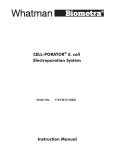
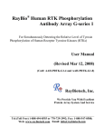
DEVELOPMENTAL DYNAMICS 229:433– 439, 2004 REVIEWS–A PEER REVIEWED FORUM A Primer on Using In Ovo Electroporation to Analyze Gene Function Catherine E. Krull* The chicken embryo has served as a classic model system for developmental studies due to its easy access for surgical manipulations and a wealth of data about chicken embryogenesis. Notably, the mechanisms controlling limb development have been explored best in the chick. Recently, the method of in ovo electroporation has been used successfully to transfect particular cells/tissues during embryonic development, without the production or infectivity associated with retroviruses. With the sequencing of the chicken genome near completion, this approach will provide a powerful opportunity to examine the function of chicken genes and their counterparts in other species. In ovo electroporation has been most effectively used to date for ectopic or overexpression analyses. However, recent studies indicate that this approach can be used successfully for loss-of-function analyses, including protein knockdown experiments with morpholinos and RNAi. Here, I will discuss parameters for using in ovo electroporation successfully to study developmental processes. Developmental Dynamics 229:433– 439, 2004. © 2004 Wiley-Liss, Inc. Key words: transfection; EGFP; chicken Received 28 August 2003; Revised 17 September 2003; Accepted 30 September 2003 BASIC PRINCIPLES Electroporation has served as an effective method for introducing DNA into bacteria, yeast, and mammalian cells (Neumann et al., 1982; Potter, 1988). This approach uses electric field pulses to disrupt plasma membrane stability transiently, creating pores in cell membranes through which DNA is driven, due to its negative charge. Usually, this type of electroporation uses high voltage and short duration electrical pulses, resulting in enhanced cell death. The application of electroporation to chicken embryos in ovo was made feasible by altering voltage and current parameters. Square wave pulses of low voltage and longer duration are used to achieve DNA delivery in the embryo, with minimal cell death and excellent survival. The first published account of electroporation in chicken tissue involved the transfection of plasmid DNA into retinal explants (Pu and Young, 1990). Shortly thereafter, this approach was applied to chicken embryos in ovo (Muramatsu et al., 1996, 1997). Since then, several investigators have made modifications to the original procedure, to enhance embryo survival, increase transfection efficiency, and target gene manipulations to particular cell types. MAKING IN OVO ELECTROPORATION WORK: THE HARDWARE REQUIREMENTS Over the past 4 years, my colleagues and I have optimized in ovo electroporation to transfect several tissues, including the neural tube, limb mesoderm, and somitic mesoderm (Swartz et al., 2001a,b; Eberhart et al., 2002). We build our own electrodes and have modified several features of our electroporation system. Our colleagues in other institutions have made additional modifications to enhance electroporation success. Below, I describe the hardware requirements for successful in ovo electroporation, defining both advantages and disadvantages of four in ovo electroporation systems. Each system delivers square wave pulses. Four in ovo electroporation systems are currently available commercially: (1) BTX electroporator, model 8300; www.btxonline.com; (2) CUY-21 electroporator, from Pro- Biological Sciences, University of Missouri-Columbia, Columbia, Missouri Grant sponsor: National Institutes of Mental Health, Muscular Dystrophy Association; Grant number: MH059894, MDA. *Correspondence to: Catherine E. Krull, Biological Sciences, 108 Lefevre, University of Missouri-Columbia, Columbia, MO 65211. E-mail: [email protected] DOI 10.1002/dvdy.10473 © 2004 Wiley-Liss, Inc. 434 KRULL TABLE 1. Comparing Electroporators ELECTROPORATOR BTX ADVANTAGES 1. Voltages can be selected easily. 2. Low or high voltage modes available, for electroporating embryos or cells in suspension. 3. Electroporation of various tissues highly efficient, after investigator-made modifications. DISADVANTAGES 1. High cost. 2. Connectors from the electroporator are flimsy and require modification by the investigator. 3. Inconsistent current delivery from pulse to pulse. 4. Electrode quality questionable. RECOMMENDATIONS Disadvantages outweigh advantages. Buy another system if possible. tech; www.protechinternational. com; (3) Intracel, TSS20 Ovodyne electroporator; www.intracel.co.uk; and (4) Grass, SD9 square pulse stimulator; www.grass-telefactor.com. If you are initiating in ovo electroporation experiments in your laboratory, investigate these electroporators first by discussing their pros and cons with investigators with expertise in electroporation (Table 1). These discussions ahead of your purchase will save your resources, including money and effort. Electrodes Electrodes can be built easily and at low cost by investigators or their local instrument shops, or purchased commercially. As one example, Protech sells high-quality electrodes for use with their CUY-21 electroporator. To construct your own electrodes, you’ll need the following equipment/supplies for electrode preparation (cost ⫽ ⬃$25 per pair): soldering iron; lead-free solder; hair dryer or heat shrink gun; black stranded 22 CUY-21 Intracel 1. Consistent current delivery, as monitored within the system. 2. High quality electrodes. 3. Effective electroporation of various tissues. 4. Voltages can be selected easily. 1. Low cost. 1. Low cost and easy to 2. Allows measurements use. of resistance, while 2. Typically available in electrodes are in most departments; place. check with local physiologists. 3. Useful for electroporating single or multiple cells. Grass stimulator 1. High cost; however, 1. Range of voltages this system does available are limited. not require the investigator to make modifications. The “Cadillac” of electroporators. Cost is high but well worth it. A very workable system that is low cost. gauge wire (Newark Electronics), catalog no. 92n5737; red stranded 22 gauge wire (Newark Electronics), catalog no. 92n5837; gold-plated jack/socket (Newark Electronics), catalog no. 40f6130; pin stamped brass (Digikey), catalog no. 82p-nd; wire insulation (Newark Electronics), catalog no. 03F3712; and for platinum electrodes, platinum rod (A-M Systems), catalog no. 711000, 0.01 inch diameter. Accessibility and low cost are strengths. Very solid performer. See Haas et al., (2001) for setup details. onto red and black stranded wire (Fig. 1, #4). Lengths of the red and black stranded wire should allow holding of the electrodes by hand or placement in a plastic electrode holder, without restriction. Red and black stranded wires are then each attached to a double banana BNC connector (Fig. 1, #2&3) with a male end. This double banana BNC connector is then connected to the electroporation system output wiring (Fig. 1, #1). Instructions Warm the soldering iron. Cut platinum wire into 6- to 7-cm lengths (yielding three sets of electrodes) or purchase precut lengths. Solder the pin stamped brass (Fig. 1, #6) onto the platinum wire (Fig. 1, #8). Cut six pieces of wire insulation (heat-shrink tubing) into 5-cm lengths. Place the tubing over the platinum wire (Fig. 1, #7), adjacent to the pin stamped brass, and apply heat from hair dryer or heat shrink gun to seal the wire insulation to the wire. Solder the gold-plated jack/socket (Fig. 1, #5) IN OVO ELECTROPORATION 101: TARGETING NEURAL TUBE CELLS Over the past few years, in ovo electroporation into the neural tube has become a routine and relatively easy approach to examine cell specification and axon guidance during chicken embryogenesis. If you are a novice using in ovo electroporation, start with neural tube electroporations. Once you become an expert in transfecting the IN OVO ELECTROPORATION IN CHICK 435 Fig. 1. Constructing electrodes. Schematic diagram showing how to build electrodes for in ovo electroporation. Refer to the Electrodes and Instructions sections for parts numbers and connections 1– 8. neural tube in this manner, electroporation of other tissues will be more straightforward. Electroporation of the chicken neural tube (Fig. 2A,B) can be subdivided into three steps: (1) preparation of the embryo and DNA for electroporation, (2) microinjection of DNA into the lumen of the neural tube, and (3) electrode placement and current application. Preparation of Embryo and DNA for Electroporation Swab eggshell with 70% ethanol, remove approximately 2–3 ml of albumen with an 18-g needle attached to a 3-cc syringe, and open a small window (1–2 cm) in the eggshell overlying the embryo with fine scissors. Enlarge window so that the embryo is in clear view. Inject a small bolus of a 10% Pelikan Drawing Ink A in Ringer’s under the blastoderm, to enhance contrast. Carefully tear off the vitelline membrane that overlies the neural tube to be electroporated, with a tungsten needle. Refer to Methods in Avian Embryology (Bronner-Fraser, 1996) for additional details. Typically, we have used two DNA constructs for in ovo electroporation: (1) pCAX, which contains a chick beta actin promoter/CMV IE enhancer to drive expression of EGFP (control; Osumi and Inoue, 2001); and (2) pMES, which contains the same promoter/enhancer and an IRES-EGFP, generating a bicistronic message (Swartz et al., 2001a,b; Eberhart et al., 2002). Constructs can be built to include other variants of GFP, including RFP. One strength of this approach is that there are no apparent limitations to the insert size that can be expressed from these plasmid constructs, in contrast to several retroviral vectors. Two, or theoretically more, constructs can be co-electroporated at the same time (Lee and Pffaff, 2003). It is important to sequence your construct to confirm expression of your DNA insert and EGFP or other marker in cell lines or primary cell cultures. Prepare DNA for electroporation by using a Qiagen Plasmid kit, to ensure high-quality DNA, and elute the DNA into water, not elution buffer. Use a spectrometer to measure DNA quality; if the OD 260/280 ⫽ 1.8, then the DNA is pure with little protein contaminants. If the OD is less than 1.6, re-prepare the DNA. Concentrate the DNA at 2–5 g/l in either PBS (1⫻ final concentration) or water, aliquot at 2– 4 l, and store at ⫺20°C to prevent degradation. Microinjection of DNA Into the Lumen of the Neural Tube If you have used vital dyes by means of a micropipette to mark small numbers of cells, this approach will be very similar. Add either a few crystals of Fast Green or 0.1% final concentration of phenol red to DNA, to verify microinjection. Backfill a micropipette with ⬃3 l of your DNA solution by using a Hamilton syringe. Place the micropipette into a needle holder, connected to a micromanipulator, which is linked to a picospritzer. Alternatively, DNA can be microinjected by using a mouth pipette or hand-held syringe. Align the DNA-loaded micropipette with the region of the neural tube to be microinjected with DNA and move the micropipette to this area by using the micromanipulator. Pierce the dorsal surface of the neural tube, where it opposes at midline. Expel DNA into the lumen; you will visualize your success immediately by the Fast Green or phenol red label. If successful, DNA will be confined to the lumen of the neural tube and spread rostrally and caudally from the injection site. If DNA instead spreads over the top of the embryo, you have not pierced the neural tube and entered its lumen. Reposition your micropipette and embryo and try again. If DNA spreads below the embryo, the micropipette has penetrated ventral to the embryo. Discard this egg and prepare a new embryo for microinjection. Electrode Placement and Current Application Shortly after microinjection of DNA into the lumen of the neural tube, place two platinum electrodes (see hardware requirements section) lateral and parallel to the embryo, on the area opaca, approximately 5– 6 mm apart. This electrode placement will result in the labeling of dorsal and ventral neural tube cells on one side of the embryo. To target a ventral quadrant of the neural tube, place the positive electrode below the embryo (see Fig. 2A) and the negative electrode dorsal to the neural tube and on the contralateral side. It is essential that your electrodes remain parallel to each other, with the same distance between them throughout your electroporations, to maintain consistency of current delivery. The territory in the middle of your electrodes should include the region of the neural tube to be electroporated and basically comprises your electric field. Do not 436 KRULL the polarity of your electrodes. Remove the electrodes, and apply a few drops of Ringer’s to cool and hydrate the embryo. Seal window in the eggshell with tape, and reincubate the egg until the desired stage of development is achieved. THE CHALLENGE: TARGETING MESENCHYME WITH IN OVO ELECTROPORATION Fig. 2. In ovo electroporation targets cells in the neural tube, limb mesoderm, and the somitic dermomyotome. A,C,E: Schematic diagrams showing how to perform in ovo electroporation, including DNA microinjection (micropipette) and electrode placement (⫹ and ⫺ electrodes), for successful transfection of a ventral quadrant of the neural tube, limb mesoderm, and lateral half of the somitic dermomyotome. In each diagram, one electrode is positioned ventral to the embryo by inserting it carefully at the interface between the embryo blastoderm and yolk. A: DNA is microinjected into the neural tube (nt) lumen and will travel into ventral neural tube cells upon current passage. som, somite; no, notochord. B: Transverse section through embryo at hindlimb level, 2.5 days after electroporation, as shown in A. EGFP signal (green) localizes to several motor neuron cell bodies in the ventral neural tube (nt) and the floor plate (fp). Islet1 antibody stains postmitotic neurons (red). C: DNA is microinjected into the coelom (co) that lies ventral to the somatopleure (sop), which will generate limb mesoderm. Injected DNA will travel into sop cells upon current passage, to transfect limb mesoderm. spl, splanchnic mesoderm. D: Transverse section through embryo at forelimb level, 1 day after electroporation, as shown in C. Limb mesoderm cells that lie at the base of the limb express EGFP (green). EphA4 protein (red) labels cells in the dermomyotome (dm) and the presumptive distal limb mesoderm (asterisk) at this stage. E: DNA is microinjected into the extracellular space between the forming dermomyotome (dm) and sclerotome (scl) and will travel into cells in the lateral half dm upon current passage. F: Transverse section through embryo at forelimb level, 1 day after electroporation, as shown in E. EGFPpositive muscle precursors (green) enter the limb from the lateral dermomyotome (dm). In this embryo, the dermomyotome of a single somite was electroporated. C-merlin protein (red) marks the myotome (m), whereas Pax7 protein (purple) binds to muscle precursors prominent in the dermomyotome. place your electrodes on embryonic tissue, to prevent injury and morphologic defects. Pass five 10- to 15-volt pulses, each of 50-msec duration. The DNA will be driven into the neural tube cells that lie adjacent to the positive electrode (anode). To transfect dorsal neural tube cells, reverse Many investigators new to in ovo electroporation have been frustrated by their attempts to transfect mesenchyme or mesenchyme-derived tissues, including limb mesoderm and somites. This frustration has left many electroporation systems gathering dust in the lab. Figure 2C–F provides schematic diagrams to illustrate in ovo electroporation of limb mesoderm and the somitic dermomyotome and images of these transfected tissues. It is imperative that investigators understand that transfecting mesenchyme with in ovo electroporation is a challenging task, due to several factors. First, as a tissue, mesenchyme typically contains cells that are loosely associated with each other. Thus, the extracellular space or matrix in mesenchyme provides a medium in which DNA can easily diffuse after microinjection. Second, luminal spaces in which to deposit DNA may not be obvious in mesenchyme. Third, it is important to remember that cells in different tissues have different resistances, based on tissue geometry and condensation. Tissue resistance affects the electric field generated during electroporation and, thus, electroporation success. This means that investigators must systematically vary electroporation parameters, including voltage, electrode placement, and pulse numbers, when attempting to transfect different tissues. A few strategies are available to deal with transfecting loosely packed mesenchyme using in ovo electroporation. One approach is to apply your DNA in a more viscous medium, such as Pluronic gel, which serves to reduce diffusion (Becker et al., 1999). A caveat of this approach is that the viscous medium may indeed have deleterious effects on IN OVO ELECTROPORATION IN CHICK 437 cells. A second approach is to follow the DNA microinjection with a small bolus of mineral oil, to localize DNA (Oberg et al., 2002). Lastly, others have reduced the time interval between DNA microinjection and electroporation by microinjecting and apply current by means of a single micropipette (see Haas et al., 2001, for details). Having a luminal surface in which to microinject DNA is a great advantage for in ovo electroporation. A lumen serves as a DNA reservoir, limiting DNA diffusion and localizing DNA to the tissue to be transfected. To determine whether the tissue you wish to transfect has a luminal surface, go to chick atlases or staging criteria (Hamburger and Hamilton, 1951; Bellairs and Osmond, 1998). Often, you will find that a luminal surface does exist at early stages of development (see Swartz et al., 2001b). Important Tips to Consider 1. Use embryos at stages 10 –20 (Hamburger and Hamilton, 1951) for whole embryo electroporation. Younger embryos tolerate less voltage than older embryos. Embryos electroporated at stages younger than stage 10 exhibit increased mortality rates and morphologic defects, due to the delicate tissue organization at early stages. Embryos older than stage 20 have more compact tissues and increased tissue layers, making microinjection and electroporation difficult. 2. If you wish to transfect tissue from older embryos, consider constructing tissue explants and performing electroporation in culture dishes. 3. Use the enclosed schematic illustrations and electroporation parameters (and other published reports) as guidelines. Each electroporation system is different and must be calibrated for each tissue to be transfected. 4. After identifying the tissue you wish to transfect, consider the electric field that you must establish to electroporate that tissue. Remember that DNA, which is negatively charged, will move into cells that lie adjacent to the positive electrode. 5. The distance between your electrodes dramatically affects your electroporation success. Increased distances between electrodes generally require increased voltages, to successfully transfect tissues that lie in the electric field generated across the electrodes, when passing current. Very small distances between electrodes narrow the electric field and can damage cells. Therefore, voltages must typically be lower. As the distance between electrodes is widened and electric field is enlarged, greater numbers of cells are transfected with in ovo electroporation. As the distance between electrodes is narrowed and the electric field is reduced, a smaller number of cells are electroporated, often resulting in focal transfections. 6. The resistance created by a tissue must impact the electric field that is generated and, thereby, influence electroporation success. 7. Voltage size and pulse number (three to five) should be altered systematically, when first attempting in ovo electroporation. Pulse duration remains typically at 50 msec, although longer pulses of 100 msec may be helpful to introduce other reagents (RNA, proteins, pharmacologic inhibitors) into embryonic cells. 8. Electrode placement will directly impact your transfection success with in ovo electroporation. Altering electrode placement by a few microns can often improve success. Again, consider the geometry of the tissue you want to transfect and whether it possesses a luminal surface; place your electrodes so that the cells you want to transfect lie within your electric field. 9. Connect your electroporator to an oscilloscope, to check current readings and verify consistency in current delivery. LOSS-OF-FUNCTION EXPERIMENTS USING IN OVO ELECTROPORATION Many investigators have routinely used in ovo electroporation for gainof-function analyses, overexpressing or ectopically expressing their gene of interest and EGFP or a variant (Arber et al., 1999; Grapin-Botton et al., 2001; Bel-Vialar et al., 2002; Eberhart et al., 2002; Bach et al., 2003; William et al., 2003). However, a few recent studies have shown that in ovo electroporation can be used effectively in loss-of-function experiments. Three approaches are available thus far: (1) proteins that act as competitive inhibitors, (2) RNAi, and (3) morpholinos (Gene Tools). These types of approaches allow investigators to disrupt protein signaling or knockdown protein levels, providing important tools for the analysis of gene function. Each of these techniques is relatively new, and only a few investigators have published studies using them successfully thus far. Expressing Proteins That Act as Competitive Inhibitors We have expressed kinase-inactive forms of the EphA4 receptor in cells that express wild-type EphA4, to disrupt EphA4 signaling (Eberhart et al., in press). kiEphA4 abolishes the phosphorylation or activation of the WT EphA4 receptor, acting as a dominant negative (Ethell et al., 2001). Recent studies have used in ovo electroporation to express mutated forms of transcriptional activators, which also serve as dominant negatives (Lee and Pfaff, 2003). The expectation is that this approach will become more widely used by investigators as comfort and expertise with in ovo electroporation develops. RNAi Stoeckli and colleagues recently demonstrated that dsRNA can be introduced into the developing chicken embryo using in ovo electroporation (Pekarik et al., 2003). Relatively long sequences (200 to 2,000 base pairs in length) interfere specifically with protein translation in chicken embryos, as seen in these studies on axon guidance across the midline. It is essential to demonstrate that protein knockdown is specific to one’s protein of interest. Typically, this task is accomplished by means of Western blot analysis, using an antibody that binds the protein of interest specifically. This approach provides a powerful method for knockdown studies and should prove extremely valuable for future functional genomics analyses. 438 KRULL Morpholinos Morpholinos have served as fantastic tools to interfere with protein translation for investigators who study development in zebrafish, Xenopus, and sea urchins (Nasevicius and Ekker, 2000; Audic et al., 2001; Howard et al., 2001). However, investigators using chickens as model systems have encountered some difficulties introducing these reagents by means of in ovo electroporation, most likely due to the reduced charge associated with these molecules, making it problematic to target them to some cell types. Erickson and colleagues were the first to demonstrate that morpholinos could be applied in chickens with in ovo electroporation (Kos et al., 2001; see Kos et al., 2003, for methods). Recently, a few investigators have used this approach to specifically knockdown proteins during chicken embryonic development (GerlachBank et al., 2004; Granata and Quaderi, 2003). As with RNAi, it is imperative that the investigator demonstrate that specific protein knockdown has been achieved. ANALYSES AFTER IN OVO ELECTROPORATION 1. After embryos develop to the appropriate stage after in ovo electroporation, confirm the success of transfection in ovo using a fluorescence dissecting microscope equipped with GFP optics. Embryos with poor transfections can be discarded; embryos that require further development before harvesting can be reincubated. 2. The percentage of transfected cells will vary with electroporation conditions. In our hands, 50 – 85% of targeted cells are transfected. Importantly, this percentage is not consistent from embryo to embryo. Therefore, it requires that investigators plan to transfect at least ⬃2– 4⫻ as many embryos as needed for their functional analysis. 3. Investigators should examine whether each construct used in in ovo electroporation results in enhanced cell death by using terminal deoxynucleotidyl transferase-mediated biotinylated UTP nick end label- ing (TUNEL) labeling or Nile blue or acridine orange stains. 4. Investigators should examine embryos carefully for nonspecific morphologic defects that are likely due to the electroporation technique. Any contact by electrodes with embryonic tissue during current passage will create damage. These nonspecific morphologic defects will complicate your analysis postelectroporation and should be avoided. 5. Embryos can be fixed and viewed as whole-mounts or prepared for cryostat, paraffin, or Vibratome sectioning. Fixation and exposure to organic solvents dampens or destroys GFP signal so the addition of a GFP antibody to restore this signal may be required. 6. Antibody staining or in situ hybridization should be performed to determine whether one’s gene of interest is expressed and the duration of expression after in ovo electroporation. Western blot analysis serves as an excellent alternative approach. COMMON PROBLEMS AND TROUBLESHOOTING A few of the typical problems encountered when using in ovo electroporation are described below, as well as troubleshooting strategies. Problem #1: None or few cells are transfected, and gene expression is short-lived. Solutions: a. Adjust electrode placement to widen the electric field and target more cells; b. A different promoter may be useful, to target gene expression more precisely; c. Replace electrodes if older than 3 months; d. Consider that the normal regulatory controls of the embryo may influence the expression of your gene of interest; e. Check that plasmid concentration is 2 g/l minimum; f. Test constructs for expression in primary cell cultures or cell lines; g. Increase voltage slightly and monitor cell death; h. Increase volume of DNA injected; and i. Apply Ringer’s solution to enhance current conduction. Problem #2: Embryos do not survive after in ovo electroporation. Solutions: a. Replace electrodes if older than 3 months; b. Determine whether electrodes are touching embryonic tissues during the electroporation, damaging the embryo; c. Confirm that DNA is in solution in water or PBS, not TE. Prepare fresh DNA if impurities or contaminants are suspect; d. Fast Green can interfere with survival of young embryos. Try phenol red (0.1% final concentration) as an alternative; and e. Reduce voltage, pulse width, and/or number of pulses. Problem #3: Embryos have major, unexpected morphologic defects. Solutions: a. Electrodes are old and must be replaced; b. Electrodes are in contact with embryonic tissues when current is being passed. Avoid contact with embryonic tissues by placing electrodes in a pool of Ringer’s solution. Electrodes placed ventral to the embryo should lie in yolk; c. Use lower voltages, to reduce cell damage. OTHER REFERENCES Several good reviews and technical papers have been published about using in ovo electroporation to analyze gene function in chickens (Itasaki et al., 1999; Momose et al., 1999; Atkins et al., 2000; Yasuda et al., 2000; Haas et al., 2001; Swartz et al., 2001a; Inoue and Krumlauf, 2001; Osumi and Inoue, 2001; Nakamura and Funahashi, 2001; Martinez and Hollenbeck, 2003). Investigators are referred to these publications for additional details. FUTURE DIRECTIONS In ovo electroporation is a relatively new approach that has been used primarily to misexpress or overexpress genes of interest during chick embryonic development. Several investigators have used this approach in chickens for their gain-of-function analyses, paired with the powerful genetics approach in mouse for lossof-function studies. In the future, RNA interference and the application by means of in ovo electroporation of other reagents (i.e., morpholinos) that knockdown protein specifically promises to enhance studies of gene function in chickens. Predictably, loss-of-function studies should become more prevalent. Another direction for the future in- IN OVO ELECTROPORATION IN CHICK 439 cludes the linking of in ovo electroporation with time-lapse imaging of cell behavior. Together, these approaches will allow investigators to exploit the advantages of the chicken embryo, which include easy access to developmental events. Novel DNA constructs that allow investigators to tightly control the temporal expression of their gene of interest should provide enormous power to this approach. Moreover, in ovo electroporation offers the unique opportunity to identify and analyze enhancer elements quickly (Timmer et al., 2001; Uchikawa et al., 2003; Ebert et al., 2003). Clearly, the sequencing of the chicken genome will provide enormous opportunities for using in ovo electroporation and the classic strengths of this model system to dissect gene function in the future. ACKNOWLEDGMENTS Thanks go to members of the Krull lab for critical discussions. REFERENCES Atkins RL, Wang D, Burke RD. 2000. Localized electroporation: a method for targeting expression of genes in avian embryos. Biotechniques 28:94–96, 98, 100. Arber S, Han B, Mendelsohn M, Smith M, Jessell TM, Sockanathan S. 1999. Requirement for the homeobox gene Hb9 in the consolidation of motor neuron identity. Neuron 23:659 –674. Audic Y, Boyle B, Slevin M, Hartley RS. 2001. Cyclin E morpholino delays embryogenesis in Xenopus. Genesis 30:107– 109. Bach A, Lallemand Y, Nicola MA, Ramos C, Mathis L, Maufras M, Robert B. 2003. Msx1 is required for dorsal diencephalon patterning. Development 130:4025– 4036. Becker DL, McGonnel I, Makarenkova HP, Patel K, Tickle C, Lorimer J, Green CR. 1999. Roles for alpha 1 connexin in morphogenesis of chick embryos using a novel antisense approach. Dev Genet 24:33–42. Bellairs R, Osmond M. 1998. The atlas of chick development. San Diego: Academic Press. Bel-Vialar S, Itasaki N, Krumlauf R. 2002. Initiating Hox gene expression: in the early chick neural tube differential sensitivity to FGF and RA signaling subdivides the HoxB genes into two distinct groups. Development 129:5103–5115. Bronner-Fraser M, Editor. 1996. Methods in avian embryology. San Diego: Academic Press. Eberhart J, Swartz ME, Koblar SA, Pasquale EB, Krull CE. 2002. EphA4 constitutes a population-specific guidance cue for motor neurons. Dev Biol 247:89–101. Eberhart J, Barr J, O’Connell S, Flagg A, Swartz ME, Cramer KS, Tosney KW, Pasquale EB, Krull CE. 2004. Ephrin-A5 exerts positive or inhibitory effects on distinct subsets of EphA4-positive motor neurons. J Neuroscience (in press). Ebert PJ, Timmer JR, Nakada Y, Helms AW, Parab PB, Liu Y, Hunsaker TL, Johnson JE. 2003. Zic1 represses Math1 expression via interactions with the Math1 enhancer and modulation of Math1 autoregulation. Development 130:1949–1959. Ethell IM, Irie F, Kalo MS, Couchman JR, Pasquale EB, Yamaguchi Y. 2001. EphB/syndecan-2 signaling in dendritic spine morphogenesis. Neuron 31:1001– 1013. Gerlach-Bank L, Cleveland AR, Barald KF. 2004. DAN directs endolymphatic sac and duct outgrowth in the avian inner ear. Dev Dyn 229:219 –230. Granata A, Quaderi NA. 2003. The Opitz syndrome gene MID1 is essential for establishing asymmetric gene expression in Hensen’s node. Dev Biol 258:397–405. Grapin-Botton A, Majithia AR, Melton DA. 2001. Key events of pancreas formation are triggered in gut endoderm by ectopic expression of pancreatic regulatory genes. Genes Dev 15:444 –454. Haas K, Sin W-C, Javaherian A, Cline HT. 2001. Single-cell electroporation for in vivo neuronal gene expression. Hamburger V, Hamilton HL. 1951. A series of normal stages in the development of the chick embryo. J Morphol 88:49–92. Howard EW, Newman LA, Oleksyn DW, Angerer RC, Angerer LM. 2001. SpKrl: a direct target of beta-catenin regulation required for endoderm differentiation in sea urchin embryos. Development 128:365–375. Inoue T, Krumlauf R. 2001. An impulse to the brain - using in vivo electroporation. Nat Neurosci 4(Suppl):1156 –1158. Itasaki N, Bel-Vialar S, Krumlauf R. 1999. Shocking developments in chick embryology: electroporation and in ovo gene expression. Nat Cell Biol 1:E203–E207. Kos R, Reedy MV, Johnson RL, Erickson CA. 2001. The winged-helix transcription factor FoxD3 is important for establishing the neural crest lineage and repressing melanogenesis in avian embryos. Development 128:1467–1679. Kos R, Tucker RP, Hall R, Duong TD, Erickson CA. 2003. Methods for introducing morpholinos into the chicken embryo. Dev Dyn 226:470 –477. Lee S-K, Pfaff SL. 2003. Synchronization of neurogenesis and motor neuron specification by direct coupling of bHLH and homeodomain transcription factors. Neuron 38:731–745. Martinez CY, Hollenbeck PJ. 2003. Transfection of primary central and peripheral nervous system neurons by electroporation. Methods Cell Biol 71:339 –351. Momose T, Tonegawa A, Takeuchi J, Ogawa H, Umesono K, Yasuda K. 1999. Efficient targeting of gene expression in chick embryos by microelectroporation. Dev Growth Differ 41:335–344. Muramatsu T, Mizutani Y, Okumura J. 1996. Live detection of the firefly luciferase gene expression by bioluminescence in incubating chicken embryos. Ann Sci Technol 67:906 –909. Muramatsu T, Mizutani Y, Ohmori Y, Okumura J. 1997. Comparison of three nonviral transfection methods for foreign gene expression in early chicken embryos in ovo. Biochem Biophys Res Commun 230:376 –380. Nakamura H, Funahashi J. 2001. Introduction of DNA into chick embryos by in ovo electroporation. Methods 24:43–48. Nasevicius A, Ekker SC. 2000. Effective targeted gene “knockdown” in zebrafish. Nat Genet 26:216 –220. Neumann E, Schaefer-Ridder M, Wang Y, Hofschneider PH. 1982. Gene transfer into mouse lyoma cells by electroporation in high electric fields. EMBO J 1:841– 845. Oberg KC, Pira CU, Revelli JP, Ratz B, Aguilar-Cordova E, Eichele G. 2002. Efficient ectopic gene expression targeting chick mesoderm. Dev Dyn 224:291–302. Osumi N, Inoue T. 2001. Gene transfer into cultured mammalian embryos by electroporation. Methods 24:35–42. Pekarik V, Bourikas D, Miglino N, Joset P, Preiswerk S, Stoeckli ET. 2003. Screening for gene function in chicken embryo using RNAi and electroporation. Nat Biotechnol 21:93–96. Potter H. 1988. Electroporation in biology: methods, applications, and instrumentation. Anal Biochem 174:361–373. Pu HF, Young AP. 1990. Glucocorticoidinducible expression of a glutamine synthetase-CAT-encoding fusion plasmid after transfection of intact chicken retinal explant cultures. Gene 89:259 – 263. Swartz M, Eberhart J, Mastick G, Krull CE. 2001a. Sparking new frontiers: using in vivo electroporation for genetic manipulations. Dev Biol 233:13–21. Swartz ME, Eberhart J, Pasquale EB, Krull CE. 2001b. EphA4/ephrin-A5 interactions in muscle precursor cell migration in the avian forelimb. Development 128: 4669 –4680. Timmer J, Johnson J, Niswander L. 2001. The use of in ovo electroporation for the rapid analysis of neural-specific murine enhancers. Genesis 29:123–132. Uchikawa M, Ishida Y, Takemoto T, Kamachi Y, Kondoh H. 2003. Functional analysis of chicken Sox2 enhancers highlights an array of diverse regulatory elements that are conserved in mammals. Dev Cell 4:509 –519. William CM, Tanabe Y, Jessell TM. 2003. Regulation of motor neuron subtype identify by repressor activity of Mnx class homeodomain proteins. Development 130:1523–1536. Yasuda K, Momose T, Takahashi Y. 2000. Applications of microelectroporation for studies of chick embryogenesis. Dev Growth Differ 42:203–206.