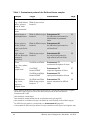
1
June 2012 QIAamp® cador® Pathogen Mini Handbook For the purification of viral RNA and DNA and bacterial DNA from animal whole blood, serum, plasma, other body fluids, swabs and washes, and tissue Sample & Assay Technologies QIAGEN Sample and Assay Technologies QIAGEN is the leading provider of innovative sample and assay technologies, enabling the isolation and detection of contents of any biological sample. Our advanced, high-quality products and services ensure success from sample to result. QIAGEN sets standards in: Purification of DNA, RNA, and proteins Nucleic acid and protein assays microRNA research and RNAi Automation of sample and assay technologies Our mission is to enable you to achieve outstanding success and breakthroughs. For more information, visit www.qiagen.com. Contents Kit Contents 4 Storage 5 Intended Use 5 Safety Information 6 Quality Control 6 Introduction 7 Principle and procedure 7 Description of protocols 7 Nucleic acid purification protocol 9 Pretreatments 9 Equipment and Reagents to Be Supplied by User 12 Important Notes 14 Protocols Purification of Pathogen Nucleic Acids from Fluid Samples Pretreatment B1 — for Difficult-to-Lyse Bacteria in Whole Blood or Pretreated Tissue 23 Pretreatment B2 — for Difficult-to-Lyse Bacteria in Cell-Free Fluids 24 Pretreatment B3 — for Easy-to-Lyse Bacteria in High-Volume Cell-Free Fluids 25 Pretreatment T1 — Mechanical Disruption of Tissue 26 Pretreatment T2 — Enzymatic Digestion of Tissue 27 Pretreatment T3 — Rapid Partial Disruption of Tissue 29 Pretreatment T4 — Organic Extraction for Difficult Tissue 30 20 Troubleshooting Guide 32 References 35 Ordering Information 36 QIAamp cador Pathogen Mini Handbook 06/2012 3 Kit Contents QIAamp cador Pathogen Mini Kit (50) (250) Catalog no. 54104 54106 Number of preps 50 250 QIAamp Mini Columns 50 250 Collection Tubes (2 ml) 200 1000 Buffer VXL* 6 ml 30 ml Buffer ACB*† (concentrate) 12 ml 60 ml QIAGEN® Proteinase K 1.25 ml 6 ml Carrier RNA (poly A) 310 μg 310 μg Buffer AW1*‡ (concentrate) 19 ml 98 ml Buffer AW2‡ (concentrate) 17 ml 81 ml Buffer AVE§ 20 ml 2 x 20 ml Quick-Start Protocol 1 1 * Contains a chaotropic salt. Take appropriate laboratory safety measures and wear gloves when handling. Not compatible with disinfectants containing bleach. See page 6 for safety information. † Before using for the first time, add isopropanol as indicated on the bottle to obtain a working solution. ‡ Before using for the first time, add ethanol (96–100%) as indicated on the bottle to obtain a working solution. § Contains sodium azide as a preservative. 4 QIAamp cador Pathogen Mini Handbook 06/2012 Storage QIAamp Mini columns and buffers can be stored dry at room temperature (15–25°C) until the expiration date on the kit box without affecting performance. Lyophilized carrier RNA can be stored at room temperature until the expiration date stated on the kit box. For use, lyophilized carrier RNA should be dissolved in Buffer AVE, and then added to Buffer VXL as described in “Preparing reagents” on page 17. This carrier RNA/Buffer AVE/Buffer VXL solution should be prepared fresh, and is stable at room temperature for up to 48 hours. Unused carrier RNA dissolved in Buffer AVE should be immediately frozen in aliquots at –20°C. Do not subject aliquots of carrier RNA to more than 3 freeze– thaw cycles. QIAGEN Proteinase K can be stored at room temperature. To store for extended periods of time, or if the ambient temperature often exceeds 25°C, we recommend storing at 2–8°C. Intended Use The QIAamp cador Pathogen Mini Kit is intended for the extraction of pathogen nucleic acids (viral RNA and DNA and bacterial DNA) from animal whole blood, serum, plasma, other body fluids, swabs, washes, and tissue. For laboratory use. Not for use in veterinary diagnostic procedures. No claim or representation is intended to provide information for the diagnosis, prevention, or treatment of a veterinary disease. All due care and attention should be exercised in the handling of the products. We recommend all users of QIAGEN products to adhere to the NIH guidelines that have been developed for recombinant DNA experiments, or to other applicable guidelines. QIAamp cador Pathogen Mini Handbook 06/2012 5 Safety Information When working with chemicals, always wear a suitable lab coat, disposable gloves, and protective goggles. For more information, please consult the appropriate safety data sheets (SDSs). These are available online in convenient and compact PDF format at www.qiagen.com/safety where you can find, view, and print the SDS for each QIAGEN kit and kit component. CAUTION: DO NOT add bleach or acidic solutions directly to the sample preparation waste. Buffer VXL, and Buffer AW1 contain guanidine hydrochloride and Buffer ACB contains guanidine thiocyanate, which can form highly reactive compounds if combined with bleach. If liquid containing these buffers is spilled, clean with suitable laboratory detergent and water. If the spilled liquid contains potentially infectious agents, clean the affected area first with laboratory detergent and water, and then with 1% (v/v) sodium hypochlorite. 24-hour emergency information Emergency medical information in English, French, and German can be obtained 24 hours a day from: Poison Information Center Mainz, Germany Tel: +49-6131-19240 Quality Control In accordance with QIAGEN’s ISO-certified Quality Management System, each lot of the QIAamp cador Pathogen Mini Kit is tested against predetermined specifications to ensure consistent product quality. 6 QIAamp cador Pathogen Mini Handbook 06/2012 Introduction The QIAamp cador Pathogen Mini Kit enables the efficient purification of viral RNA and DNA and bacterial DNA from a broad range of animal samples including whole blood, serum, plasma, swabs, washes, and tissue (see “Starting material” on page 14). The extracted nucleic acids are free of proteins, nucleases, and other impurities, and are ready for use in downstream applications, such as real-time PCR-based pathogen identification. The kit is not intended for host RNA or host DNA preparation. Principle and procedure Samples are lysed under highly denaturing conditions at room temperature (15–25°C) in the presence of proteinase K and Buffer VXL, which together ensure the inactivation of nucleases. Adding Buffer ACB adjusts the binding conditions for the copurification of DNA and RNA. The lysate is then transferred to a QIAamp Mini column. During centrifugation, nucleic acids are adsorbed onto the silica membranes while contaminants pass through. Two efficient wash steps remove the remaining contaminants and enzyme inhibitors, and nucleic acids are eluted in Buffer AVE. Performance is not guaranteed for every combination of starting material and pathogen species and must be validated by the user. Some samples may require a pretreatment (see Table 1, page 8). Description of protocols There are two types of protocol in this handbook. Samples will either directly undergo nucleic acid purification, or undergo pretreatment followed by nucleic acid purification. Most sample types can be directly processed without pretreatment. However, depending on the starting material and the target pathogen, one of the pretreatment protocols may be needed. Table 1 on page 8 provides an overview of which pretreatment protocols are suited to which starting material and pathogen combinations. Nucleic acid purification protocol (page 20) Pretreatment protocols (pages 23–31) QIAamp cador Pathogen Mini Handbook 06/2012 7 Table 1. Pretreatment protocols for fluid and tissue samples Sample Target Fluids Viral RNA and DNA, e.g., whole blood, DNA of easy-to-lyse bacteria* serum, plasma, swab or wash fluid, pretreated tissue Pretreatment Page – 20 Whole blood or pretreated tissue DNA of difficult-to-lyse Pretreatment B1 bacteria* for difficult-to-lyse bacteria in whole blood or pretreated tissue 23 Serum, plasma, swabs, washes, body cavity fluids, urine DNA of difficult-to-lyse Pretreatment B2 bacteria* for difficult-to-lyse bacteria in cell-free fluids† 24 High volume cell-free fluids (for increased sensitivity) DNA of easy-to-lyse bacteria* Pretreatment B3 for easy-to-lyse bacteria in high-volume cell-free fluids 25 Tissue Viral RNA and DNA‡ 26 e.g., liver, spleen, kidney, lymph node Pretreatment T1 mechanical disruption of tissue Viral DNA§, bacterial DNA¶ Pretreatment T2 enzymatic digestion of tissue 27 Viral RNA and DNA, bacterial DNA¶ Pretreatment T3 rapid partial disruption of tissue 29 Viral RNA and DNA, DNA of easy-to-lyse bacteria*, ** Pretreatment T4 organic extraction for difficult tissue 30 Difficult tissue e.g., brain, pancreas, adipose tissue * Gram-positive bacteria are difficult to lyse due to their rigid cell wall. Many Gram-negative bacteria are easy to lyse, but some are difficult to lyse and will also benefit from Pretreatment B1 or B2 † Not suitable for whole blood. ‡ Not suitable for bacterial DNA due to centrifugation step (see page 26). § Not suitable for viral RNA as the lysis conditions do not sufficiently conserve RNA integrity. ¶ For difficult-to-lyse bacteria, subsequently use Pretreatment B1 (page 23). ** Not suitable for difficult-to-lyse bacteria as they would be lost during the centrifugation step. 8 QIAamp cador Pathogen Mini Handbook 06/2012 Nucleic acid purification protocol The protocol “Purification of Pathogen Nucleic acids from Fluid Samples” (page 20) is optimized for purification of viral RNA and DNA and the DNA of easy-to-lyse bacteria from up to 200 μl of fluid material. Suitable starting materials for direct processing using this method include: Whole blood Serum Plasma Body cavity fluids (e.g., peritoneal, synovial, cerebrospinal) Liquid extracts from swabs (e.g., nasal, pharyngeal, and cloacal swabs) Wash fluids (e.g., from bronchoalveolar lavages) Other fluids, such as urine* Pretreatments The various pretreatments included in this handbook are optimized for specific combinations of starting material and target pathogen. The choice of pretreatment depends on the workflow focus, and is to be followed by nucleic acid purification. Table 1 on page 8 summarizes the pretreatments and their applications. Some of the pretreatments may require additional components (see page 12) Pretreatment for extraction of DNA of difficult-to-lyse bacteria Treatment with chemicals and proteinase K is sufficient for complete lysis in the case of many Gram-negative bacteria, but the cell walls of Gram-positive bacteria and some Gram-negative bacteria must be disrupted by additional methods. For maximal lysis efficiency when working with difficult-to-lyse bacteria, we recommend mechanical disruption using Pathogen Lysis Tubes, which contain glass beads. The tubes must be ordered separately (for ordering information, see page 36). For pretreatment of difficult-to-lyse bacteria in whole blood, use Pretreatment B1 (page 23). For pretreatment of difficult-to-lyse bacteria from cell-free fluid samples such as serum, plasma, swabs, washes, body cavity fluids and urine, use Pretreatment B2 (page 24). * The processing of samples with high inhibitor contents, such as urine, may require a reduction in sample input volume. QIAamp cador Pathogen Mini Handbook 06/2012 9 For pretreatment of difficult-to-lyse bacteria in tissue samples, first use one of the tissue pretreatments (see pages 9 and 23–31), followed by Pretreatment B1 (page 23). Optional pretreatment for easy-to-lyse bacteria in high-volume cell-free fluids Optionally, to increase the sensitivity of detection for DNA of easy-to-lyse bacteria from fluid samples containing no cells or a low amount of cells, such as serum, plasma, other cell-free body fluids, swabs, and washes, the sample input volume can be increased. Subsequently, the bacteria can be concentrated by pelleting. To use an increased input volume before the extraction of DNA of easy-to-lyse bacteria, use Pretreatment B3 (page 25). Pretreatment of tissue samples Mechanical or enzymatic disruption of tissue structure is a prerequisite to enable the extraction and purification of nucleic acids. The protocols listed in this section are for tissue pretreatment in workflows focused on viral RNA and DNA and bacterial DNA. The starting material is up to 25 mg of tissue such as liver, lymph node, spleen, or kidney. The choice of pretreatment depends on the various workflow demands: the targeted pathogen, type of tissue, anticipated sensitivity of the application, available laboratory equipment, and acceptable time and effort. The pretreatment protocols are suited for most types of tissue and pathogens. However, we recommend evaluating the suitability of these approaches for each new combination of tissue type and pathogen. To extract viral RNA and viral DNA from tissue, use Pretreatment T1 (page 26). This pretreatment is not suitable for the extraction of bacterial DNA due to the centrifugation step, which may result in a substantial loss of bacterial DNA with the pelleted material. To extract bacterial or viral DNA from tissue, use Pretreatment T2 (page 27). This pretreatment is not suitable for the extraction of viral RNA because the lysis conditions do not sufficiently conserve RNA integrity. As mentioned on page 9, depending on the bacteria, an additional pretreatment may be needed after Pretreatment T2. To extract the DNA of difficult-to-lyse bacteria from tissue, after Pretreatment T2, use Pretreatment B1 (page 23). 10 QIAamp cador Pathogen Mini Handbook 06/2012 For some applications, partial tissue disruption by vigorous shaking may be sufficient to release an adequate amount of cells from the tissue structure for reliable pathogen identification. This can be the case for tissue with weak cell adhesion, for pathogens located in the extracellular space or in easily detached cells, or for applications with high pathogen loads. In this case, Pretreatment T3 (page 29) may be suitable. For difficult-to-lyse bacteria, subsequently use Pretreatment B1 (page 23). Some tissue types, such as pancreas or brain tissue, can be very difficult to process due to their high lipid and/or nuclease content. To extract viral RNA and DNA and bacterial DNA of easy-to-lyse bacteria from these tissues, use Pretreatment T4 (page 30). QIAamp cador Pathogen Mini Handbook 06/2012 11 Equipment and Reagents to Be Supplied by User When working with chemicals, always wear a suitable lab coat, disposable gloves, and protective goggles. For more information, consult the appropriate safety data sheets (SDSs), available from the product supplier. For all protocols Pipettors and disposable pipet tips with aerosol barriers (20–1000 μl) Isopropanol Ethanol (96–100%)* † Phosphate-buffered saline (PBS) may be required for sample dilution Microcentrifuge 2 ml microcentrifuge tubes Vortexer Pretreatment B1 — for difficult-to-lyse bacteria in whole blood or pretreated tissue Vortexer with Microtube foam insert (Scientific Industries, cat. no. 5040234-00) or TurboMix Attachment (Scientific Industries, cat. no. SI-0564); or FastPrep®-24 (MP Biomedicals, cat. no. 6004500), or TissueLyser II (cat. no. 85300) with a TissueLyser II Adapter Set 2 x 24 (cat. no. 69982) or 2 x 96 (cat. no. 69984), or TissueLyser LT (cat. no. 85600) with the TissueLyser LT Adapter for 12 tubes (cat. no. 69980)† Pathogen Lysis Tubes L (cat. no. 19092) containing 50 Pathogen Lysis Tubes with glass beads and 1 vial Reagent DX (cat. no. 19088) for bead-beating of bacteria Buffer ATL (cat. no. 19076) * Denatured alcohol is not suitable as it contains other substances such as methanol or methylethylketone. † This is not a complete list of suppliers and does not include many important vendors of biological supplies. 12 QIAamp cador Pathogen Mini Handbook 06/2012 Pretreatment B2 — for difficult-to-lyse bacteria in cell-free fluids Vortexer with Microtube foam insert (Scientific Industries, cat. no. 5040234-00) or TurboMix Attachment (Scientific Industries, cat. no. SI-0564); or FastPrep-24 (MP Biomedicals, cat. no. 6004500), or TissueLyser II (cat. no. 85300) with a TissueLyser II Adapter Set 2 x 24 (cat. no. 69982) or 2 x 96 (cat. no. 69984), or TissueLyser LT (cat. no. 85600) with the TissueLyser LT Adapter for 12 tubes (cat. no. 69980)* Pathogen Lysis Tubes L (cat. no. 19092) or S (cat. no. 19091) containing 50 Pathogen Lysis Tubes with glass beads and 1 vial Reagent DX (cat. no. 19088) for bead-beating of bacteria Buffer ATL (cat. no. 19076) Pretreatment T1 — mechanical disruption of tissue TissueLyser II (cat. no. 85300) with a TissueLyser II Adapter Set 2 x 24 (cat. no. 69982), or TissueLyser LT (cat. no. 85600) with the TissueLyser LT Adapter for 12 tubes (cat. no. 69980), or other bead-mill homogenizer* Note: A Vortexer with microtube foam insert (Scientific Industries, cat. no. 504-0234-00) can be used instead. 5 mm stainless steel beads (cat. no. 69989) PBS, pH 7.2 (50 mM potassium phosphate, 150 mM NaCl) or NaCl 0.9% Pretreatment T2 — enzymatic digestion of tissue Thermoshaker suitable for 2 ml collection tubes Buffer ATL (cat. no. 19076) * This is not a complete list of suppliers and does not include many important vendors of biological supplies. QIAamp cador Pathogen Mini Handbook 06/2012 13 Important Notes Starting material Do not overload the QIAamp Mini column, as this can lead to impaired nucleic acid extraction and/or performance in downstream assays. For samples with very high host nucleic acid contents (e.g., for certain tissues, such as spleen or blood samples with highly increased cell counts), use less than the maximum amount of sample recommended in the protocol or pretreatments. In some downstream applications such as PCR and RT-PCR, very high background concentrations of nucleic acids may impair the reaction. Use appropriate controls (e.g., an internal control) to verify successful PCR amplification. Avoid transferring material to the QIAamp Mini column that could subsequently clog the membrane (e.g., blood clots, solid tissue, swab fibers). Highly viscous fluids may require a treatment to reduce their viscosity to allow for efficient extraction of pathogen nucleic acids. Please contact QIAGEN Technical Service for recommendations. Avoid repeated thawing and freezing of samples since this may reduce nucleic acid yield and quality. Animal whole blood Blood samples treated with EDTA, citrate, or heparin as anticoagulant can be used for nucleic acid purification. Samples can be either fresh or frozen, provided that they have not been frozen and thawed more than once. After collection, whole blood samples can be stored at 2–8ºC for up to 6 hours. For longer storage, we recommend freezing aliquots at –20ºC or –80ºC. We recommend using 50–200 μl blood per sample. Typically, 200 μl of blood can be used with most blood samples. However, highly elevated cell counts due to inflammatory or neoplastic diseases may strongly increase the host nucleic acid content of a sample. In this case, reduction of sample input to 50 μl may improve results in downstream assays, particularly in RT-PCR. If using less than 200 μl blood, adjust the sample volume to 200 μl with PBS or 0.9% NaCl. For blood samples containing nucleated erythrocytes (e.g., samples from bird and fish), use 5–25 μl blood and adjust the sample volume to 200 μl with PBS or 0.9% NaCl. 14 QIAamp cador Pathogen Mini Handbook 06/2012 Animal serum, plasma, other body fluids, swab, and wash specimens Frozen plasma or serum must not be thawed more than once before processing. We recommend storing swabs in transport media; for example, viral transport media (VTM) or brain–heart infusion broth (BHI). Remove the swab and squeeze out the liquid by pressing the swab against the inside of the storage tube. For extraction of viral RNA or DNA, we recommend centrifuging the swab media briefly to ensure any residual solid materials are removed. Note: Solid pieces remaining in the sample fluid may aggregate on the QIAamp Mini column, which may decrease nucleic acid yield. Up to 200 μl serum, plasma, other body fluid, swab media supernatant, or wash fluid can be processed. Carrier RNA must be used in the nucleic acid purification protocol to prevent the loss of nucleic acids during the procedure (see page 16 for information on the use of carrier RNA). The processing of samples with very high inhibitor contents, such as urine or feces suspensions, may require a reduction in sample input volume and/or an extra pretreatment to remove inhibitors. To reduce the input volume, use 25–50 μl of the sample and adjust the volume to 200 μl with PBS or 0.9% NaCl. For extraction of bacterial DNA, the input volume can be increased to more than 200 μl, e.g., 1.5 ml for increased sensitivity of bacterial detection. See Pretreatment B2 (page 24) for extraction of DNA from difficult-to-lyse bacteria and Pretreatment B3 (page 25) for extraction of DNA from easy-to-lyse bacteria. Animal tissues When working with tissue samples, mechanical or enzymatic disruption of the tissue structure is the prerequisite for liberation of cells, subsequent release of nucleic acids, and membrane permeability of the material. Different tissue types can vary widely with regard to texture and rigidity, cell types, and content of host nucleic acids and inhibitory substances. In addition, the localization of pathogen nucleic acids in the tissue may vary depending on tissue type, pathogen, and stage of infection. Therefore, suitability of the pretreatment protocols in this handbook should be evaluated for each new combination of tissue and pathogen. Up to 25 mg of fresh or frozen tissue can be used as a starting amount. For tissues with a very high number of cells for a given mass of tissue, such as spleen, a reduced amount of starting material (5–10 mg) should be used. QIAamp cador Pathogen Mini Handbook 06/2012 15 Yields of nucleic acids For samples containing a low amount of cells (e.g., serum), the yield of viral or bacterial nucleic acids obtained can be below 1 μg and is therefore difficult to quantify using a spectrophotometer. In addition, eluates prepared with carrier RNA may contain much more carrier RNA than target nucleic acids. The QIAamp cador Pathogen Mini protocol recovers total nucleic acids. Therefore, cellular DNA and RNA will be copurified from any cells in the sample along with viral RNA and DNA and bacterial DNA and cannot be distinguished using spectrophotometric measurements. We recommend using quantitative amplification methods such as quantitative real-time PCR or real-time RT-PCR to determine pathogen nucleic acid yields. Using carrier RNA and internal controls Carrier RNA We recommend adding carrier RNA to fluids containing a low amount of cells such as serum, plasma, swabs media, and wash fluid as it enhances adsorption of viral RNA and DNA and bacterial DNA to the silica membranes, which is especially important when the target molecules are not abundant. In addition, an excess of carrier RNA reduces the chances of viral RNA degradation in the rare event that RNases are not denatured by the chaotropic salts and detergents in the lysis buffer. Not using carrier RNA may decrease the recovery of pathogen nucleic acids. Do not add carrier RNA to whole blood and tissue samples, or other samples containing a high amount of cells. Internal control Use of an internal control, such as the QIAGEN Internal Control (to be used with QuantiFast Pathogen +IC Kits, see page 37 for ordering information), is optional, depending on the amplification system used. Using the QIAamp cador Pathogen Mini Kit in combination with amplification systems that use an internal control may require introduction of these internal controls during the purification procedure to monitor the efficiency of sample preparation and downstream assay. Add unprotected internal control nucleic acids (e.g., plasmid DNA or in-vitro transcribed RNA) to the sample lysate or Buffer VXL only. Do not add these internal control nucleic acids directly to the sample. The amount of internal control added depends on the assay system and the elution volume. Evaluation of the correct amount of internal control nucleic acid must be performed by the user. Refer to the manufacturer’s instructions to determine the optimal concentration of internal control. 16 QIAamp cador Pathogen Mini Handbook 06/2012 Storing nucleic acids For short-term storage of up to 24 hours, we recommend storing the purified viral RNA and DNA or bacterial DNA at 2–8°C. For storage longer than 24 hours, we recommend storing purified nucleic acids at –20ºC, or even –80°C in the case of RNA. Handling RNA RNases are very stable and active enzymes that generally do not require cofactors to function. Since RNases are difficult to inactivate and only minute amounts are sufficient to destroy RNA, do not use any plasticware or glassware without first eliminating possible RNase contamination. Great care should be taken to avoid inadvertently introducing RNases into the RNA sample during or after the purification procedure. Preparing reagents Carrier RNA stock solution For use, lyophilized carrier RNA should first be dissolved in Buffer AVE. Add 310 μl Buffer AVE to the tube containing 310 μg lyophilized carrier RNA to obtain a stock solution of 1 μg/μl. Add this solution to Buffer VXL as described below. Unused carrier RNA dissolved in Buffer AVE should be frozen in aliquots at –20°C. Aliquots of carrier RNA should not be subjected to more than 3 freeze–thaw cycles. Adding carrier RNA to Buffer VXL We recommend adding carrier RNA to fluids containing a low amount of cells such as serum, plasma, swabs media, and wash fluid. Do not add carrier RNA to samples with a high cell content such as whole blood and tissue because high amounts of background nucleic acids may negatively influence downstream applications such as RT-PCR. Carrier RNA dissolved in Buffer AVE is added to Buffer VXL (see protocols starting on page 20) so that 1 μg carrier RNA is present in each sample. Note: 100 μl of Buffer VXL containing dissolved carrier RNA is used per prep. Note: Carrier RNA does not dissolve in Buffer VXL. It must first be dissolved in Buffer AVE. The Buffer VXL solution containing dissolved carrier RNA should be prepared fresh, and is stable at room temperature (15–25ºC) for up to 48 hours. QIAamp cador Pathogen Mini Handbook 06/2012 17 QIAGEN Proteinase K The QIAamp cador Pathogen Mini Kit contains ready-to-use proteinase K supplied in a specially formulated storage buffer. The activity of the proteinase K solution is 600 mAU/ml. QIAGEN Proteinase K is stable for at least 1 year after delivery when stored at room temperature (15–25°C). To store for more than 1 year or if ambient temperature often exceeds 25°C, we recommend storing proteinase K at 2–8°C. Buffer ACB Buffer ACB is supplied as a concentrate. Before using for the first time, the appropriate amount of isopropanol (100%) must be added, as indicated on the bottle and in Table 2. Tick the check box on the bottle label to indicate that isopropanol has been added. Mix well after adding isopropanol. Table 2. Preparation of Buffer ACB No. of preps ACB concentrate Isopropanol Final volume 50 12 ml 8 ml 20 ml 250 60 ml 40 ml 100 ml Buffer AW1 Buffer AW1 is supplied as a concentrate. Before using for the first time, the appropriate amount of ethanol (96–100%) must be added to Buffer AW1, as indicated on the bottle and in Table 3. Tick the check box on the bottle label to indicate that ethanol has been added. Mix well after adding ethanol. Table 3. Preparation of Buffer AW1 No. of preps AW1 concentrate Ethanol Final volume 50 19 ml 25 ml 44 ml 250 98 ml 130 ml 228 ml 18 QIAamp cador Pathogen Mini Handbook 06/2012 Buffer AW2 Buffer AW2 is supplied as a concentrate. Before using for the first time, the appropriate amount of ethanol (96–100%) must be added, as indicated on the bottle and in Table 4. Tick the check box on the bottle label to indicate that ethanol has been added. Mix well after adding ethanol. Table 4. Preparation of Buffer AW2 No. of preps AW2 concentrate Ethanol Final volume 50 17 ml 40 ml 57 ml 250 81 ml 190 ml 271 ml Handling Buffer AVE Buffer AVE is RNase-free upon delivery. It contains sodium azide, an antimicrobial agent that prevents growth of RNase-producing organisms. However, as this buffer does not contain any RNase-degrading chemicals, it will not actively inhibit RNases introduced by inappropriate handling. For RNA applications, when handling Buffer AVE, avoid contamination with RNases. Follow general precautions for working with RNA, such as frequent change of gloves and keeping tubes closed whenever possible. QIAamp cador Pathogen Mini Handbook 06/2012 19 Protocol: Purification of Pathogen Nucleic Acids from Fluid Samples This protocol is for the purification of viral RNA and DNA and the DNA of easyto-lyse bacteria from fluid samples or pretreated tissue samples. The sample volumes can be up to 200 μl. Important points before starting Before beginning the procedure, read “Important Notes” (page 14). Check that Buffer ACB, Buffer AW1, and Buffer AW2, and carrier RNA have been prepared according to the instructions in “Preparing reagents” (page 17). Check that Buffer VXL or Buffer ACB does not contain a precipitate. If necessary, incubate Buffer VXL or ACB for 30 minutes at 37ºC with occasional shaking to dissolve precipitate. Things to do before starting If necessary, thaw and equilibrate the samples at room temperature (15–25°C). If the volume of the samples is less than 200 μl, add PBS or 0.9% NaCl to a final volume of 200 μl. If necessary, prepare a mixture of Buffer VXL and carrier RNA according to Table 3 for use in step 3 of the procedure. Note: Prepare a volume of the Buffer VXL/Carrier RNA/internal control mixture that is 10% greater than that required for the total number of sample purifications to be performed. Table 5. Preparation of a Buffer VXL/carrier RNA mixture Number of samples Reagent 1 12* 30* Buffer VXL 100 μl 1.32 ml 3.3 ml 1 μl 13 μl 33 μl Carrier RNA (1 μg/μl) * The volume prepared is 110% of the required volume to compensate for pipetting error and possible evaporation. 20 QIAamp cador Pathogen Mini Handbook 06/2012 Procedure 1. Pipet 20 μl proteinase K into a 2 ml microcentrifuge tube (not provided). 2. Add 200 μl fluid sample to the proteinase K. Note: If processing lower sample volumes, adjust the volume to 200 μl with PBS or 0.9% NaCl. 3. Add 100 μl Buffer VXL. Close the cap and mix by pulse vortexing. To ensure sufficient lysis, thoroughly mix the sample and Buffer VXL to yield a homogenous solution. If using sample fluid containing Buffer ATL, e.g., after enzymatic digestion of tissue, precipitates may form. Precipitates can be dissolved by brief incubation at 56°C. However, they have no influence on subsequent protocol steps. Note: If processing cell-free samples, ensure that 1 μg Carrier RNA is added per 100 μl of Buffer VXL before use. Do not add Carrier RNA if processing cell-rich samples, such as whole blood and tissue. 4. Incubate at 20–25°C for 15 min. 5. Briefly centrifuge the 2 ml tube to remove drops from the inside of the lid. 6. Add 350 μl Buffer ACB to the sample, close the cap, and mix thoroughly by pulse-vortexing. Ensure that isopropanol was added to the Buffer ACB concentrate before use. 7. Briefly centrifuge the 2 ml tube to remove drops from inside the lid. 8. Transfer the lysate from step 7 to the QIAamp Mini column placed in a 2 ml collection tube without wetting the rim. Close the cap, and centrifuge at 6000 x g (8000 rpm) for 1 min. Place the QIAamp Mini column into a clean 2 ml collection tube, and discard the collection tube containing the filtrate. If the lysate has not completely passed through the column after centrifugation, centrifuge again at a higher speed (up to 20,000 x g; 14,000 rpm) until the QIAamp Mini column is empty. 9. Open the QIAamp Mini column, and add 600 μl Buffer AW1 without wetting the rim. Close the cap, and centrifuge at 6000 x g (8000 rpm) for 1 min. Place the QIAamp Mini column in a clean 2 ml collection tube, and discard the tube containing the filtrate. 10. Open the QIAamp Mini column, and add 600 μl Buffer AW2 without wetting the rim. Close the cap, and centrifuge at 6000 x g (8000 rpm) for 1 min. Place the QIAamp Mini column in a clean 2 ml collection tube, and discard the tube containing the filtrate. 11. Centrifuge at full speed (20,000 x g; 14,000 rpm) for 2 min to dry the membrane. QIAamp cador Pathogen Mini Handbook 06/2012 21 12. Place the QIAamp Mini column in a clean 1.5 ml microcentrifuge tube (not provided), and discard the collection tube containing the filtrate. Open the QIAamp Mini column and add 50–150 μl Buffer AVE to the center of the membrane. Close the cap, and incubate at room temperature (15–25°C) for 1 min. Centrifuge at full speed (20,000 x g; 14,000 rpm) for 1 min. Important: Ensure that the elution buffer is equilibrated to room temperature. If elution is performed with a small volume (<75 μl), the elution buffer must be dispensed onto the center of the membrane for complete elution of bound RNA and DNA. Elution volume is flexible and can be adapted according to requirements of the downstream application. To reduce noise, the centrifugation speed for elution can be set to 6000 x g. If this is done, the recovered eluate volume will be approximately 5 μl less than elution buffer volume applied onto the column. 22 QIAamp cador Pathogen Mini Handbook 06/2012 Pretreatment B1 — for Difficult-to-Lyse Bacteria in Whole Blood or Pretreated Tissue This pretreatment is for the extraction of DNA of difficult-to-lyse bacteria from whole blood or pretreated tissue. Important points before starting Buffer ATL and Pathogen Lysis Tubes L with glass beads (including Reagent DX) must be ordered separately (for ordering information, see page 36). Buffer ATL may form precipitates upon storage. If necessary, warm to 56°C until the precipitates have fully dissolved. Things to do before starting Before use, add 100 μl Reagent DX to 15 ml Buffer ATL. If smaller amounts are needed, transfer 1.5 ml of Buffer ATL into a sterile 2 ml vial and add 10 μl Reagent DX. Mix well after addition of Reagent DX. After preparation, the mixture is stable for 6 months at room temperature (15–25°C). Procedure 1. Add 100 μl Buffer ATL (containing Reagent DX) into a fresh Pathogen Lysis Tube. 2. Add 400 μl blood or other sample fluids. If using less starting material, adjust the volume to 400 μl with PBS or 0.9% NaCl. 3. Place the Pathogen Lysis Tube on a vortexer with a Microtube Foam Insert and vortex for 10 min at maximum speed. Alternatively, the Pathogen Lysis Tube may be processed on a TissueLyser LT for 10 min at 50 Hz, or on a FastPrep-24 by applying a velocity of 6.5 m/s for two 45 s periods with a 5 min resting time between them. 4. Remove the Pathogen Lysis Tube from the vortexer and briefly centrifuge the tube to remove drops from the inside of the lid. Use 200 μl of the supernatant as starting material for the protocol “Purification of Pathogen Nucleic acids from Fluid Samples” (page 20). QIAamp cador Pathogen Mini Handbook 06/2012 23 Pretreatment B2 — for Difficult-to-Lyse Bacteria in Cell-Free Fluids This pretreatment is for the extraction of bacterial DNA of difficult-to-lyse bacteria from cell-free fluids, such as serum. Important points before starting Buffer ATL and Pathogen Lysis Tubes L or S with glass beads (including Reagent DX) must be ordered separately (for ordering information, see page 36). Note: The choice of L or S tubes depends on the bacterial target. Buffer ATL may form precipitates upon storage. If necessary, warm to 56°C until the precipitates have fully dissolved. Things to do before starting Before use, add 100 μl Reagent DX to 15 ml Buffer ATL. If smaller amounts are needed, transfer 1.5 ml of Buffer ATL into a sterile 2 ml vial and add 10 μl Reagent DX. Mix well after addition of Reagent DX. After preparation, the mixture is stable for 6 months at room temperature (15–25°C). Procedure 1. Add up to 1.5 ml fluid sample to the Pathogen Lysis tube and centrifuge the tube for 5 min at maximum speed (>14,000 x g). 2. Remove and discard the supernatant. Repeat steps 1 and 2, if necessary. Use a pipet to remove the supernatant being careful not to remove any glass beads. 3. Add 500 μl Buffer ATL (containing Reagent DX) and resuspend the pellet. 4. Place the Pathogen Lysis Tube on a vortexer with a Microtube Foam Insert and vortex for 10 min at maximum speed. Alternatively, the Pathogen Lysis Tube may be processed on a TissueLyser LT for 10 min at 50 Hz, or on a FastPrep-24 by applying a velocity of 6.5 m/s for two 45 s periods with a 5 min resting time between them. 5. Remove the Pathogen Lysis Tube from the vortexer and briefly centrifuge the tube to remove drops from the inside of the lid. Use 200 μl of the supernatant as starting material for the protocol “Purification of Pathogen Nucleic acids from Fluid Samples” (page 20). 24 QIAamp cador Pathogen Mini Handbook 06/2012 Pretreatment B3 — for Easy-to-Lyse Bacteria in HighVolume Cell-Free Fluids This optional pretreatment is designed to increase sensitivity using a high sample volume for the extraction of bacterial DNA of easy-to-lyse bacteria from cell-free fluids, such as serum. Procedure 1. Add up to 1.5 ml fluid sample to a 2 ml microcentrifuge tube and centrifuge for 5 min at maximum speed (>14,000 x g). 2. Remove and discard the supernatant. Repeat steps 1 and 2, if necessary. 3. Resuspend the pellet in 200 μl PBS by vigorous vortexing. Use 200 μl from step 3 as starting material for the protocol “Purification of Pathogen Nucleic acids from Fluid Samples” (page 20). QIAamp cador Pathogen Mini Handbook 06/2012 25 Pretreatment T1 — Mechanical Disruption of Tissue This pretreatment is for the extraction of viral RNA and viral DNA from most types of tissue. It is not suitable for bacterial DNA due to the centrifugation step. Important point before starting Stainless steel beads must be ordered separately (for ordering information, see page 36). Procedure 1. Place up to 25 mg tissue in 2 ml microcentrifuge tubes each containing 1 stainless steel bead (5 mm mean diameter). For tissues with a very high number of cells for a given mass of tissue (e.g., spleen), a reduced amount of starting material (5–10 mg) should be used. If working with fibrous tissue, cutting the tissue into smaller pieces before starting disruption will improve disruption efficiency. 2. Add 300 μl PBS or 0.9% NaCl solution to each tube. 3. Place the tubes in the TissueLyser II Adapter Set. 4. Operate the TissueLyser II for 2 min at 25 Hz. Optional: If working with fiber-rich tissue, disassemble the adapter set, rotate the rack of tubes so that the tubes nearest to the TissueLyser II are now outermost, and reassemble the adapter set. Operate the TissueLyser II for a further 2 min at 25 Hz. 5. Disassemble the adapter set. Centrifuge the samples at 14,000 x g for 2 min at room temperature (15–25C). 6. Use 200 μl of the supernatant from step 5 as the starting material for the protocol “Purification of Pathogen Nucleic acids from Fluid Samples” (page 20). For fiber-rich tissues, complete disruption may not always be possible. Ensure that no solid particles are transferred to the purification protocol. 26 QIAamp cador Pathogen Mini Handbook 06/2012 Pretreatment T2 — Enzymatic Digestion of Tissue This pretreatment is for the extraction of bacterial and viral DNA from most types of tissue. It is not suitable for viral RNA because the lysis conditions do not sufficiently conserve RNA integrity. Important point before starting Buffer ATL must be ordered separately (for ordering information, see page 36). Things to do before starting Buffer ATL may form precipitates upon storage. If necessary, warm to 56°C until the precipitates have fully dissolved. Heat a thermomixer block, shaking water bath, or rocking platform to 56°C for use in step 3 of the pretreatment protocol. Procedure 1. Cut up to 25 mg tissue into small pieces and place in a 2 ml microcentrifuge tube. Add 180 μl Buffer ATL. For tissues with a very high number of cells for a given mass of tissue (e.g., spleen) a reduced amount of starting material (5–10 mg) should be used. We recommend cutting the tissue into small pieces for efficient lysis. 2. Add 20 μl proteinase K. Close the cap and mix thoroughly by vortexing. Briefly centrifuge the tube to collect any solution from the cap. Note: When using this pretreatment, do not use proteinase K in step 1 of the subsequent nucleic acid purification protocol (“Purification of Pathogen Nucleic acids from Fluid Samples” on page 20). This digestion eliminates the need for the use of this reagent in that later step. QIAamp cador Pathogen Mini Handbook 06/2012 27 3. Incubate at 56°C with constant agitation until the tissue is completely lysed. Lysis time varies depending on the type of tissue processed. Lysis is usually complete in 1–3 h. If more convenient, overnight lysis is possible but should be evaluated for specific sample types. After incubation, the lysate may appear viscous, but should not be gelatinous. If a substantial gelatinous pellet remains after incubation and vortexing, extend incubation time at 56°C for proteinase K digest and/or increase amount of proteinase K to 40 μl. Reduce the amount of starting material in future preparations of this tissue type. If no thermomixer, shaking water bath, or rocking platform is available, incubate in a heating block or water bath and vortex occasionally during incubation to disperse the sample. 4. Optional (for viral DNA or DNA of easy-to-lyse bacteria; not suitable for difficult-to-lyse bacteria): If solid tissue or debris remains in the tubes after lysis, add 50 μl Buffer ATL. Mix by vortexing and centrifuge at 6,000 x g (8,000 rpm) for 1 min. Use 200 μl of the supernatant in step 5. 5. Use 200 μl lysate as starting material for step 5a or 5b. Important: Ensure that no solid particles are transferred to the next protocol. 5a. For isolation of viral DNA or DNA from easy-to-lyse bacteria, proceed directly with the protocol “Purification of Pathogen Nucleic Acids from Fluid Samples” (page 20). Note: As mentioned above, do not use proteinase K in step 1 of the purification protocol. 5b. For isolation of DNA from difficult-to-lyse bacteria, proceed with Pretreatment B1 (page 23). 28 QIAamp cador Pathogen Mini Handbook 06/2012 Pretreatment T3 — Rapid Partial Disruption of Tissue This pretreatment is suitable for applications that do not require complete tissue disruption. It is for the extraction of viral RNA and DNA and bacterial DNA from tissues. Important points before starting For some tissues and pathogens, vigorous shaking is sufficient to disassemble cells and/or release pathogens from the tissue structure. Typically, released cells will not be disrupted by this pretreatment. Cell lysis is performed subsequently in the protocol “Purification of Pathogen Nucleic acids from Fluid Samples” (page 20). The suitability of this protocol depends on the strength of cell adhesion and/or pathogen location within the tissue, and on the required sensitivity of pathogen detection and must be evaluated for each new combination of tissue and pathogen. Procedure 1. Place up to 25 mg tissue in a 2 ml microcentrifuge tube. 2. Add 500 μl PBS to the sample and close the lid. If a larger amount of tissue is used, adapt the volume of PBS and size of tube accordingly. Keep a ratio of approximately 1:20 (m/v) for tissue and PBS. Ensure that the tube volume is sufficient to allow vigorous shaking. 3. Place the tube onto a vortexer fixed in a foam adapter in vertical or horizontal orientation. 4. Vortex for 5 min at full speed. Proceed with step 5. Important: Do not allow the sample to stand for more than 1 min after vortexing. If the sample has stood for more than 1 min, agitate again by vortexing for a few seconds. 5. Use 200 μl of the supernatant as starting material for the protocol “Purification of Pathogen Nucleic acids from Fluid Samples” (page 20). Ensure that no solid particles are transferred to the purification protocol. 5a. For isolation of viral DNA or DNA from easy-to-lyse bacteria, proceed directly with the protocol “Purification of Pathogen Nucleic Acids from Fluid Samples” (page 20). 5b. For isolation of DNA from difficult-to-lyse bacteria, proceed with Pretreatment B1 (page 23). QIAamp cador Pathogen Mini Handbook 06/2012 29 Pretreatment T4 — Organic Extraction for Difficult Tissue This pretreatment is for the extraction of viral RNA and DNA and DNA of easy-to-lyse bacteria from tissues containing a high amount of lipids and/or nucleases, such as brain or pancreas. Important point before starting Phenol and chloroform must be purchased separately. We recommend using equilibrated phenol solution; pH 8, (e.g., Sigma, cat. no. P4557). Ensure you are familiar with the safety requirements for these chemicals. For more information, please consult the appropriate safety data sheets (SDSs) available from the product supplier. Use appropriate tubes with a safe-lock lid. Things to do before starting Cool a microcentrifuge to 4°C for use in step 8. Procedure 1. Place up to 25 mg tissue in 2 ml microcentrifuge tubes each containing 1 stainless steel bead (5 mm mean diameter). For tissues with a very high number of cells for a given mass of tissue (e.g., spleen), a reduced amount of starting material (5–10 mg) should be used. If working with fibrous tissues, cutting the tissue into smaller pieces before starting disruption will improve disruption efficiency. 2. Add 400 μl PBS or 0.9% NaCl solution and 200 μl phenol (TE saturated, pH 8) to each tube and close the lid tightly. 3. Place the tubes in the TissueLyser II Adapter Set. 4. Operate the TissueLyser II for 2 min at 25 Hz. Optional: If working with fiber-rich tissue, disassemble the adapter set, rotate the rack of tubes so that the tubes nearest to the TissueLyser II are now outermost, and reassemble the adapter set. Operate the TissueLyser II for a further 2 min at 25 Hz. 5. Disassemble the adapter set. Place the tubes containing the homogenates on the bench top at room temperature (15–25°C) for 2–3 min. This step promotes dissociation of nucleoprotein complexes. 6. Add 100 μl chloroform. Securely cap each tube containing the homogenate, and shake vigorously for 15 s by vortexing or repeated inversion of the tube. Thorough mixing is important for subsequent phase separation. 30 QIAamp cador Pathogen Mini Handbook 06/2012 7. Place the tube containing the homogenate on the bench top at room temperature for 3 min. 8. Centrifuge at 12,000 x g for 15 min at 4°C. After centrifugation, the sample separates into 3 phases: an upper, colorless, aqueous phase containing nucleic acids; a white interphase; and a lower, organic phase. For tissues with an especially high fat content, an additional clear phase may be visible below the organic phase. The volume of the aqueous phase should be approximately 300 μl. Important: If the same centrifuge is to be used the protocol “Purification of Pathogen Nucleic acids from Fluid Samples” (page 20), heat the centrifuge to room temperature. 9. Use 200 μl of the upper phase from step 7 as starting material for the protocol “Purification of Pathogen Nucleic acids from Fluid Samples” (page 20). QIAamp cador Pathogen Mini Handbook 06/2012 31 Troubleshooting Guide This troubleshooting guide may be helpful in solving any problems that may arise. For more information, see also the Frequently Asked Questions page at our Technical Support Center: www.qiagen.com/FAQ/FAQList.aspx. The scientists in QIAGEN Technical Services are always happy to answer any questions you may have about either the information and protocols in this handbook or sample and assay technologies (for contact information, see back cover or visit www.qiagen.com). Comments and suggestions Little or no pathogen DNA or RNA in the eluate a) Buffer ACB prepared incorrectly Check that Buffer ACB concentrate was diluted with the correct volume of isopropanol, as indicated on the bottle. Use 100% isopropanol. Repeat the purification protocol with new samples. b) Buffer AW1 or Buffer AW2 prepared incorrectly Check that Buffer AW1 or Buffer AW2 concentrate was diluted with the correct volume of ethanol, as indicated on the bottle. Use 96–100% ethanol. Do not use denatured alcohol, which contains other substances such as methanol or methylethylketone. Repeat the purification protocol with new samples. c) Insufficient sample lysis Proteinase K was stored at elevated temperatures for too long. Repeat the purification procedure using new samples and fresh proteinase K (see storage recommendations on page 5). For some DNA viruses and bacteria, heated lysis may improve lysis efficiency. In this case, mix the sample thoroughly after addition of proteinase K and incubate for 15 min at 70°C. d) QIAamp Mini column not incubated with Buffer AVE before elution 32 After addition of Buffer AVE, the QIAamp Mini column should be incubated at room temperature (15–25°C) for 1 min. QIAamp cador Pathogen Mini Handbook 06/2012 Comments and suggestions e) Carrier RNA not added to Buffer VXL For samples containing a low number of cells, not using carrier RNA may decrease the recovery of pathogen nucleic acids. For these samples, reconstitute carrier RNA in Buffer AVE and add reconstituted carrier RNA to Buffer VXL as described on page 17. Repeat the purification procedure with new samples. f) Degraded carrier RNA Carrier RNA reconstituted in Buffer AVE was not stored at –20°C or underwent multiple freeze–thaw cycles. Alternatively, Buffer VXL–carrier RNA mixture was stored for more than 48 h at 2–8°C. Prepare a new tube of carrier RNA dissolved in Buffer AVE and mix with Buffer AL. Repeat the purification procedure with new samples. g) Buffer VXL–carrier RNA mixture mixed insufficiently Mix Buffer AL with carrier RNA by gently inverting the tube of Buffer AL–carrier RNA at least 10 times. h) RNase contamination in Buffer AVE If tubes containing Buffer AVE are accessed repeatedly, be careful to not introduce RNases which can degrade viral RNA. In case of RNase contamination, replace the open vial of Buffer AVE with a new vial. Repeat the purification procedure with new samples. i) Nucleic acids in samples already degraded prior to purification Samples were frozen and thawed more than once or stored at room temperature for too long. Always use fresh samples or samples thawed only once. Repeat the purification protocol with new samples. DNA or RNA does not perform well in downstream applications a) Little or no DNA or RNA in the eluate See “Little or no pathogen DNA or RNA in the eluate” (above) for possible reasons. QIAamp cador Pathogen Mini Handbook 06/2012 33 Comments and suggestions b) Too much eluate in the amplification reaction Some sample types may contain high amounts of background nucleic acids (e.g., animal whole blood, tissue) or PCR inhibiting substances (feces). High amounts of background nucleic acids may inhibit amplification reactions, and removal of inhibitors might not be complete without special treatment. Reduce the amount of sample input or/and the amount of eluate added to the amplification reaction. c) Too much carrier RNA in the eluate Determine the maximum amount of carrier RNA suitable for your amplification reaction. Adjust the concentration of carrier RNA solution added to the Buffer VXL accordingly. d) Performance of purified nucleic acids in assays varies with aging of reconstituted wash buffers Salt and ethanol components of Buffer AW1 downstream or Buffer AW2 may have separated out after being left for a long period between preparations. Always mix buffers thoroughly before each preparation. e) Residual ethanol in the eluate Use the drying step (step 11) in the protocol: “Purification of Pathogen Nucleic acids from Fluid Samples” (page 20). Precipitate in buffers a) Precipitate in Buffer VXL Precipitate may form after storage at low or Buffer ACB temperature or prolonged storage. To dissolve precipitate, incubate Buffer VXL or ACB for 30 min at 37ºC with occasional shaking. b) Precipitate in sample– Buffer VXL mixture 34 If using sample fluid containing Buffer ATL, e.g., after enzymatic digestion of tissue, precipitate may form after addition of Buffer VXL to the sample (step 3 of the protocol “Purification of Pathogen Nucleic acids from Fluid Samples”). The precipitate does not influence subsequent protocol steps. However, the precipitate can be dissolved by brief incubation at 56°C. QIAamp cador Pathogen Mini Handbook 06/2012 References QIAGEN maintains a large, up-to-date online database of scientific publications utilizing QIAGEN products. Comprehensive search options allow you to find the articles you need, either by a simple keyword search or by specifying the application, research area, title, etc. For a complete list of references, visit the QIAGEN Reference Database online at www.qiagen.com/RefDB/search.asp or contact QIAGEN Technical Services or your local distributor. QIAamp cador Pathogen Mini Handbook 06/2012 35 Ordering Information Product Contents Cat. no. QIAamp cador Pathogen Mini Kit (50) For 50 preps: QIAamp Mini Columns, QIAGEN Proteinase K, Carrier RNA, Buffers, Collection Tubes (2 ml) 54104 QIAamp cador Pathogen Mini Kit (250) For 250 preps: QIAamp Mini Columns, QIAGEN Proteinase K, Carrier RNA, Buffers, Collection Tubes (2 ml) 54106 Buffer ATL (200 ml) 200 ml Tissue Lysis Buffer for 1000 preps 19076 TissueLyser II Bead mill, 100-120/220-240 V, 50/60 Hz; requires the TissueLyser Adapter Set 2 x 24 or TissueLyser Adapter Set 2 x 96 (available separately)* 85300 TissueLyser Adapter Set 2 x 24 2 sets of adapter plates and 2 racks for use with 2 ml microcentrifuge tubes on the TissueLyser II 69982 TissueLyser Adapter Set 2 x 96 2 sets of adapter plates for use with Collection Microtubes (racked) on the TissueLyser II 69984 TissueLyser LT Compact bead mill, 100-240 V AC, 50–60 Hz; requires the TissueLyser LT Adapter, 12-Tube (available separately)† 85600 TissueLyser LT Adapter, 12-Tube Adapter for disruption of up to 12 samples in 2 ml microcentrifuge tubes on the TissueLyser LT 69980 Pathogen Lysis Tubes L 50 Pathogen Lysis Tubes and 1 vial of Reagent DX 19092 Pathogen Lysis Tubes S 50 Pathogen Lysis Tubes and 1 vial of Reagent DX 19091 Stainless Steel Beads, 5 mm (200) 200 stainless steel beads (5 mm diameter), suitable for use with TissueLyser systems 69989 * The TissueLyser II must be used in combination with the TissueLyser Adapter Set 2 x 24 or TissueLyser Adapter Set 2 x 96. † The TissueLyser LT must be used in combination with the TissueLyser LT Adapter, 12-Tube. 36 QIAamp cador Pathogen Mini Handbook 06/2012 Product Contents Cat. no. Internal Control RNA (High conc.) For approximately 200 sample preps (depending on elution volume): Lyophilized Internal Control RNA, Nucleic Acid Dilution Buffer 211492 Internal Control DNA (High Conc.) For approximately 200 sample preps (depending on elution volume): Lyophilized Internal Control RNA, Nucleic Acid Dilution Buffer 211392 QuantiFast Pathogen PCR +IC Kit (100) For 100 x 25 μl reactions: Master Mix, lyophilized Internal Control Assay, lyophilized Internal Control DNA, ROX Dye Solution, High-ROX Dye Solution, RNase-Free Water, Nucleic Acid Dilution Buffer, Buffer TE 211352 QuantiFast Pathogen RT-PCR +IC Kit (100) For 100 x 25 μl reactions: Master Mix, RT Mix, lyophilized Internal Control Assay, lyophilized Internal Control RNA, ROX Dye Solution, High-ROX Dye Solution, RNaseFree Water, Nucleic Acid Dilution Buffer, Buffer TE 211452 For up-to-date licensing information and product-specific disclaimers, see the respective QIAGEN kit handbook or user manual. QIAGEN kit handbooks and user manuals are available at www.qiagen.com or can be requested from QIAGEN Technical Services or your local distributor. QIAamp cador Pathogen Mini Handbook 06/2012 37 Notes 38 QIAamp cador Pathogen Mini Handbook 06/2012 Trademarks: QIAGEN®, QIAamp®, cador® (QIAGEN Group); FastPrep® (MP Biomedicals LLC). Limited License Agreement for QIAamp cador Pathogen Mini Kit Use of this product signifies the agreement of any purchaser or user of the product to the following terms: 1. The product may be used solely in accordance with the protocols provided with the product and this handbook and for use with components contained in the kit only. QIAGEN grants no license under any of its intellectual property to use or incorporate the enclosed components of this kit with any components not included within this kit except as described in the protocols provided with the product, this handbook, and additional protocols available at www.qiagen.com. Some of these additional protocols have been provided by QIAGEN users for QIAGEN users. These protocols have not been thoroughly tested or optimized by QIAGEN. QIAGEN neither guarantees them nor warrants that they do not infringe the rights of third-parties. 2. Other than expressly stated licenses, QIAGEN makes no warranty that this kit and/or its use(s) do not infringe the rights of third-parties. 3. This kit and its components are licensed for one-time use and may not be reused, refurbished, or resold. 4. QIAGEN specifically disclaims any other licenses, expressed or implied other than those expressly stated. 5. The purchaser and user of the kit agree not to take or permit anyone else to take any steps that could lead to or facilitate any acts prohibited above. QIAGEN may enforce the prohibitions of this Limited License Agreement in any Court, and shall recover all its investigative and Court costs, including attorney fees, in any action to enforce this Limited License Agreement or any of its intellectual property rights relating to the kit and/or its components. For updated license terms, see www.qiagen.com. © 2011–2012 QIAGEN, all rights reserved. www.qiagen.com Australia [email protected] Austria [email protected] Belgium [email protected] Brazil [email protected] Canada [email protected] China [email protected] Denmark [email protected] Finland [email protected] France [email protected] Germany [email protected] Hong Kong [email protected] India [email protected] Ireland [email protected] Italy [email protected] Japan [email protected] Korea (South) [email protected] Luxembourg [email protected] Mexico [email protected] The Netherlands [email protected] Norway [email protected] Singapore [email protected] Sweden [email protected] Switzerland [email protected] UK [email protected] USA [email protected] 1072905 06/2012 Sample & Assay Technologies