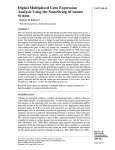
1
Chapter 1
RNA Isolation from Xenopus Inner Ear Sensory Endorgans
for Transcriptional Profiling and Molecular Cloning
Casilda Trujillo-Provencio, TuShun R. Powers, David R. Sultemeier,
and Elba E. Serrano
Abstract
The amphibian Xenopus offers a unique model system for uncovering the genetic basis of auditory
and vestibular function in an organism that is well-suited for experimental manipulation during animal development. However, many procedures for analyzing gene expression in the peripheral auditory
and vestibular systems mandate the ability to isolate intact RNA from inner ear tissue. Methods presented
here facilitate preparation of high quality inner ear RNA from larval and post-metamorphic Xenopus specimens that can be used for a variety of purposes. We demonstrate that RNA isolated with these protocols
is suitable for microarray analysis of inner ear organs, and for cloning of large transcripts, such as those
for ion channels. Genetic sequences cloned with these procedures can be used for transient transfection
of Xenopus kidney cell lines with GFP fusion constructs.
Key words: RNA, auditory, vestibular, Xenopus laevis, Xenopus tropicalis, microarray, cloning,
transcriptional profiling, heterologous gene expression.
1. Introduction
Xenopus laevis (X. laevis) is a widely used and well-established
organism that has contributed to our understanding of embryogenesis and cellular development for well over 50 years (1). In
large part, X. laevis is a popular experimental species because the
stages of X. laevis development have been richly detailed from
the fertilized egg to the mature adult, and because the organism is extremely easy to breed and maintain in the laboratory
(2). However, the relatively long generation time (∼18 months)
and allotetraploid genome have been an impediment to genetic
Bernd Sokolowski (ed.), Auditory and Vestibular Research: Methods and Protocols, vol. 493
C 2009 Humana Press, a part of Springer Science+Business Media
DOI 10.1007/978-1-59745-523-7 1 Springerprotocols.com
3
4
Trujillo-Provencio et al.
studies with this species. In recent years, Xenopus tropicalis
(X. tropicalis) has emerged as another member of the genus Xenopus that is a superior alternative for molecular genetic analysis
and large scale sequencing efforts due to its diploid genome and
shorter (∼5 months) generation time (3, 4).
Using X. laevis and X. tropicalis as our experimental systems,
we aim to understand the developmental mechanisms that give
rise to the uniquely-shaped auditory and vestibular endorgans
of the inner ear with their characteristically patterned epithelia
comprised of specialized mechanosensory hair cells (5–7). We are
especially interested in the specification of the electrical phenotype of hair cells and in determining the cadre of ion channels that
typify endorgans of the inner ear (8–10). Our target for molecular
investigations is the inner ear of both Xenopus species. However,
the diminutive inner ears that reside in the Xenopus otic capsule
provide limited amounts of tissue for RNA isolation. This restriction has posed an additional challenge for the identification of
genes that are expressed in the inner ear (6, 9).
The protocols that we have developed and that are presented
here permit isolation of high quality RNA from larval and postmetamorphic Xenopus inner ears of both species. Furthermore,
methods that are applicable for RNA isolation from the Xenopus
inner ear have been validated as suitable for RNA isolation from
other Xenopus organs such as brain and kidney, as well as Xenopus
cell lines. Since the inner ear originates from a neurogenic placode
(11), many genes expressed in the inner ear are also expressed in
nervous tissue. Inner ears also share common sensitivities with
kidneys to antibiotics and other drugs (12). Therefore, these
RNA isolation protocols can be used to design experiments that
identify genes expressed during development of various Xenopus
organs, and in the response of organs and cell lines to chemical
challenges. The total RNA isolated with our procedures was used
in two downstream applications, transcriptional profiling (13) and
molecular cloning (9). Ion channel genes cloned with these methods can be fused to sequences for a fluorescent reporter molecule
such as green fluorescent protein (GFP) and expressed in Xenopus kidney cell lines using lipid-mediated transient transfection
protocols (14).
2. Materials
2.1. Tissue
Preparation
1. Xenopus laevis and Xenopus tropicalis are obtained from
Nasco (Fort Atkinson, WI). X. laevis larvae are purchased as
a unit of Stage 48–55 tadpoles (cat. no. LM00450MX) and
maintained in aquaria until they reach developmental Stage
55–56. Juvenile animals (X. laevis, cat. no. LM00453MX and
X. tropicalis, cat. no. LM00821MX) are used within a week
RNA Isolation from Xenopus Inner Ear
2.
3.
4.
5.
6.
7.
5
of arrival. All procedures involving animals are approved by
the New Mexico State University Institutional Animal Care
and Use Committee.
Ethyl 3-aminobenzoate methanesulfonate salt: Prepare a
2% (w/v) stock solution in water (see Notes 1 and 2).
Store the stock solution at 4 ◦ C and discard after one
month. Working solutions of 0.2% (larvae) and 0.5% (juvenile animals) are prepared the day of use by diluting
the stock solution with 0.01 M phosphate buffered saline:
1. 5 mM KH2 PO4 , 8. 1 mM Na2 HPO4 , 138 mM NaCl,
2. 7 mM KCl, pH 7.4 (PBS, cat. no. P3813, Sigma-Aldrich,
St. Louis, MO).
Water treated with 0.1% (v/v) diethyl pyrocarbonate (DEPC,
cat. no. D5758, Sigma-Aldrich) (see Notes 1 and 3).
R
R
RNaseZap
and RNAlater
(Ambion, Austin, TX).
Dissecting tools: Dumont #5 forceps, standard straight fine
Iris scissors, scalpel handle #3 (all from Fine Sciences Tools,
Foster City, CA); BD Bard-Parker Sterile Scalpel Blades
#11 (VWR, West Chester, PA); 60 × 15 mm glass Petri dish
(VWR) filled with black wax (Nasco, Fort Atkinson, WI).
Olympus SZ61 dissecting stereomicroscope (Leeds Precision
Instruments, Minneapolis, MN).
Microcentrifuge tubes sterilized by autoclaving, 1.7 mL and
2.0 mL; and 22 gauge needles (VWR).
R
RNeasy
Mini Kit (Qiagen, Valencia, CA).
14. 3 M β-mercaptoethanol.
R
Kimwipes
(VWR).
Brinkmann Polytron PT1200 handheld homogenizer with
sawtooth 0.5 mm generator (VWR).
2.2. Total RNA
Isolation
1.
2.
3.
4.
2.3. Total RNA
Clean-up
1. The DNA-freeTM kit, 5 M ammonium acetate, 5 mg/mL linear acrylamide, and THE RNA Storage Solution (all from
Ambion, Austin, TX).
2. 100% ethanol (Aaper Alcohol, Shelbyville, KY).
3. 70% ethanol is prepared by diluting 100% ethanol with
DEPC-treated water.
4. Refrigerated centrifuge (e.g., Beckman Coulter AllegraTM
21R Refrigerated Centrifuge; VWR).
2.4. Determination of
Total RNA Quality and
Quantity
1. Agilent 2100 Bioanalyzer and Agilent RNA 6000 Nano Kit
(Agilent Technologies, Palo Alto, CA).
2.5. Microarray
Analysis
R
R
1. GeneChip
Xenopus laevis Genome Array and GeneChip
One-Cycle Target Labeling Kit (Affymetrix, Santa Clara,
CA).
6
Trujillo-Provencio et al.
2. gcRMA (Robust multichip averaging) summarization
method downloaded from bioconducter.org and executed in
the R package (http://www.r-project.org/).
R
R
3. Spotfire
DecisionSite
9.0 for Functional Genomics
(Spotfire, Inc., Somerville, MA).
2.6. Molecular
Cloning of Genes
Identified with
Transcriptional
Profiling
1. SMARTTM RACE cDNA Amplification Kit (Clontech Laboratories, Inc., Mountain View, CA).
R
2. PicoMaxx
High Fidelity PCR System (Stratagene, La Jolla,
CA).
3. Thermal cycler (e.g., TC-512, Techne Inc., Burlington, NJ).
4. Lyophilized gene specific primers, 10 nmole (Operon,
Huntsville, AL).
R
5. SeaKem
LE Agarose (Cambrex Bio Science Rockland, Inc.,
Rockland, ME).
6. Tris-acetate/EDTA electrophoresis buffer (TAE 1X): 0.04 M
Tris base, 0.001 M EDTA, 1.0 M glacial acetic acid. A 50X
stock solution is prepared in a glass bottle and stored at room
temperature (22–26 ◦ C).
7. Agarose gel with ethidium bromide: Prepare by diluting 10 mg/mL ethidium bromide stock solution (cat.
no. E1510, Sigma-Aldrich) to a final concentration of
0. 25 μg/mL in melted agarose gel. Do not include ethidium
bromide in the running buffer.
8. 1 kb DNA ladder (New England Biolabs, Inc., Ipswich, MA).
R
9. S.N.A.P.TM UV-Free Gel Purification Kit and TOPO
XL
R
PCR Cloning Kit with One Shot TOP10 ElectrocompTM
E. coli (Invitrogen, Carlsbad, CA).
10. Lambda Bio UV/Vis Spectrometer (Perkin Elmer Corp.,
Norwalk, CT).
11. Electroporator 2510 and electroporation cuvettes (Eppendorf, Westbury, NY).
R
12. BigDye
Terminator v3.1 Cycle Sequencing Kit (Applied
Biosystems, Foster City, CA).
R
13. ABI PRISM
3100 Genetic Analyzer (Applied Biosystems).
2.7. Heterologous
Inner Ear Gene
Expression
1. pAcGFP1-C1 expression vector (Clontech Laboratories, Inc.).
2. XbaI and SpeI restriction enzymes; Antarctic phosphatase; and
Quick LigationTM Kit (all from New England Biolabs).
3. X. laevis A6 kidney cell line (American Type Culture Collection, Manassas, VA).
4. Cell culture medium per 100 mL: 75 mL of NCTC-109 (cat.
no. N1140, Sigma-Aldrich), 10 mL Newborn Calf Serum (cat.
no. N4637, Sigma-Aldrich), 2 mL of 200 mM L-glutamine
(cat. no. G7513, Sigma-Aldrich), and 13 mL of Milli-Q
water. Combine medium components, then filter sterilize
with a 250 mL StericupTM -GP Filter Unit (Millipore Cor-
RNA Isolation from Xenopus Inner Ear
7
poration, Billerica, MA). Passage reagent: Filter sterilized
0.25% Trypsin-EDTA (cat. no. T4049, Sigma-Aldrich). Store
aliquots consisting of 10 mL of Newborn Calf Serum, 2 mL of
L-glutamine, and 10 mL of 0.25% Trypsin-EDTA at −20 ◦ C.
R
5. Labconco
PurifierTM Class II Safety Cabinet and VWR Water
Jacketed CO2 Incubator Model 2310 (VWR).
6. BD Falcon culture slides, two- or four-chamber (cat. no.
53106-302 or 53106-304, VWR).
7. LipofectamineTM 2000 Transfection Reagent (cat. no. 11668027, Invitrogen).
2.8. Epifluorescence
Imaging
1. 3.7% paraformaldehyde is prepared by diluting 10%
paraformaldehyde in PBS. The paraformaldehyde should
be prepared fresh for each experiment.
2. Hoechst 33342, 10 mg/mL.
R
3. SlowFade
Antifade Kit (Invitrogen).
R
4. VWR micro cover glasses No.1, 24 × 50 mm.
5. Clear fingernail polish.
6. CoolSNAPTM HQ CCD camera (Photometrics, Tucson, AZ).
7. Nikon TE2000 epifluorescence microscope equipped with a
UV-2A filter cube (UV excitation) and a B-2E/C filter cube
(Blue excitation) (A.G. Heinze Inc., Lake Forest, CA).
R
8. MetaVueTM Imaging System Version 6.0r5 and MetaMorph
Offline Imaging System Version 6.0r5 (Molecular Devices
Corporation, Sunnyvale, CA).
R
R
9. Adobe
Photoshop
Version 7.0.1 (Adobe Systems Incorporated, San Jose, CA).
3. Methods
Many methods requiring total RNA as a starting material are significantly affected by the quality of the RNA isolated from the
tissue of interest. Furthermore, when tissue is limited, the success of methods that dictate a specific amount of starting RNA is
hindered because the amount of total RNA that can be retrieved
is diminished. Isolation of total RNA from the minuscule inner
ear of Xenopus (5, 7), for molecular pursuits such as transcriptional profiling and molecular cloning, presents such a challenge.
In addition, the total RNA must be of very high quality, with a
demonstrable lack of degradation and the presence of transcripts
greater than 3 kb.
After numerous attempts to isolate a sufficient amount of
high quality total RNA from the inner ear of Xenopus, we were
able to optimize the method by working quickly through tissue
dissections, meticulously cleaning the tools used in the procedure
8
Trujillo-Provencio et al.
R
R
with RNaseZap
, bathing the exposed tissue with RNAlater
,
R
and incorporating the use of the Qiagen RNeasy Mini Kit.
Additionally, the use of the Agilent 2100 Bioanalyzer for
RNA characterization greatly enhanced our capacity to detect
and analyze isolated total RNA and to standardize protocols (see
Fig. 1.2). Previously, the use of denaturing gel electrophoresis
and a spectrophotometer was standard for assessing the quality
and quantity of RNA. This procedure resulted in the loss of μg
amounts of total RNA. In contrast, the Agilent 2100 Bioanalyzer
can detect and assess the quantity of as little as 25 ng/μL total
RNA and the quality of as little as 5 ng/μL total RNA (15).
The Bioanalyzer characterizes the sample with an RNA integrity
number (RIN) that can be used to establish a quantitative
standard for RNA quality for each application. We consider this
type of equipment and quantitative analysis a major contributor
to the success and replicability of our methods, especially for
transcriptional profiling.
High quality RNA enhances the success of RT-PCR Rapid
Amplification of cDNA Ends (RACE) reactions, especially when
long transcripts are sought. With our protocols, we typically
visualize one sharp band of a PCR product when gene specific
R
primers and the PicoMaxx
High Fidelity PCR system are used
R
to amplify a gene of interest (see Fig. 1.3C). The PicoMaxx
High Fidelity PCR system uses a blend of Taq and Pfu DNA polyR
polymerase-enhancing
merases and an exclusive ArchaeMaxx
factor. The system provides high PCR sensitivity and fidelity
which facilitates the detection and amplification of low copy number targets up to 10 kb in size. For example, we routinely obtain
R
target PCR products in the 3–4 kb range (9). Using the TOPO
R
XL PCR Cloning Kit in conjunction with the PicoMaxx High
Fidelity PCR system enabled the cloning of transcripts greater
than 3 kb. It is crucial to confirm that clones are full length and
of the expected identity by sequencing prior to other downstream
applications such as heterologous expression in a cell culture system (see Fig. 1.3D).
We enlisted the transcriptional profiling approach, using
microarray analysis, in order to better understand global gene
expression patterns that underlie X. laevis inner ear function
(see Fig. 1.3A). Using microarray methods, we identified several
R
genes on the GeneChip
X. laevis genome array that are differentially expressed in X. laevis inner ear as compared with brain
(13). For example, we detected a gene which has been implicated in hereditary deafness, the gap junction protein β2 (GJB2)
(16), during analysis of our microarray data. The difference in
GJB2 expression levels in X. laevis inner ear was nearly 4X that of
the brain (see Fig. 1.3B). The relative GJB2 abundances detected
through microarray analysis of inner ear and brain RNA are also
apparent when inner ear and brain RNA are used as template in
RNA Isolation from Xenopus Inner Ear
9
RT-PCR RACE reactions with GJB2 gene specific primers (see
Fig. 1.3C). Thus, RT-PCR RACE reactions replicate the gene’s
transcriptional profile between organs as detected with microarrays. We interpret these findings as an indication of the indispensable contribution of high quality RNA to the consistency between
replicates and the identification of inner ear specific genes.
In summary, we recommend:
• Work quickly and follow established best practices for handling RNA (17)
R
• Clean tools with RNaseZap
• Bathe tissue with RNAlater R during surgery
R
• Use Qiagen RNeasy
Mini Kit
• High quality RNA (RIN>8) is essential; use the Agilent Bioanalyzer or similar equipment to assess RNA quality
• At least 1 μg of high quality RNA (RIN>8) is optimal
• Use total RNA within 72 h for microarray experiments
• 18–20 μg of biotin-labeled cRNA are required before proceeding to the fragmentation and hybridization steps
• Review electropherograms for size distributions of cRNA/
mRNA samples; transcripts greater than 3 kb should be
present in the sample, especially if ion channels and other
membrane transporters and receptors are target genes
R
• Use the PicoMaxx
High Fidelity PCR System or equivalent
R
• Use the TOPO XL PCR Cloning Kit or equivalent
As per standard laboratory procedures, investigators are
reminded that they should review chemical MSDS sheets prior to
use and ensure that reagents are handled and discarded in compliance with all federal and state regulations. Protocols that use
animals in research must be approved by the Institutional Animal
Care and Use Committee prior to initiating experiments. Recommended guidelines for the use of Xenopus in research are available
online (18–20).
3.1. Tissue
Preparation
1. Take precautions to eliminate sources of RNase contamination by following rigorous laboratory procedures for working with RNA. Minimal practices include a clean bench, strict
adherence to the use of gloves throughout the procedure, a
dedicated set of pipettors, and disposable sterile plasticware.
Prior to dissections, clean all dissection tools and the Polytron
R
generator with RNaseZap
and rinse thoroughly with DEPCtreated water (see Note 4).
2. Partially immerse stage 55–56 larvae and juvenile (2.5–3.5 cm,
1.0–3.0 g) X. laevis or juvenile X. tropicalis (1.5–2.5 cm,
0.8–2.0 g) in a solution of ethyl 3-aminobenzoate methanesulfonate salt (0.2% for Stage 55–56 larvae and 0.5% for juveniles)
for 10–30 min at room temperature (22–26 ◦ C). The solution
should cover the torso of the animal but leave the nostrils of
the animal exposed to air.
10
Trujillo-Provencio et al.
3. Work quickly during the dissections to minimize tissue exposure to RNases. Pierce larval hearts and decapitate juvenile animals prior to aseptic removal of inner ear tissue. Also, extract
brain and kidney tissue from juvenile animals.
4. Use a dissecting stereomicroscope (e.g., Olympus SZ61) for
the dissection of tissues.
5. Remove larval inner ears by approaching the tissue from the
dorsal side of the animal (see Fig. 1.1A). Peel the skin away
from the area above the brain and otic capsules and remove
the soft bone of the skull and otic capsules, using a surgical scalpel and fine forceps. Once the tissue is exposed (see
R
Fig. 1.1B), bathe it with RNAlater
to prevent degradation
of RNA. Extract the inner ear by grasping the eighth cranial
nerve and withdrawing the nerve and its target sensory organs
from the otic capsule.
6. Remove juvenile inner ears and brains by approaching the
tissue ventrally from the roof of the mouth after removing
the lower jaw. To facilitate the dissection, pin the top of the
head and the exposed spinal cord to the dissecting dish with
22 gauge needles. Despite the animal size difference, X. tropicalis inner ears are only slightly smaller than those of X. laevis,
and overall the inner ears of the two species are of comparable dimensions (see Fig. 1.1C,D). Remove the skull and otic
capsule bone with a surgical scalpel and fine forceps. Bathe
R
the exposed tissue (see Fig. 1.1D) copiously with RNAlater
.
Then, extract the inner ear tissue as described in the larval
Fig. 1.1. Xenopus inner ear and brain dissections. (A) Dorsal view of X. laevis Stage 56
larva prior to dissection. (B) Dorsal view of exposed X. laevis Stage 56 inner ear (E) and
brain (Br) after the tissue and bone are removed. (C) Ventral view of X. laevis juvenile
upper jaw prior to dissection. (D) Ventral view of exposed X. tropicalis juvenile inner ear
(E) and brain (Br) after tissue and bone are removed. Arrows point to the 8th cranial
nerve (N). Anterior (top), Posterior (bottom). Scale bar = 1 mm.
RNA Isolation from Xenopus Inner Ear
11
preparation. After removal of inner ear tissue, sever the brain
from all nerve branches and carefully remove with fine forceps.
7. The dissected tissue is placed in a 2.0 mL microcentrifuge
R
tube, containing 200–400 μL of RNAlater
, immediately
after removal from the animal.
8. Use tissue immediately for RNA isolation or store at −20 ◦ C
for several months or at −80 ◦ C indefinitely prior to RNA isolation.
3.2. Total RNA
Isolation
3.3. Total RNA
Clean-up
R
1. Use the RNeasy
Mini Kit from Qiagen for total RNA
isolation.
R
, blot quickly
2. Remove the dissected tissue from the RNAlater
R
on a Kimwipe (from a clean box reserved for RNA work),
and immediately place in a pre-weighed 2.0 mL microcentrifuge tube containing 600 μL of Buffer RLT and 6 μL of
14. 3 M β-mercaptoethanol.
3. Determine the tissue weight by subtracting the weight of
the tube plus Buffer RLT and β-mercaptoethanol from the
weight of tube containing the tissue plus Buffer RLT and
β-mercaptoethanol.
4. Extract no more than 30 mg of tissue, as recommended by the
vendor, for each total RNA isolation (see Note 5).
5. Disrupt the tissue using a homogenizer such as a Brinkman
Polytron with a 0.5 mm generator set at the highest setting.
Pulse samples 3–5 times for ∼5–10 s until the tissue is completely homogenized and the solution is uniform in texture
and color. Cool the tube on ice between pulses.
6. After the tissue is uniformly homogenized, follow the
R
RNeasy
Mini Kit protocol as described in the manual.
7. Elute total RNA in two steps, first with 50 μL and then with
40 μL of RNase-free water provided with the kit.
1. Use the DNA-freeTM kit from Ambion to purify total RNA
sample.
2. Treat the total RNA obtained from step 7, Section 3.2 with
DNaseI to degrade any DNA in the sample. Follow the protocol manual with a DNaseI incubation time of 30 min.
3. Following the DNase inactivation step, transfer the total RNA
solution to a clean 1.7 mL microcentrifuge tube and precipitate by adding 0.1X volume of 5 M ammonium acetate,
0.015X volume of 5 mg/mL linear acrylamide, and 2X volume of 100% ethanol.
4. Incubate the total RNA precipitation reaction overnight at
−20 ◦ C.
5. Recover the total RNA pellet by centrifugation at 10,000 g for
20 min at 4 ◦ C. Discard the supernatant, and wash the pellet
with 300 μL of 70% ethanol. Repeat centrifugation for 10 min.
12
Trujillo-Provencio et al.
6. Discard the wash supernatant, and air dry the pellet at room
temperature for 15 min.
7. Resuspend total RNA in 8–15 μL of THE RNA Storage Solution and incubate in a 37 ◦ C water bath for 5 min.
8. Store the sample at −80 ◦ C or place on ice and analyze for
quality and recovery.
3.4. Determination of
Total RNA Quality and
Quantity
1. Dilute the total RNA sample 1:3 in THE RNA Storage Solution and use the Agilent RNA 6000 Nano Kit to prepare the
RNA for the determination of quality and quantity.
2. Follow the vendor’s instructions of the Agilent 2100 Bioanalyzer to generate an electropherogram and an RNA Integrity
Number (RIN) for each RNA sample. An electropherogram
displaying two well-defined peaks of 1.8 kb (18S rRNA) and
4.0 kb (28S rRNA) and a RIN above 8 are indicative of
high quality RNA (see Fig. 1.2B1). For typical yields of total
RNA isolated from Xenopus tissues see Note 6. Figure 1.2
shows representative electropherograms generated by the
2100 expert software.
3.5. Microarray
Analysis
1. Use total RNA, obtained in Section 3.3, step 8, from juvenile and larval X. laevis in microarray experiments within 72 h
of tissue dissection. At least 1 μg (5–10 μL) of high quality
RNA with a RIN above 8 is required for cRNA synthesis (see
Note 7).
2. Prepare biotin-labeled cRNA for hybridization to the
R
GeneChip
Xenopus laevis Genome Array by using the
GeneChip R One-Cycle Target Labeling kit, following
the vendor’s instructions.
3. Determine the amount of biotin-labeled cRNA by analyzing
diluted cRNA (1:10 or 1:20) on the Agilent 2100 Bioanalyzer in order to ensure that 18–20 μg of biotin-labeled cRNA
are available for the fragmentation and hybridization steps (see
Note 8). Review electropherograms, generated by using the
2100 expert software from Agilent, to evaluate the size distributions of the cRNA samples to be used for transcriptional
profiling. For inner ear and brain, expect the sample to contain cRNA that is greater than 3 kb. (see Fig. 1.2C for a cRNA
sample prepared by the BioMicro Center at the Massachusetts
Institute of Technology, Cambridge, MA, using the aforementioned technology).
4. After image acquisition, normalize the raw data using
the gcRMA (Robust multichip averaging) summarization
method. After normalization, analyze all microarray data using
R
R
the Spotfire
DecisionSite
9.0 for Functional Genomics
software. A heat map that graphically represents the levels of
gene expression from one array of X. laevis inner ear tissue is
shown in Fig. 1.3A.
RNA Isolation from Xenopus Inner Ear
13
Fig. 1.2. X. laevis inner ear total RNA and cRNA evaluated with an Aligent 2100 Bioanalyzer. (A1) Electropherogram
of degraded total RNA. This electropherogram is typical of RNA isolated from X. laevis juvenile inner ear tissue prior to
R
Mini Kit. The RNA sample had a low (5.2) RNA Integrity Number (RIN). (A2)
implementation of the Qiagen RNeasy
Gel representation of total RNA shown in A1. (B1) Electropherogram of high quality (non-degraded) total RNA. This
R
electropherogram is typical of RNA extracted from X. laevis Stage 56 larvae inner ear tissue with the Qiagen RNeasy
Mini Kit using the procedures described in Sections 3.2–3.4. The RNA sample had a high (9.0) RNA Integrity Number
(RIN). (B2) Gel representation of total RNA shown in B1. (C) Electropherogram of biotin-labeled cRNA synthesized from
the total RNA shown in B1. The cRNA of inner ear samples typically includes sequences greater than 3000 nt. Axis units:
Abscissa A1 and B1, migration time (seconds, s); Abscissa C, nucleotides (nt); Ordinate, fluorescence units (FU).
14
Trujillo-Provencio et al.
Fig. 1.3. Applications of RNA for transcriptional profiling (A, B), molecular cloning (C), and heterologous gene expression
in cell culture (D). (A) Heat map of inner ear microarray data. X. laevis juvenile inner ear total RNA ( ∼ 1 μg) was
R
R
Xenopus laevis Genome Array. The heat map was constructed using Spotfire
labeled and hybridized to the GeneChip
R
DecisionSite 9.0 software. The data are organized numerically by probe set ID with the lowest ID number at the top.
In this sample, the normalized intensity values (log base 2) for the 15,491 probe sets ranged from 0.8 (low) to 15.22
(high). The intensity color code for expression: L – low, M –intermediate, H – high. (B) Microarray detection of the
expression of the gap junction protein beta 2 (GJB2) in inner ear and brain. The average and standard deviation values in
the histograms were calculated from three different microarray experiments. Low standard deviations characterized the
sample replication. (C) RT-PCR RACE detection of GJB2 transcript in X. laevis juvenile inner ear and brain. Lane 1 – inner
ear, minus RT control, GJB2 primer pair 1; Lane 2 – brain, minus RT control, GJB2 primer pair 1; Lane 3 – PCR reaction
control, minus cDNA template, GJB2 primer pair 1; Lane 4 – inner ear, GJB2 primer pair 1; Lane 5 – brain, GJB2 primer
pair 1; Lane 6 – inner ear, GJB2 primer pair 2; Lane 7 – brain, GJB2 primer pair 2. Lane L − 5 μL of NEB 1 kb DNA
ladder. cDNA is detected in lanes 4–7. Expression levels correlate with the microarray data shown in B. (D) GFP-positive
Xenopus A6 kidney cells transfected with the GFP-BK clone. Dashed lines encircle transfected A6 cells (GFP) and dotted
lines encircle stained nuclei (Hoechst). Scale bar = 50 μm.
RNA Isolation from Xenopus Inner Ear
3.6. Molecular
Cloning of Genes
Identified with
Transcriptional
Profiling
15
1. The expression of genes detected with microarray analysis can
be confirmed with molecular cloning using RT-PCR RACE
protocols.
2. Use X. laevis and X. tropicalis inner ear or brain total RNA,
acquired in Section 3.3, step 8, as a template for RT synthesis of first strand cDNA. Prepare 5 -RACE-Ready first
strand cDNA in separate reactions, according to the instruction manual of the SMARTTM RACE cDNA Amplification
Kit, using 1 μg of control RNA from the kit and 1 μg of the
following experimental samples: X. laevis inner ear or brain
total RNA, X. tropicalis inner ear or brain total RNA.
3. Run control PCR reactions that amplify the first strand
cDNA, prepared from the kit RNA according to the vendor’s
protocols, to evaluate the success of the 5 RACE reactions.
Analyze control PCR products by agarose gel electrophoresis, as described in the kit manual, before proceeding with
the experimental PCR reactions with Xenopus samples. If
the expected products are observed, use the Xenopus first
strand cDNA samples for PCR reactions. If products are not
detected, the first strand synthesis reactions must be repeated
following recommendations outlined in the troubleshooting
section of the user manual.
4. Use the 5 -RACE-Ready inner ear or brain first strand cDNA
samples (5–10 μL in a 50 μL reaction volume) to amplify secR
ond strand cDNA according to the PicoMaxx
High Fidelity
PCR System user manual. The thermal cycler parameters are
as follows: initial denaturation, 95 ◦ C for 2 min; 35 cycles of
95 ◦ C for 40 s, Tm minus 3 ◦ C for 30 s, 72 ◦ C for 3 min; final
extension 72 ◦ C for 10 min. Design gene specific primers to
the gene of interest and obtain from Operon (Huntsville,
AL). The primer Tm’s are specified in the Oligonucleotide
Data Sheet provided by Operon and vary by primer pair.
5. Analyze 7 μL of the PCR reaction on a 1.2% agarose gel
(0. 25 μg/mL ethidium bromide) in 1X TAE buffer.
6. Isolate the resultant PCR fragments using the S.N.A.P.TM
UV-Free Gel Purification Kit, according to the vendor’s
instructions, until the quantification step. For quantification,
analyze 10 μL of the isolated fragment on a Lambda Bio
UV/Vis Spectrometer.
R
7. Clone the purified PCR product using the TOPO
XL PCR
R
Cloning Kit with One Shot TOP10 ElectrocompTM E. coli,
according to the kit manual with a 3:1 molar ratio of insert
to vector. After the ligation step, use the Electroporator
2510 to transform the electrocompetent cells with a setting
of 1400 V.
8. Analyze between 5–10 positive clones by restriction enzyme
digestion to confirm the presence of an insert. Sequence
16
Trujillo-Provencio et al.
9.
10.
11.
12.
13.
14.
R
clones with inserts following the BigDye
Terminator v3.1
Cycle Sequencing Kit protocol and analyze sequencing reacR
tions using the ABI PRISM
3100 Genetic Analyzer.
Molecular cloning, using the RT-PCR RACE procedures
described above, can be used to confirm the differential
expression of genes such as the X. laevis EST, GenBank
Accession no. BJ076720 (National Institute for Basic Biology Mochii normalized Xenopus tailbud library, Clone ID#
XL058il6 (3 )). This gene is detected with Affymetrix (Santa
R
Clara, CA) probe set Xl.8924.1.A1 at using the GeneChip
Xenopus laevis Genome Array. The Affymetrix annotation
states that this probe set targets X. laevis connexin 29 (gap
junction protein β2, GJB2). Microarray analysis suggests that
this gene is expressed at higher levels in the inner ear than in
the brain (see Fig. 1.3B).
Design primers to amplify two regions within the 3,292 bp
Affymetrix consensus sequence for the Xl.8924.1.A1 at
probe set. Use the HMMgene v. 1.1 program (21) to determine the putative coding sequence within the consensus
sequence for the design of primer pair 1. Use the sequence
for the X. laevis EST (GenBank Accession #BJ076720) that
is arrayed on the chip for the design of primer pair 2.
Primer pair 1 amplifies an 804 bp product that includes
the entire GJB2 coding sequence. The forward primer
(5 -AGTCAGCGCACAGAGACCAA-3 ) aligns with the
5 UTR sequence (31 bp upstream of the translational start site) and the reverse primer (5 -AGCTGACC
TGCCACAGTAAC-3 ) aligns 7 bp downstream of the stop
codon.
Primer pair 2 amplifies an 859 bp product within the
EST sequence. The forward primer (5 -CGGTCATCATT
CAGAGTT-3 ) aligns 71 bp upstream of the start of
the EST sequence and the reverse primer (5 -ACACTC
CAGGAAAACAC-3 ) is 24 bp downstream from the end of
the EST sequence.
Prepare the first strand cDNA from X. laevis inner ear and
brain total RNA as described previously. The second strand
PCR reactions follow the steps outlined in Section 3.6, step
4 with the following annealing temperatures (Tm): 60 ◦ C for
primer pair 1 and 53 ◦ C for primer pair 2.
Clone and sequence the resultant PCR products (see
Fig. 1.3C) following the methods described in Section 3.6,
steps 6–7. Confirm that cloned inserts correspond to the
expected GJB2 and EST targets by sequencing. The relative
product intensities on the gel parallel the relative transcript
levels detected by microarray analysis.
RNA Isolation from Xenopus Inner Ear
3.7. Heterologous
Inner Ear Gene
Expression
3.8. Epifluorescence
Imaging
17
R
1. Subclone a TOPO
XL cloned calcium-activated potassium
channel (BK) isoform from X. laevis inner ear tissue (9, 14)
that shares 99% nucleotide identity with a posted Xenopus
spinal cord GenBank sequence (Accession no. AF274053) into
the pAcGFP1-C1 expression vector.
2. Use XbaI and SpeI restriction enzymes to digest 5 μg of the
R
BK TOPO
XL clone, following the vendor’s recommended
reaction conditions. Isolate the insert using the S.N.A.P.TM
UV-Free Gel Purification Kit as described in Section 3.6,
step 6.
3. Prepare the vector, pAcGFP1-C1, by digesting 1 μg of plasmid DNA with XbaI restriction enzyme and then dephosphorylating with Antarctic phosphatase, following the vendor’s
protocol.
4. Use the Quick LigationTM Kit to clone the BK isoform fragment into the GFP expression vector, following the vendor’s
protocol. Confirm positive clones by restriction enzyme analR
ysis and subsequent sequencing reactions using the BigDye
Terminator v3.1 Cycle Sequencing Kit.
5. Culture the X. laevis A6 kidney cell line according to the vendor’s recommendations and maintain in antibiotic-free media
at 26 ◦ C in 5% CO2 .
6. Passage A6 cells at a plating density of 4. 5 × 10⁴ cells/cm2
on two- or four-chamber culture slides 24 h prior to transient
transfections.
7. Transfect cells with a final concentration of 2 μg/mL of Lipofectamine 2000 Transfection Reagent and 1 μg/mL of plasmid
diluted in serum free NCTC-109 medium, following the vendor’s protocol. Transfect cultures with Lipofectamine without
plasmid in control experiments.
1. After 24–48 h, wash cells gently in serum free NCTC-109
medium.
2. Fix cells in 3.7% paraformaldehyde for 10 min at room temperature and wash with PBS (pH 7.4).
3. Expose cells to 1 μg/mL Hoechst 33342 diluted in PBS
(pH 7.4) for 10 min at room temperature followed by a PBS
(pH 7.4) wash.
4. Prepare slides for imaging by adding AntiFade Solution C
R
from the SlowFade
Antifade Kit to the chambers for 15 min
at room temperature. Remove the AntiFade Solution C and
detach the side walls of the culture chambers.
R
5. Add a few drops of AntiFade Solution A from the SlowFade
Antifade Kit to the slide prior to applying a cover-slip. Seal the
cover-slip with clear fingernail polish.
6. Capture images using a CCD camera connected to an epifluorescent microscope (e.g., CoolSNAPTM HQ CCD camera
18
Trujillo-Provencio et al.
connected to a Nikon TE2000 epifluorescence microscope
with UV-2A, Hoechst and B-2E/C, GFP filter cubes) and the
MetaVueTM Imaging System software (see Fig. 1.3D).
7. Process images offline using appropriate software (e.g.,
R
R
MetaMorph
Offline Imaging System software and Adobe
R
Photoshop Version 7.0.1 for figure preparation).
4. Notes
1. Prepare all solutions in Milli-Q water (18 M-cm) with a total
organic content of less than five parts per billion.
2. Solutions of ethyl 3-aminobenzoate methanesulfonate salt
are buffered to pH 7.0 with sodium bicarbonate (cat. no.
S5761, Sigma-Aldrich). Use gloves when preparing and handling solutions of ethyl 3-aminobenzoate methanesulfonate
salt. Prepare the stock solution in a chemical fume hood to
avoid inhalation of dust.
3. WARNING: DEPC is a combustible explosive and a toxic
chemical. It is recommended that users purchase small
amounts of DEPC and use all the DEPC as soon as possible. Take appropriate precautions when handling: e.g., always
wear personal protective equipment and work in an approved
chemical fume hood. Dilute DEPC in water to a final concentration of 0.1%. After adding the DEPC, close the container tightly and vigorously shake. Allow the solution to
sit overnight at room temperature and autoclave for 20 min
before use. The DEPC-treated water can be stored at room
temperature after autoclaving. Solutions requiring DEPCtreated water are so indicated in the text.
4. The Polytron blade can be removed carefully from the outer
shaft of the generator, and both the blade and outer shaft
R
must be thoroughly cleaned. RNaseZap
is copiously applied
and the surfaces are lightly scrubbed with Kimwipes. Rinse the
blade and outer shaft several times with DEPC-treated water,
rinse once with 70% ethanol, and allow to air dry. (Reminder:
70% ethanol should be made with DEPC-treated water).
This cleaning procedure should be repeated between tissue
samples.
5. Typically, the amount of tissue recovered from Xenopus juvenile animals is 1 mg per inner ear and 10 mg per brain. The
amount of tissue recovered from Stage 56 X. laevis is 0.7 mg
per inner ear. Total RNA is isolated by combining tissue from
at least 3 animals.
R
6. The average yields of total RNA using the Qiagen RNeasy
kit are as follows: X. laevis juvenile −1. 8 μg per brain and
0. 3 μg per inner ear; X. laevis Stage 56 − 0. 3 μg per inner
RNA Isolation from Xenopus Inner Ear
19
ear; X. tropicalis juvenile −1. 9 μg per brain and 0. 2 μg per
inner ear. Downstream protocols (i.e. Microarray analysis and
RT-PCR) typically require at least 1 μg of total RNA.
7. The Agilent 2100 Bioanalyzer allows precise optimization of
total RNA isolation procedures (see Fig. 1.2). The Bioanalyzer can detect ng quantities of nucleic acids in 1 μL and also
quantifies RNA integrity with a number (RIN). Therefore,
this instrument is superior to spectrophotometer quantification (260/280 ratios) for evaluating RNA quality.
R
8. The GeneChip
One-Cycle Target Labeling Kit procedure
can successfully label synthesized cRNA from 1–8 μg of total
RNA. However, in one reaction of 12 completed with these
protocols, a smaller amount (0. 8 μg) of X. laevis inner ear
total RNA produced 22 μg of biotin-labeled cRNA.
Acknowledgments
The authors thank Joanna Beeson for technical support with
cell culture experiments, and Alicia Arguelles and Erica Koval
for their assistance with manuscript preparation. We are grateful to Dr. Charlie Whittaker, of the MIT Center for Cancer
Research, and to Manlin Luo and Dr. Rebecca Fry, of the MIT
BioMicro Center, for their technical assistance and generous
advice. We also acknowledge Dr. Peter Sorger and members of
the MIT Cell Decision Processes Center for stimulating intellectual interactions and for facilitating access to essential equipment
and computing resources. Funding for this research was provided by awards from the National Institutes of Health (NIGMSMORE S06GM008136, NIDCD R01DC003292, and NIGMS
P50GM068762).
References
1. Tinsley, R. C. and Kobel, H. R. (ed.) (1996)
Biology of Xenopus. Oxford University Press,
Oxford.
2. Nieuwkoop, P. D. and Faber, J. (ed.) (1967)
Normal Table of Xenopus laevis (Daudin). A
Systematical and Chronological Survey of the
Development from the Fertilized egg Till the
End of Metamorphosis. 2nd Edition. NorthHolland Publishing, Co., Amsterdam.
3. Amaya, E. (2005) Xenomics. Genome Res. 15,
1683–1691.
4. Showell, C. and Conlon, F. L. (2007) Decoding development in Xenopus tropicalis. Genesis
45, 418–426.
5. Diaz, M. E., Varela, A., and Serrano, E. E.
(1995) Quantity, bundle types, and distribution of hair cells in the sacculus of Xenopus laevis during development. Hear. Res. 9,
33–42.
6. Serrano, E. E., Trujillo-Provencio, C., Sultemeier, D., Bullock, W. M., and Quick, Q.
A. (2001) Identification of genes expressed
in the Xenopus inner ear. Cell. Mol. Biol. 47,
1229–1239.
7. Quick, Q. A. and Serrano, E. E. (2005) Inner
ear formation during the early larval development of Xenopus laevis. Dev. Dyn. 234,
791–801.
20
Trujillo-Provencio et al.
8. Varela-Ramirez, A., Trujillo-Provencio, C.,
and Serrano, E. E. (1998) Detection of transcripts for delayed rectifier potassium channels
in the Xenopus laevis inner ear. Hear. Res. 119,
125–134.
9. Sultemeier, D. R., Provencio-Trujillo, C., and
Serrano, E. E. (2007) Cloning and characterization of Xenopus inner ear calcium-activated
potassium channel α- and β-subunits. ARO.
Abstr. 30:740.
10. Gabashvili, I. S., Sokolowski, B. H.,
Morton, C. C., and Giersch, A. B. (2007)
Ion channel gene expression in the inner ear.
J. Assoc. Res. Otolaryngol. 8, 305–328.
11. Schlosser, G. and Northcutt, R. G. (2000)
Development of neurogenic placodes in Xenopus laevis. J. Comp. Neurol. 418, 121–146.
12. Humes, H. D. (1999) Insights into ototoxicity: Analogies to nephrotoxicity. Ann. N. Y.
Acad. Sci. 884, 15–18.
13. Powers,
T.,
Trujillo-Provencio,
C.,
Whittaker, C., and Serrano, E. E. (2007)
Gene expression profiling of Xenopus organs
yields insight into the Xenopus inner ear
transcriptome. ARO. Abstr. 30:741.
14. Sultemeier, D. R., Knight, V. B.,
Manuelito, S. J., Hopkins, M., and Serrano,
15.
16.
17.
18.
19.
20.
21.
E. E. (2005) Heterologous and homologous
expression systems for functional analysis
of Xenopus inner ear genes. ARO. Abstr.
28:28.
Agilent Technologies, Inc. (2003) Reagent Kit
Guide: RNA 6000 Nano Assay. November
2003 Edition, Germany.
Petersen, M. B. and Willems, P. J. (2006)
Non-syndromic, autosomal-recessive deafness.
Clin. Genet. 69, 371–392.
Ambion Inc. (2008) The basics: RNase
control. http://www.ambion.com/techlib/
basics/rnasecontrol/index.html
Nasco. (2008). Animal Protocol for Xenopus
laevis, African Clawed Frog Colony. http://
www.enasco.com/Static.do?page=xen rcare
Grainger Lab. (2001) Grainger Lab X.
tropicalis
Adult
Husbandry
Protocol.
http://faculty.virginia.edu/xtropicalis/
husbandry/TropadultcareNew.htm
University of Arizona: IACUC Learning
Module – Xenopus laevis. (2008) Care
and Handling of Xenopus laevis. http://
www.iacuc.arizona.edu/training/xenopus/.
Center for Biological Sequence Analysis.
(2007) HMMgene v. 1.1 Program. http://
www.cbs.dtu.dk/services/HMMgene/