1
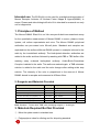
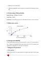
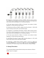
ELISA kit User Manual
Mouse Receptor Activator Of Nuclear Factor Kappa B
Ligand(RANKL)ELISA Kit
Catalog No.: 27853
96T
For Research Use Only
Not for In Vitro Diagnostic Use
1
Intended use: The ELISA Kit is to be used for quantitative determination of
Mouse Receptor Activator Of Nuclear Factor Kappa B Ligand(RANKL) in
serum, Plasma and other biological fluids. Kit is intended for research use only
not for diagnostics.
1. Principles of Method
The Mouse RANKL Elisa Kit is an Vitro enzyme-linked immunosorbent assay
for the quantitative measurement of Mouse RANKL in serum, plasma, tissue
lysates, cell culture supernatants and urine. The Mouse RANKL polyclonal
antibodies are pre-coated onto 96-well plate. Standard and samples are
aspirated into the wells and Mouse RANKL present in a sample is bound to the
wells by the immobilized antibody. The biotinylated detection antibodies are
added to the wells and then followed by washing with PBS or TBS buffer. After
washing away unbound biotinylated antibody, Avidin-Biotin-Peroxidase
Complex is added to the wells. The wells are washed again, a TMB substrate
solution is added to the wells and the color changes after adding acidic stop
solution. The intensity of the color is proportional to the amount of Mouse
RANKL bound in samples and measured at 450nm±10nm.
2. Reagents and Materials Provided
Composition
Quantity
Composition
Quantity
Pre-coated Micro titer plate, 96 wells
1
Sample Diluent Buffer
2 X 15ml
Standards
2
ABC Diluent Buffer
1 X 12ml
Detection Antibody
1 X 120 ul
Antibody Diluent Buffer
1 X 12ml
Avidin-Biotin-Peroxidase Complex(ABC)1:100
1 X 120 ul
Stop solution
1 X 10ml
TMB color developing reagent A
1 X 10ml
TBS Diluent/Wash Buffer(25X)
1 X 20ml
TMB color developing reagent B
1 X 1.5 ml
Instruction manual
1
Note: Standard:Frozen dried, TMB color developing reagent A:Avoid light
3. Materials Required But Not Provided
① Micro titer plate reader in standard size.
② Polypropylene tubes for diluting and for aliquoting standard.
2
③ Distilled or de-ionized water.
④ Calibrated, adjustable precision pipettes, preferably with disposable plastic
tips.
4. Performance Characteristic
Normal Range: 1000pg/ml-15.6pg/ml
Sensitivity: <5pg/ml
Specificity: The ELISA Kit shows no cross reactivity with any of the cytokines.
5. Reference curve
6. Storage and stability
All the components in the kit can be stored up to 1 year at -20℃and 4 weeks at
2-8℃. Please avoid repeated freeze-thaw cycles and do not mix reagents from
different kits unless they have the same lot numbers.
7. Reagents Preparation
① Plate washing
Discard the solution in the plate without touching the side walls. Blot the plate
3
onto paper towels or other absorbent material. Soak each well with at least
0.35 ml PBS or TBS buffer for 1~2 minutes, then discard the rinse solution.
Repeat this process for several times.
② Sample Preparation and Storage
Stored samples to be assayed within 24 hours at 2-8°C. For long-term storage,
aliquot and freeze samples at -20°C. Avoid repeated freeze-thaw cycles.
Cell culture supernatant, tissue lysates or body fluids: Remove
particulates by centrifugation, analyze immediately or aliquot and store at
-20°C.
Serum: Allow the serum to clot in a serum separator tube (about 2 hours or 4℃)
at room temperature. Centrifuge at approximately 1000 X g for 10 min.
Analyze the serum immediately or aliquot and store frozen at -20°C.
Plasma: Collect plasma using heparin, EDTA, citrate as an anticoagulant.
Centrifuge for 15 minutes at 1000 x g within 30 min of collection. Analyze
immediately or aliquot and store frozen at -20°C.
Sample Dilution Guideline
User needs to estimate the concentration of the target protein in the
samples and select proper dilution factor so that the diluted target
protein concentration falls near the middle of the linear regime in the
standard curve.
③ Reagent Preparation and Storage
A. Preparation of the standard: Standard solution should be prepared no more
than 2 hours prior to the experiment. Add 1 ml sample diluent buffer into one
tube,
4
dissolve
the
standard
thoroughly
and
make
times
dilution.
B. Preparation of biotinylated anti-Mouse RANKL antibody working solution:
The solution should be prepared no more than 2 hours prior to the experiment.
Note: please make centrifuging for the vial before preparing.
a. The total volume should be (100ul) or 0.1ml/well x (the number of wells).
(Allowing 0.1-0.2ml more than total volume)
b. Biotinylated anti-Mouse RANKL antibody should be diluted in 1:100(1+99)
with the antibody diluent buffer and mixed thoroughly.
C. Preparation of Avidin-Biotin-Peroxidase Complex (ABC) working solution:
The solution should be prepared no more than 1 hour prior to the experiment.
Note: please make centrifuging for the vial before preparing.
a. The total volume should be: (100µl) or 0.1ml/well x (the number of wells).
(Allowing 0.1-0.2ml more than total volume)
b. Avidin-Biotin-Peroxidase Complex (ABC) should be diluted in 1:100 (1+99)
with the ABC dilution buffer and mixed thoroughly.
D. Preparation of TMB working solution: transfer 9 volumes of TMB color
developing Reagent A in one volume of TMB color developing Reagent B for
30 minutes in 37℃before using to make TMB substrate, mixing thoroughly.
8. Assay Procedure
The user should decide sample dilution fold by crude estimation of Mouse
RANKL amount in samples.
5
① Aliquot 100µl per well of the grades Mouse RANKL standard solutions into
the pre-coated 96-well plate. Add 100ul of the sample diluent buffer into
the control wells. Add 100µl of each properly diluted sample of sera,
plasma, body fluids, tissue lysates or cell culture supernatants to each
empty well.
② Seal the plate with the cover and incubate at 37°C for 90 min.
③ Remove the cover, discard plate content, and blot the plate onto paper
towels or other absorbent material. Do NOT let the wells completely dry at
any time, washing twice with 300µl of wash buffer.
④ Add 100µl of biotinylated anti-Mouse RANKL antibody working solution
into each well and incubate the plate at 37°C for 60 min.
⑤ Wash the plate three times with 0.01M TBS and each time let washing
buffer stay in the wells for 1 min.
⑥ Add 100µl of prepared ABC working solution into each well and incubate
the plate at 37°C for 30 min.
⑦ Wash plate 5 times with 0.01M TBS and each time let washing buffer stay
in the wells for 1-2 min.
⑧ Add 100µl of prepared TMB color developing agent into each well and
incubate plate at 37°C for 10-15 minutes away from light, observe the color
at all times, when shades of blue can be seen in the wells with the
three-four most concentrated Mouse RANKL standard solutions; the other
wells show no obvious color.
⑨ Add 100µl of prepared stop solution into each well to stop the reaction. The
color changes into yellow immediately.
6
⑩ Read the O.D. absorbance at 450nm in a micro plate reader within 30 min
after adding the stop solution or even 620-630nm Secondary wavelength.
⑪ The standard curve can be plotted as the relative O.D.450 of each
standard solution (Y) vs. the respective concentration of the standard
solution (X). The Mouse RANKL concentration of the samples can be
interpolated from the standard curve.
Summary
Prepare reagents, samples and standards.
Add 100µl prepared samples or standards and incubate the plate at 37℃
for 90min, wash plate twice with 300 µl TBS.
Add 100µl biotinylated antibodies and incubate the plate at 37℃ for 60min,
wash plate 3 times with TBS.
Add 100µl ABC working solution and incubate the plate at 37℃ for 30 min,
wash plate five times with TBS.
Add 90µl TMB color developing agent and incubate at 37℃.
Add 100µl stop solution.
Read the O.D. absorbance at 450nm within 30 min.
Calculate the Mouse RANKL concentration in the samples by plotting
graph between Concentrations and corresponding absorbencies of
Standards.
7
8