1
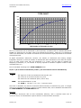
Cellecta CellTracker™ Lentiviral Barcode Library User Manual V3, 6/6/15 CellTracker Lentiviral Barcode Library User Manual www.cellecta.com Table of Contents A. Background 3 B. Required Materials 3 C. Packaging Protocol 4 D. Transduction and Lentiviral Titer Estimation 6 E. Genomic DNA Extraction for Barcode Amplification and HT Sequencing 10 F. Amplification of Barcodes from Genomic DNA 11 G. HT sequencing of Barcodes 13 H. Barcode Enumeration (Conversion of raw HT Seq data to number of reads for each barcode) 13 I. Troubleshooting 14 J. Technical Support 15 K. Safety Guidelines 16 L. Appendix 17 1. Map of CellTracker Lentiviral Barcode Library Vector 17 2. Barcode Library Vector Cassette 17 3. HT Sequencing Primers 18 4. Barcode Library Vector Features 18 M. Terms and Conditions Cellecta, Inc. 19 2 of 19 v3, 6/6/15 CellTracker Lentiviral Barcode Library User Manual www.cellecta.com A. Background The protocols below provide guidelines for packaging the vector library into VSV-g pseudotyped lentiviral particles, transduction of target cells, and the preparation of barcodes derived from transduced cells for high-throughput (HT) sequencing and analysis. Ensure that you have the latest version of this user manual. http://www.cellecta.com/resources/literature/. Please check the Cellecta website at B. Required Materials: The CellTracker Barcode Library is provided as plasmid DNA or as packaged VSV-g pseudotyped viral particles: CellTracker Barcode Library, plasmid (Cat.# BC13X13-30M-P) 200 μg CellTracker Barcode Library, packaged (Cat.# BC13X13-30M-V) >1 × 10⁸ TU Additional Materials Required to Package the Plasmid Library: Ready-to-use Lentiviral Packaging Plasmid Mix (Cat.# CPCP-K2A). Libraries can be packaged into lentiviral particles with nearly any 2nd or 3rd generation HIV-based lentiviral packaging mix. Cellecta’s lentiviral packaging mix contains two plasmids: psPAX2 and pMD2.G, pre-mixed in an appropriate ratio. 293T/17 Cell Line (ATCC, Cat.# CRL-11268™) Dulbecco's Modified Eagle Medium (D-MEM) (1X) (Mediatech CellGro, Cat.# 15-013-CV) - See General Note in Packaging Protocol Glutamine (L-Alanyl-L-Glutamine, Dipeptide L-glutamine) (Mediatech, Cat.# 25-015-CI) Fetal Bovine Serum (recommended: Mediatech, Cat.# MT 35-010-CV) Trypsin-EDTA Lipofectamine™ Reagent (Invitrogen, Cat.# 18324-020) 500 ml, 0.2 μm filter units (Fisher Scientific Cat.# 09-741-05 or Thermo Scientific Cat.# 5690020) D-PBS Tissue Culture Plates and Related Tissue Culture Supplies Plus™ Reagent (Invitrogen, Cat.# 11514-015) Additional Materials Required to Transduce Cells with Packaged Lentiviral Particles: Polybrene® (hexadimethrine bromide) (Sigma-Aldrich, Cat.# 107689) Puromycin Additional Materials Required to Amplify and Sequence Barcodes: 15-ml BD FALCON screw-cap centrifuge tubes (12,000 RCF rated, PP, P:CHCl3-resistant, BD Biosciences, Cat.# 352196) Buffer P1 (50mM Tris-HCl pH 8.0, 10mM EDTA) (QIAGEN, Cat.# 19051) Sonicator for Genomic DNA Shearing DNase I, RNase-free (Epicentre, Cat.# D9905K) dNTP Mix (10 mM each) RNase A (QIAGEN, Cat.# 19101) Phenol:Chloroform pH 8.0 (Sigma-Aldrich, Cat.# P3803) Titanium Taq DNA polymerase with PCR buffer (Clontech-Takara, Cat.# 639242) QIAquick PCR purification kit (QIAGEN, Cat.# 28106) Cellecta, Inc. 3 of 19 v3, 6/6/15 CellTracker Lentiviral Barcode Library User Manual www.cellecta.com QIAquick Gel Extraction Kit (QIAGEN, Cat.# 28706) Primer for sequencing barcodes in barcode constructs (IDT)*: See Appendix, Section I.3 HT sequencing primers (IDT)*: See Appendix, Section I.3 PCR primers for barcode amplification from genomic DNA (IDT)*: See Appendix, Section I.3 HT Sequencing Kits (Illumina): Platform GAIIx HiSeq Kit Type Sequencing Cluster Generation Sequencing Cluster Generation Illumina Cat.# FC-104-5001 GD-203-5001 FC-401-3002 GD-401-3001 Description TruSeq SBS Kit v5 – GA (36-cycle) TruSeq SR Cluster Kit v5 – CS – GA TruSeq SBS Kit v3 – HS (50 cycle) TruSeq SR Cluster Kit v3-cbot-HS Related Products and Services from Cellecta HT Barcode Sequencing of Transduced Cell Pellets from Genetic Screen, Cat.# CANA-SQ C. Packaging Protocol The following protocol describes the generation of packaged lentiviral particles from the plasmid CellTracker Barcode Library. The yield of recombinant lentiviral particles typically produced with this procedure is 1-10 × 106 TU/ml. The protocol is written for packaging with 10 x 15-cm plates to produce at least 3 × 10⁸ TU of total lentiviral particles which can then be optionally concentrated. It is recommended to package at least this much lentivirus at a time to maintain barcode diversity in the library above 10 million. General Note on Packaging ADD FRESH GLUTAMINE (1X) to Dulbecco's Modified Eagle Medium (D-MEM) at the time a sealed bottle of D-MEM is opened, even if the label indicates glutamine has already been added. Glutamine in solution at +4°C has a half-life of 1 – 2 months, so glutamine(+) D-MEM purchased “off-the-shelf” from a supplier is to be regarded as glutamine(-). In our experience, the addition of glutamine increases titer approximately 2-fold. If D-MEM comes supplemented with stable LAlanyl-L-Glutamine dipeptide, addition of fresh glutamine is not necessary. 2-3 Days Prior to Starting Packaging 1. Start growing 293T cells in D-MEM medium plus glutamine (see Required Materials), supplemented with 10% FBS without antibiotics. Day 0 – Plate Cells 2. Twenty four (24) hours prior to transfection, plate 12.5 × 10 6 293T cells in each of ten (10) 15-cm plates (or 150 cm2 flasks). Use 30 ml of media per plate. Disperse the cells and ensure even distribution. At the moment of transfection, the cells should have reached ~80% confluency. Increase or decrease the number of 293T cells seeded if optimal confluency is not achieved in 24 hours. Incubate at 37°C in a CO2 incubator for 24 hours. Day 1 – Transfection (Ten 15-cm plates*) 3. In sterile 50-ml polypropylene tube, mix 600 μl (300 μg) of the Ready-to-use Packaging plasmid mix (see Required Materials for formulation) with 60 μg** of the plasmid library and add the plasmid mixture to 12 ml D-MEM medium without serum or antibiotics. Add 600 μl of Plus Reagent, mix, and incubate at room temperature for 15 min. 10X 15-cm plates 600 μl 60 μl 12,000 μl Cellecta, Inc. Component Ready-to-use Packaging Plasmid Mix (0.5 μg/μl) * Plasmid CellTracker Barcode Library (1 μg/μl) ** D-MEM, no FBS, no antibiotics 4 of 19 v3, 6/6/15 CellTracker Lentiviral Barcode Library User Manual 600 μl 13,260 μl www.cellecta.com Plus Reagent Total volume * IMPORTANT: DO NOT use less than 60 μg (ten 15-cm plates) to package a batch of CellTracker Barcode library. Some barcodes may be lost if the yield is less than 1 × 10⁸ TU. 4. Add 900 μl of Lipofectamine Reagent to 12 ml of D-MEM medium without serum or antibiotics in order to make a convenient master mix. Mix gently. 10X plates 12,000 μl 900 μl 12,900 μl Component D-MEM, no FBS, no antibiotics Lipofectamine Total volume 5. Add the diluted Lipofectamine Reagent (from step 4) to the DNA / Plus Reagent complex (from step 3), mix gently by flicking the tube or vortexing and incubate at room temperature for 15 min. 6. Add 2.5 ml of the DNA / Plus Reagent / Lipofectamine Reagent complex (from step 5) to each 15cm plate from step 2, and mix complexes with medium by gentle rotation. Take care not to dislodge cells from the plate. Incubate at 37°C in the CO2 incubator for 24 hours. Day 2 – DNase I Treatment 7. At 24 hours post-transfection, replace the medium containing complexes with fresh 30 ml D-MEM medium supplemented with 10% FBS, DNase I (1 U/ml), MgCl 2 (5 mM), 20mM HEPES pH7.4. Continue incubation in the CO2 incubator at 37°C overnight. Overnight DNase I treatment before harvesting virus does not negatively affect lentiviral titer or infectivity and helps prevent undesirable carryover of plasmid library into the virus prep. NOTE: Failure to change the media the day after transfection results in large carryover of plasmid (free and/or Lipofectamine-bound) in your lentiviral prep. This may cause problems with most downstream molecular biology applications, especially whenever there is a PCR step involved. Day 3 – Collect Lentiviral Supernatant 8. At 48 hours post-transfection, collect all 30 ml of the virus-containing medium from each plate and filter the supernatant (300 ml) through a Nalgene 0.2 μm PES filter (a low protein binding filter) to remove debris and floating packaging cells. Failure to filter supernatant could result in carry-over of cells into your lentiviral prep. NOTE: Usually, the peak of virus production is Supernatant can also be collected again at 72 hours hour supernatant with 30 ml of fresh D-MEM medium pH7.4 and continue incubation in the CO2 incubator at achieved at 48 hours post-transfection. post-transfection—replace the collected 48supplemented with 10% FBS, 20mM HEPES 37°C for 24 hours. CAUTION: You are working with infectious lentiviral particles at this stage. Please follow the recommended guidelines for working with BSL-2 safety class materials (see Safety Guidelines). 9. Aliquot and store the supernatant at –80°C. lentiviral titer with each cycle. Freezing and thawing usually results in ~20% loss of Cellecta offers lentiviral packaging services. Please contact us at [email protected] or visit our website at http://www.cellecta.com for more information. Cellecta, Inc. 5 of 19 v3, 6/6/15 CellTracker Lentiviral Barcode Library User Manual www.cellecta.com D. Transduction Protocols and Lentiviral Titer Estimation Transduction Lentiviral transductions are performed by mixing cells and virus in culture media supplemented with Polybrene®. For both adherent and suspension cells, transductions are initiated in suspension and carried out overnight. Adherent cells are allowed to adhere to substrate during transduction and are transduced at a cell density that allows for 2-3 population doublings before reaching confluence. Suspension cells are typically transduced at higher density than standard growth density, and then they are diluted to standard growth density 18-24 hours after transduction. Check Toxicity of Polybrene Polybrene is a polycation that neutralizes charge interactions to increase binding between the lentiviral envelope and the plasma membrane. The optimal concentration of Polybrene depends on cell type and may need to be empirically determined. Excessive exposure to Polybrene can be toxic to some cells. Before conducting the titer estimation experiment, we recommended performing a Polybrene toxicity titration in target cells. Grow cells in complete culture medium with a range of Polybrene concentrations (0 μg/ml, 1 μg/ml, 2 μg/ml, 3 μg/ml, 4 μg/ml, 5 μg/ml) for 24 hours, and then replace old medium with Polybrene-free complete culture medium. Grow cells for an additional 72 hours, and then check toxicity by counting viable cells. For your experiments, use the highest concentration of Polybrene that results in less than 10% cell toxicity compared to no Polybrene (typically, 5 μg/ml is recommended). For some cell types, you cannot use Polybrene. Protocol For Titering lentiviral stock (RFP assay) The CellTracker Barcode Library vector expresses the fluorescent protein TagRFP (excitation ~560nm emission ~590nm), allowing lentiviral titer estimation by flow cytometry (RFP assay) or by a combined flow cytometry/puromycin resistance assay (RFP/PuroR assay). To check lentiviral titer, we recommend always using the same cells you will use in the screen. Most of the commonly used mammalian cell lines can be effectively transduced by lentiviral constructs. Relative titers can vary up to 50-fold depending on the chosen cell line. Transduction (HEK293 cells): The following protocol has been optimized for HEK293 cells. For other adherent cell types, parameters such as media, growth surface, time of detection, etc. will have to be adjusted. Day 1 1. Quickly thaw the lentiviral particles in a water bath at 37°C. Transfer the thawed particles to a laminar flow hood, gently mix by rotation, inversion, or gentle vortexing, and keep on ice. CAUTION: Only open the tube containing the lentiviral particles in the laminar flow hood. NOTE: Unused lentiviral stock may be refrozen at –80°C, but it will typically result in a loss of about ~20% in titer. 2. Trypsinize and resuspend HEK293 cells to a density of 1 × 10⁵ cells/ml in D-MEM supplemented with 10% FBS and 5 μg/ml Polybrene. Aliquot 1 ml/well in a 12-well plate and add 0 μl, 3 μl, 10 μl, 33 μl, and 100 μl of lentiviral stock (supernatant filtered to remove cells and cell debris, not concentrated) to six different wells. If concentrated virus is used, scale down virus volumes accordingly. Mix and return cells to CO2 incubator. Grow cells under standard conditions for 24 hours. NOTE: It is important to accurately record the original # of cells at Time of Transduction, as this is critical in titer calculation. For adherent cells other than HEK293, choose a different # of cells at time of transduction, depending on cell size. As a rule of thumb, cells should be transduced at such a density such that they would become confluent in ~48 hours. For example, for HeLa cells, the suggested cell # is 50,000 cells/well in a 12-well plate. Cellecta, Inc. 6 of 19 v3, 6/6/15 CellTracker Lentiviral Barcode Library User Manual www.cellecta.com Day 2 3. At 24 hours post-transduction, replace media with fresh D-MEM supplemented with 10% FBS and without Polybrene. Return cells to CO2 incubator, and grow under standard conditions for additional 48 hours. Avoid confluence: trypsinize and re-plate cells if needed. Day 4 (72 hours after transduction) 4. Detach cells from the plate by trypsin treatment, block trypsin with FBS/media, centrifuge, resuspend in 1X D-PBS, and determine the % of transduced (RFP-positive) cells by flow cytometry. NOTE: Attempting to determine the % of transduced cells by fluorescence microscopy is NOT RECOMMENDED. IMPORTANT: Flow cytometry settings to detect RFP-positive cells are the following: Excitation: 561nm (530nm laser is still acceptable), Emission: 600/20 band-pass filter, or similar (for TagRFP). 5. Proceed to Lentiviral Titer estimation (RFP assay). Alternative Transduction protocol (spinoculation) for hard to transduce cells The following protocol has been optimized for K-562 cells. For other cell types, parameters such as media, growth surface, time of detection, etc. will have to be adjusted. 1. K-562 cells are transduced (“infected”) using spinoculation. This is performed using multi-well tissue culture plates and a tabletop centrifuge capable of 1,200 × g and centrifugation of multiwell plates. 2. Grow K-562 cells and maintain them between 2 × 10⁵ and 1 × 10⁶ cells/ml. become too dense or let the medium become yellow at any point. Do not let them 3. For lentiviral library titration, K-562 cells are resuspended at 2 × 10⁶ cells per ml in RPMI 10%FBS supplemented with 20mM HEPES pH7.4 and Polybrene 5 μg/ml. 0.5-ml aliquots are placed into each well in a 24-well plate (1 × 10⁶ cells/well total). This cell density has proven effective for many suspension cell lines in-house at Cellecta. To each cell-containing well, add increasing amounts of lentiviral stock to be titered. For 100-fold concentrated lentiviral stock, for example, add 0 μl, 0.3 μl 1 μl, 3 μl, and 10 μl virus. Close the plate, mix by gentle agitation, wrap the perimeter with parafilm, and place the plate into centrifuge with an appropriate balance and centrifuge at 1,200 × g at +25°C for 2 hours. 4. Following centrifugation, remove plate(s) from centrifuge, carefully remove parafilm, and place in incubator. After 3 hours, “feed” cells with 0.5 ml additional complete medium per well (no Polybrene). 5. 24 hours after spinoculation, resuspend cells at 2 × 10⁵ cells/ml in RPMI 10% FBS in the appropriate culture vessel and grow for additional 48 hours. 6. 72 hours after spinoculation, perform titer as previously described. NOTE: Use larger vessels for large-scale genetic screen transductions. Scale up all volumes accordingly. Lentiviral Titer estimation (RFP assay) Lentiviral titer is measured as Transduction Units/ml (TU/ml). One TU produces one integration event (i.e., one infection) in target cells. Infections (i.e., transductions) can be calculated from observed % of transduced cells according to the graph below. Cellecta, Inc. 7 of 19 v3, 6/6/15 CellTracker Lentiviral Barcode Library User Manual www.cellecta.com TITER CHART 100 90 80 % infected cells 70 60 50 40 30 20 10 0 0 0.5 1 1.5 2 2.5 MOI (number of TU/number of cells) The % of transduced cells is determined by flow cytometry (excitation=561nm emission=600/20 for TagRFP) by observing the % of RFP+ cells in the transduced cell sample. When the % of transduced cells is at or below 20%, the number of infections can be considered roughly equivalent to the number of transduced cells. At higher transduction efficiencies, however, the fraction of transduced integrations increases, so that the number of infected cells after transduction of TU. In other words, some cells are transduced 2 or 3 times. Use the Multiplicity of Infection (i.e., the number of TU/cell) required to transduce a with MOIs above 0.2. cells bearing multiple is less than the number graph to calculate the specific number of cells Titer is calculated according to the TITER FORMULA below: TU/ml = (# of cells at Transduction) x MOI / (ml of Viral Stock used at Transduction) Example: IF: The original # of cells at Transduction was 100,000, and The volume of virus stock used was 10 μl, and The observed % of transduced (RFP+) cells is 25%, THEN: The calculated MOI is 0.3, and The TITER is: 100,000 × 0.3 / 0.01 = 3,000,000 TU/ml Once titer is estimated, the amount of Lentiviral Stock necessary to transduce any given # of target cells at any transduction efficiency (range of 10-80% infected cells) can be backward calculated from the TITER FORMULA and TITER CHART above. Example: To transduce 20,000,000 cells at 50% transduction efficiency, with a Lentiviral Stock titer of 3,000,000 TU/ml, we calculated the required amount of Lentiviral Stock as follows: Cellecta, Inc. 8 of 19 v3, 6/6/15 CellTracker Lentiviral Barcode Library User Manual www.cellecta.com 1. We calculate the required MOI to achieve 50% transduction efficiency, using the TITER CHART: 50% transduction efficiency = 0.7 MOI 2. We calculate the volume of Lentiviral Stock required using the TITER FORMULA: TU/ml = (# of cells at Transduction) x MOI / (ml of Viral Stock used at Transduction) 3,000,000 = 20,000,000 × 0.7 / (ml Viral Stock) Viral Stock = 20,000,000 × 0.7 / 3,000,000 = 4.67 ml Transduction of Founder Cells By transducing the CellTracker Barcode Library into a large pooled cell population, you can create a founder population in which each cell contains a unique integrated barcode. During transduction, the library of lentiviral constructs carrying each barcode enter the cells and stably integrate into the genomic DNA. Each lentiviral construct also has an RFP marker and puromycin selection to help maintain the barcode cassette. Notes on Transducing Founder Cells Cell transduction is a random process following a statistical distribution. Therefore, if too high an MOI is used, many cells will take up more than one barcode. Cells with more than one barcode show up as more than one population in the final analysis. For example, if two barcodes integrate into one founder cell, and the cell produces 50 progeny, the data (after harvesting the cells and HT sequencing of the genomic DNA) will show two clonal populations with two different barcodes, each having the same number of cells. It will not be obvious that these two populations are from the same founder cell. For this reason, we typically recommend using low MOIs of <0.3 so that >90% of the transduced cells only contain one barcode (see graph above). Transducing larger populations of cells increases the frequency of having more than one founder cell with the same barcode. Since the CellTracker Barcode library has several million unique barcodes, a majority of cells will contain unique barcodes, even with library transductions of a million or more. However, with larger transductions, two or more founder cells can receive the same barcode. To minimize this, we recommend starting screens with less than a million cells if possible. For more details on the complexity and representation of barcodes in the library, and estimates of the number of barcodes repeats you should expect with transductions of different size founder cell populations, please refer to the QC information. Since the purpose of using this complete barcode library is to track the fate of cells from a founder population, we do not recommend splitting and discarding any cells during the course of an experiment: Discarding any portion of cells will eliminate some of the barcodes unless the cell population has expanded to several orders of magnitude over the founder population size. Even if the population has expanded many fold over, splitting or otherwise discarding cells may skew the clonal population sizes for different barcodes (i.e., clone that grow slowly versus cells that grow quickly may be differentially affected). If you must discard cells, it is crucial to design the experiment to minimize the impact on the barcode representation in your samples. Following transduction, you may want to select cells with barcodes using Puromycin. For this, you should calculate a Puromycin Kill Curve using the following procedure: 1. Aliquot cells in a 12-well plate, at such a density so they are at 72 hours from confluency. 2. Add puromycin at 0 μg/ml, 0.5 μg/ml, 1 μg/ml, 2 μg/ml, 5 μg/ml, and 10 μg/ml in six different wells. 3. Mix and return cells to incubator. 4. Grow cells under standard conditions for 42-72 hours. Use the lowest concentration of Puromycin that kills >90% of cells in 42-72 hours. Cellecta, Inc. 9 of 19 v3, 6/6/15 CellTracker Lentiviral Barcode Library User Manual www.cellecta.com E. Genomic DNA Extraction for Barcode Amplification and HT Sequencing Identification of barcodes in the experimental samples requires amplification of the barcode portion of the integrated lentiviral constructs from sample genomic DNA. Subsequent high-throughput sequencing of barcodes by the Illumina GAIIx or HiSeq is done to quantify each barcode and, based on this, determine the relative size of each clonal population derived from a founder cell. Notes on Genomic DNA Extraction and Amplification If the experiment is intended to track the fate all the cells derived from each clone in a founder cell population (e.g., you want to determine the growth rates of all the founder clones): The best approach is to isolate genomic DNA and amplify pooled barcodes from the whole population of progeny cells (e.g. the whole tumor in a xenograft model). If it is not possible to use all the progeny cells, you should start isolation with a minimum of 200-fold the number cells as were in the initial founder population If the experiment is intended to just identify which fraction of cells from a founder cell population that are still present in a population (e.g., survive some sort of selection that eliminates a portion of the initial founder population), you should start isolation with a minimum of 10-fold the number cells as were in the initial founder population. Pooled barcodes should be amplified by two rounds of PCR using Titanium Taq DNA polymerase mix (Clontech-Takara, see Required Materials). The protocol was optimized using an ABI GeneAmp PCR System 9700. Use of other PCR enzymes and/or thermal cyclers may require additional optimization. Recommended Protocol NOTE: Use of disposable tubes is highly recommended in order to avoid contamination. 1. Suspend cell pellet in 5 ml QIAGEN buffer P1 (with RNaseA) in 15 ml POLYPROPYLENE (phenol/chloroform resistant), BD FALCON screw-cap centrifuge tube (12,000 RCF rated, BD Biosciences Cat.# 352196). 2. Add 0.25 ml 10%SDS, mix and incubate 5 minutes at RT. 3. Using an ultrasonic homogenizer (see Required Materials), sonicate to shear DNA into 10-100 kb sized fragments. To prevent cross-contamination, thoroughly wash the ultrasound head with running water and dry-up with clean paper towel between samples. 4. Add 5 ml phenol/chloroform pH8.0 solution, vortex hard and spin down 60 min, +20°C at 8,000 rpm in JA-14 or equivalent rotor (Beckman). 5. You should have about 5 ml of clear upper phase. Transfer 4 ml of upper phase to new 15 ml DISPOSABLE screw cap tube (same as in Step 1). 6. Add 0.5 ml 3M Sodium Acetate, 4 ml isopropanol, mix well, and spin down 30 min, +20°C at 8,000 rpm in JA-14 or equivalent rotor. 7. In order to have a more visible pellet, compacted at the bottom of the tube, it is recommended to incubate overnight at RT before centrifugation. IMPORTANT: If starting material is less than 5 million cells, add carrier before centrifugation (linear polyacrylamide, 25 μg/ml final) and spin down for a longer time (60 min). 8. Discard supernatant, add 10 ml 70% ethanol, spin down 5 min, +20°C at 8,000 rpm in JA-14 or equivalent rotor. 9. Discard supernatant and air-dry pellet. Cellecta, Inc. 10 of 19 v3, 6/6/15 CellTracker Lentiviral Barcode Library User Manual www.cellecta.com 10. Dissolve DNA pellet in appropriate volume of dH2O to a concentration of approximately 2 mg/ml. Expected yield is about 10 μg per 1 million cells. 11. Incubate 30 minutes at +80°C before spectrophotometer reading. F. Amplification of Barcodes from Genomic DNA The lentiviral library and PCR primer designs include sequences complementary to the sequences of the immobilized primers necessary for generating amplification clusters in Illumina’s GAIIx or HiSeq Flow Cells. Our library design is only compatible with Single-Read Flow Cells (in the SingleRead Cluster Generation Kit), because our primers are not complementary to the sequences immobilized on Paired-End flow cells (in the Paired-End Cluster Generation Kit). See Required Materials for the appropriate Illumina catalog numbers. Use 10 ng of plasmid library as an amplification control in the first round of PCR, and use the subsequent PCR products in all remaining steps. The protocol below is for 200 ug of genomic DNA. For whole amount of genomic DNA use proportionally more tubes. First Round of PCR 1. For each sample, prepare 4 × 100 µl reactions containing 200 µg of genomic DNA: ___ 3 3 2 10 ___ 1 100 µl µl µl µl µl µl µl µl 94°C, 94°C, 65°C, 72°C, 68°C, Genomic DNA (50 µg) FwdHTS3 primer* (10 µM) R2 primer* (10 µM) 50X dNTP Mix (10 mM each) 10X Titanium Taq Buffer Deionized water 50X Titanium Taq Total volume 3 minutes 30 seconds 10 seconds 20 seconds 2 min 1 cycle 16 cycles 1 cycle * Please see Appendix for primer sequences. Second Round of PCR The second round of PCR—nested PCR—is required in order to significantly reduce genomic DNA carryover into the samples used for HT sequencing. 1. Combine together the 4 × 100 µl First Round PCR reactions and use a 2 µl aliquot in the second round of analytical PCR with nested primers in each 100 µl reaction: 2 5 5 2 10 75 1 100 µl µl µl µl µl µl µl µl First Round PCR Product Gex1-Bpi primer* (10 µM) NR2 primer* (10 µM) 50X dNTP Mix (10 mM each) 10X Titanium Taq Buffer Deionized water 50X Titanium Taq Total volume 94°C, 3 minutes Cellecta, Inc. 1 cycle 11 of 19 v3, 6/6/15 CellTracker Lentiviral Barcode Library User Manual 94°C, 65°C, 72°C, 68°C, 30 seconds 10 seconds 10 seconds 2 min www.cellecta.com 10, 12, or 14 cycles 1 cycle * Please see Appendix for primer sequences. NOTE: Avoid overcycling of PCR reactions—this will usually result in the generation of a longer fragment that corresponds to a fusion double barcode product. The amplified pooled barcode cassettes are then analyzed on a 3.5% agarose-1XTAE gel (load 5 µl/lane). The results should reveal a bright band at 267 bp for the Gex1-Bpi/NR2 amplicon. The goal of this analytical PCR step is to optimize the starting amount of First Round PCR product and the number of cycles (if necessary) in order to achieve equal intensities of a single 267-bp barcode band across all DNA samples from the genetic screen. Select optimal amount of starting amount and cycle number which produce bright single band without overcycling. Repeat second-round amplification of barcodes from each sample using the optimized volume of First Round PCR product, 2 × 100 µl of Second Round PCR product per sample, and 12-14 cycles of PCR. Set up 2 × 100 µl reactions for each sample containing an adjusted “equal” amount of First Round PCR product (2 µl or more): 2 5 5 2 10 75 1 100 µl µl µl µl µl µl µl µl 94°C, 94°C, 65°C, 72°C, 68°C, First Round PCR Product Gex1-Bpi primer* (10 µM) NR2 primer* (10 µM) 50X dNTP Mix (10 mM each) 10X Titanium Taq Buffer Deionized water 50X Titanium Taq Total volume 3 minutes 30 seconds 10 seconds 10 seconds 2 min 1 cycle 12 or 14 cycles 1 cycle * Please see Appendix for primer sequences. Analyze the PCR products by gel-electrophoresis on a 3.5% agarose-1XTAE gel in order to ensure equal yields of amplified barcodes for all samples. Combine amplified barcodes from the 2 × 100 µl Second Round PCR reactions and purify the samples as follows: 1. Purify the each specific PCR product with the single column from QIAquick PCR purification kit (QIAGEN) following the manufacturer’s protocol, 2. Separate by electrophoresis in a preparative 3.5% agarose-1XTAE gel, 3. Cut out band and extract DNA from the gel using the QIAquick gel purification kit (QIAGEN), and 4. Quantitate using A260 nm measurement using NanoDrop spectrophotometer (or equivalent) and adjust concentration to 10nM (~0.75 ng/µl). G. HT Sequencing of Barcodes HT sequencing of pooled amplified barcodes can be performed on the Illumina GAIIx (~20-30 million reads per sample) or HiSeq (~80-100 million reads per sample) using the GexSeqS sequencing primer and following the manufacturer’s protocol. The final concentration of GexSeqS primer in the reaction should be 500 nM. For the cluster generation step, use 20 fmoles (2 µl of 10 nM PCR product) of the gel-purified band from the 2nd round of PCR. The number of cycles (read length) required is 44. Cellecta, Inc. 12 of 19 v3, 6/6/15 CellTracker Lentiviral Barcode Library User Manual www.cellecta.com The Barcode library and PCR primer designs include sequences complementary to the sequences of the immobilized primers necessary for generating amplification clusters in Illumina’s GAIIx or HiSeq flow cells. Our design is only compatible with Single-Read Flow Cells (in the Single-Read Cluster Generation Kit), because our primers are not complementary to the sequences immobilized on Paired-End flow cells (in the Paired-End Cluster Generation Kit). See Required Materials for a list of recommended Illumina kits for HT Sequencing of Barcode Library samples. HT sequencing of samples on the Illumina MiSeq is not supported. Please contact us at [email protected] for information on our HT sequencing and data analysis services. * Please see Appendix for HT sequencing primer sequences. H. Barcode Enumeration (Conversion of raw HT sequencing data to number of reads for each barcode) For help with Barcode Enumeration, please contact Cellecta Technical Support at [email protected]. I. Troubleshooting Low Lentiviral Titer (<106 TU/ml in supernatant) 1. Poor transfection efficiency (48 hour post-transfection, less than 80% of 293T cells are very brightly fluorescent) Problem: 293T Cells have too high or too low density Solution: Plate fewer or more cells in order to have about 80% confluency at time of transfection. Problem: Plasmid DNA/Lipofectamine/Plus Reagent ratios are incorrect Solution: Optimize the ratios using the guidelines provided in the Lipofectamine protocol. 2. Inefficient production of the virus Problem: 293T Cells are of poor quality Solutions: Optimize growth conditions, check growth medium, and don’t grow 293T cells for more than 20 passages. Check for mycoplasma contamination. Do not overgrow the cells (do not allow the cells to reach more than 90% confluency in order to keep the culture continuously in logarithmic growth phase). Problem: Lentiviral supernatant harvested too early or too late Solution: Harvest supernatant 48 hours and 72 hours after transfection. Problem: 293T cell media is too acidic at time of virus harvesting Solution: Make sure to replace media 24 hours before harvesting, and make sure to supplement media with HEPES pH 7.4 20mM final. 3. Inefficient transduction of titering cells See below. Inefficient Transduction of Packaged Library Cellecta, Inc. 13 of 19 v3, 6/6/15 CellTracker Lentiviral Barcode Library User Manual www.cellecta.com 1. Poor transduction efficiency Problem: Target cells have too high or too low density Solution: Plate fewer or more cells in order to have 20-50% confluency at transduction stage. Problem: Target cell line may be difficult to transduce Solutions: Use a higher concentration of lentiviral particles. Perform “Spinoculation” to improve transduction efficiency. Check to see if Polybrene was added at 5 µg/ml. Problem: Wrong amount of Polybrene added during transduction stage Solution: If Polybrene is toxic to the target cells, optimize Polybrene concentration in the range of 0 – 5 µg/ml by performing a toxicity titration as described in Transduction Protocols and Lentiviral Titer Estimation. Problem: Loss of lentiviral titer during storage Solution: Ensure storage of aliquoted packaged library at –80°C. Each freeze-thaw cycle typically causes reduction of the titer by ~20%. Use a fresh stock for transduction. Problem: The RFP assay is performed too early Solution: Normally, the maximal expression of RFP from the integrated provirus is expected to develop by 72 hours after transduction. However, some cells exhibit delayed expression. Try the assay at a later time, such as 96 hours. Problem: The RFP assay is performed with the wrong flow cytometry settings. Solution: RFP+ cells are to be detected using a 561nm laser for excitation (530nm still acceptable) and 600/20 band-pass filters (or similar) for detection (for TagRFP). Using blue laser (488nm) for excitation leads to gross underestimation of viral titer. Problem: In the RFP assay, the % of transduced cells is determined by fluorescence microscopy instead of flow cytometry. Solution: Use flow cytometry. 2. Transduction affects target cell viability Problem: Polybrene is toxic for target cells Solution: Optimize the concentration and exposure time to Polybrene during the transduction step. For some sensitive cells, Polybrene should not be used. Problem: Virus-containing conditioned media is toxic to target cells. Solution: Concentrate and resuspend the virus in target cell growth media, PBS 10% FBS, or PBS 1% BSA. Difficulties with Probe Preparation and HT Sequencing 1. No PCR Product Problem: Incorrect primers or bad reagents used, or missing reagents. Solutions: Include 10 ng of plasmid library DNA as a positive control. If it produces the correct amplification product, the problem lies with the genomic DNA or previous PCR prep. If not, confirm use of the correct primers and reagents. Verify that primer sequences are correct. Please see Appendix, Section I.3. 2. No barcodes present in HT Sequencing results Cellecta, Inc. 14 of 19 v3, 6/6/15 CellTracker Lentiviral Barcode Library User Manual www.cellecta.com Problem: Incorrect primer used in Illumina-Solexa Cluster Generation step. Solution: Ensure that you or the HT Sequencing core facility uses the GexSeq Sequencing primer (see Appendix, Section I.3), NOT the Sequencing primer that comes with the Illumina Cluster Generation Kit. Problem: Incorrect Cluster Generation kit used. Solution: Ensure that you or the HT Sequencing core facility uses the proper Single-Read Cluster Generation Kit (see Required Materials). J. Technical Support For additional information or technical assistance, please contact us by phone or email: Phone: Toll-Free: Fax: +1 (650) 938-3910 +1 (877) 938-3910 +1 (650) 938-3911 E-mail: Technical Support: General Information: Sales: Orders: Blog: [email protected] [email protected] [email protected] [email protected] http://www.cellecta.com/blog/ Postal Mail: Cellecta, Inc. 320 Logue Ave. Mountain View, CA 94043 USA For more information about Cellecta’s products and services, please visit our web site at http://www.cellecta.com. Cellecta, Inc. 15 of 19 v3, 6/6/15 CellTracker Lentiviral Barcode Library User Manual www.cellecta.com K. Safety Guidelines The HIV-based lentivector system is designed to maximize its biosafety features, which include: A deletion in the enhancer of the U3 region of 3’ΔLTR ensures self-inactivation of the lentiviral construct after transduction and integration into genomic DNA of the target cells. The RSV promoter upstream of 5’LTR in the lentivector allows efficient Tat-independent production of lentiviral RNA, reducing the number of genes from HIV-1 that are used in this system. Number of lentiviral genes necessary for packaging, replication and transduction is reduced to three (gag, pol, rev). The corresponding proteins are expressed from different plasmids lacking packaging signals and share no significant homology to any of the expression lentivectors, pVSV-G expression vector, or any other vector to prevent generation of recombinant replication-competent virus. None of the HIV-1 genes (gag, pol, rev) will be present in the packaged lentiviral genome, as they are expressed from packaging plasmids lacking packaging signal—therefore, the lentiviral particles generated are replication-incompetent. Lentiviral particles will carry only a copy of your expression construct. Despite the above safety features, use of HIV-based vectors falls within NIH Biosafety Level 2 criteria due to the potential biohazard risk of possible recombination with endogenous lentiviral sequences to form self-replicating virus or the possibility of insertional mutagenesis. For a description of laboratory biosafety level criteria, consult the Centers for Disease Control Office of Health and Safety Web site at: http://www.cdc.gov/od/ohs/biosfty/bmbl4/bmbl4s3.htm It is also important to check with the health and safety guidelines at your institution regarding the use of lentiviruses and follow standard microbiological practices, which include: Wear gloves and lab coat at all times when conducting the procedure. Always work with lentiviral particles in a Class II laminar flow hood. All procedures are performed carefully to minimize the creation of splashes or aerosols. Work surfaces are decontaminated at least once a day and after any spill of viable material. All cultures, stocks, and other regulated wastes are decontaminated before disposal by an approved decontamination method such as autoclaving. Materials to be decontaminated outside of the immediate laboratory area are to be placed in a durable, leakproof, properly marked (biohazard, infectious waste) container and sealed for transportation from the laboratory. Cellecta, Inc. 16 of 19 v3, 6/6/15 CellTracker Lentiviral Barcode Library User Manual www.cellecta.com L. Appendix 1. Map of CellTracker Lentiviral Barcode Library Vector * All Cellecta lentiviral vectors are covered by a lentiviral expression system license owned by Life Technologies Corporation (LTC). See Terms and Conditions. 2. Barcode Library Vector Cassette CellTracker Library in pRSI16 vector: - size of Gex1-Bpi/NR2 amplicon is 267 bp. ClaI-U6-FwdU6-1>FwdU6-2>EcoRI>FwdHTS3>Gex1-Bpi>BarCode18TTCG BarCode18<GexSeqS<XhoI<cPPT<Gex2-NR2<R2<RevUbiC1 FwdHTS3 TCGGATTCAAGCAAAAGACGGCATA Gex1-Bpi Bpi TCAAGCAGAAGACGGCATACGAAGACA BC18 BC18 XhoI U6-ACCGGAGTCTTCTTTTTTGAATTCAAGCAAAAGACGGCATACGAAGACAGTTCG-NNNNNNNNNNNNNNNNNTTCGNNNNNNNNNNNNNNNNN-TTCGGACTGTAGAACTCTGAACCTCGAGCAA U6-TGGCCTCAGAAGAAAAAACTTAAGTTCGTTTTCTGCCGTATGCTTCTGTCAAGC-NNNNNNNNNNNNNNNNNAAGCNNNNNNNNNNNNNNNNN-AAGCCTGACATCTTGAGACTTGGAGCTCGTT AAGCCTGACATCTTGAGACTTGGAGA cPPT GexSeqS TTTAAAAGAAAAGGGGGGATTGGGGGGTACAGTGCAGGGGAAAGAATAGTAGACATAATAGCAACAGACATACAAACTAAAGAATTACAAAAACAAATTACAAAAATTCAAAATT AAATTTTCTTTTCCCCCCTAACCCCCCATGTCACGTCCCCTTTCTTATCATCTGTATTATCGTTGTCTGTATGTTTGATTTCTTAATGTTTTTGTTTAATGTTTTTAAGTTTTAA EcoRI AgeI BstBI BspEI UbiC promoter TCTGCGTTGTTGTCGGTGCTCGTTCTCTGCTCTTCACGCTACTGAATTCATCACCGGTTCTTCGAAGGCCTCCGCGCCGGGTTTTGGCGCCTCCCGCGGGCGCCCCCCTCCTCACGGCG AGACGCAACAACAGCCACGAGCAAGAGACGAGAAGTGCGATGACTTAAGTAGTGGCCAAGAAGCTTCCGGAGGCGCGGCCCAAAACCGCGGAGGGCGCCCGCGGGGGGAGGAGTGCCGC AGACGCAACAACAGCCACGAGAGCCACCAGCGGCATAGTAA AGGAGTGCCGCTCGCGACGGA Gex2-NR2 AAGAGACGAGAAGTGCGATGA RevUbiC1 R2 Cellecta, Inc. 17 of 19 v3, 6/6/15 CellTracker Lentiviral Barcode Library User Manual www.cellecta.com 3. HT Sequencing Primers Primer Name FwdHTS3 R2 Gex1-Bpi Gex2-NR2 GexSeqS FwdU6-1 FwdU6-2 Used for 1st Round 1st Round 2nd Round 2nd Round HT Sequencing Standard sequencing Standard sequencing Sequence (IDT preferred) 5’-TCGGATTCAAGCAAAAGACGGCATA-3’ 5’-AGTAGCGTGAAGAGCAGAGAA-3’ 5’-TCAAGCAGAAGACGGCATACGAAGACA-3’ 5’-AATGATACGGCGACCACCGAGAGCACCGACAACAACGCAGA-3’ 5’-AGAGGTTCAGAGTTCTACAGTCCGAA-3’ (HPLC Purified) 5’-CAAGGCTGTTAGAGAGATAATTGGAA-3’ 5’-CCTAGTACAAAATACGTGACGTAGAA-3’ 4. Barcode Library Vector Features Feature Rous Sarcoma Virus (RSV) enhancer/promoter HIV-1 truncated 5′ LTR HIV-1 psi (ψ) packaging signal HIV-1 Rev response element (RRE) U6 promoter cPPT UbiC promoter TagRFP 2A (T2A) PuroR WPRE ∆U3/HIV-1 truncated 3′ LTR SV40 polyadenylation signal SV40 Ori AmpR pUC ori Cellecta, Inc. Function Allows Tat-independent production of viral mRNA (Dull et al., 1998). Source Rous sarcoma virus Permits viral packaging and reverse transcription of the viral mRNA (Luciw, 1996). HIV-1 Allows viral packaging (Luciw, 1996). HIV-1 Permits Rev-dependent nuclear export of unspliced viral mRNA (Kjems et al., 1991; Malim et al., 1989). Human U6 promoter drives RNA Polymerase III transcription for generation of shRNA transcripts. Central polypurine tract, cPPT, improves transduction efficiency by facilitating nuclear import of the vector's preintegration complex in the transduced cells. Ubiquitin C promoter drives expression of TagRFP and PuroR. TagRFP fluorescent protein (Evrogen) serves as an indicator of successful transduction. Thosea asigna virus 2A translational cleavage site containing 18 amino acid residues. Cleavage occurs via a co-translational ribosome skipping mechanism between the C-terminal glycine and proline residues, leaving 17 residues attached to the end of TagRFP and 1 residue to the start of the puromycin resistance marker. Puromycin-resistant marker for selection of the transduced cells. Woodchuck hepatitis virus posttranscriptional regulatory element— enhances the stability of viral transcripts. 3' Self-inactivating long terminal repeat. Allows viral packaging but self-inactivates the 5′ LTR for biosafety purposes (Dull et al., 1998). The element also contains a polyadenylation signal for transcription termination and polyadenylation of mRNA in transduced cells. Required for viral reverse transcription; selfinactivating 3' LTR with deletion in U3 region prevents formation of replication-competent viral particles after integration into genomic DNA. Allows transcription termination and polyadenylation of mRNA. Allows for episomal replication of plasmid in eukaryotic cells. Ampicillin resistance gene (β-lactamase) for selection of plasmid in bacterial cells. pUC bacterial origin of replication. 18 of 19 HIV-1 Human HIV-1 Human sea anemone Entacmaea quadricolor Thosea asigna virus Streptomyces alboniger Woodchuck hepatitis virus HIV-1 SV40 SV40 bacterium Salmonella paratyphi pUC v3, 6/6/15 CellTracker Lentiviral Barcode Library User Manual www.cellecta.com M. Terms and Conditions Cellecta, Inc. Limited License Cellecta grants the end user (the “Recipient”) of the CellTracker™ Pooled Lentiviral Barcode Library (the “Product”) a non-transferable, non-exclusive license to use the reagents for internal research use only as described in the enclosed protocols; in particular, research use only excludes and without limitation, resale, repackaging, or use for the making or selling of any commercial product or service without the written approval of Cellecta, Inc. -separate licenses are available for non-research use or applications. The Product is not to be used for human diagnostics or included/used in any drug intended for human use. Care and attention should be exercised in handling the Product by following appropriate research laboratory practices. Cellecta’s liability is expressly limited to replacement of Product or a refund limited to the actual purchase price. Cellecta’s liability does not extend to any damages arising from use or improper use of the Product, or losses associated with the use of additional materials or reagents. This limited warranty is the sole and exclusive warranty. Cellecta does not provide any other warranties of any kind, expressed or implied, including the merchantability or fitness of the Product for a particular purpose. Use of the Product for any use other than described expressly herein may be covered by patents or subject to rights other than those mentioned. Cellecta disclaims any and all responsibility for injury or damage that may be caused by the failure of the Recipient or any other person to use the Product in accordance with the terms and conditions outlined herein. The Recipient may refuse these licenses by returning the enclosed Product unused. By keeping or using the enclosed Product, you agree to be bound by the terms of these licenses. The laws of the State of California shall govern the interpretation and enforcement of the terms of these Licenses. Limited Use Label Licenses The Recipient acknowledges that the Product has been developed by Cellecta based on licenses from Third Parties and agrees with the Terms of Limited Use for the Recipient provided by the Third Parties: Life Technologies Corporation End-User Label License for the use of Lentiviral Expression System: “This product or service (based upon the Lentiviral Expression System) is sublicensed from Life Technologies Corporation under U.S. Patent Nos. 5,686,279; 5,834,256; 5,858,740; 5,994,136; 6,013,516; 6,051,427; 6,165,782; 6,218,187; 6,428,953; 6,924,144; 7,083,981 and 7,250,299 and corresponding patents and applications in other countries for internal research purposes only. Use of this technology for gene therapy applications or bioprocessing other than for nonhuman research use requires a license from GBP IP, LLC. Please contact GBP IP, LLC 537 Steamboat Road, Suite 200, Greenwich, CT 06830. Use of this technology to make or sell products or offer services for consideration in the research market requires a license from Life Technologies Corporation, 5791 Van Allen Way, Carlsbad, CA 92008.” Evrogen IP JSC End-User Label License for the use of lentiviral expression constructs comprising TagRFP-encoded gene: “This product is for internal non-commercial research use only. No rights are conveyed to modify or clone the gene encoding fluorescent protein contained in this product. The right to use this product specifically excludes the right to validate or screen compounds. For information on commercial licensing, contact Evrogen Licensing Department, email: [email protected]”. © 2015 Cellecta, Inc. All Rights Reserved. Trademarks CELLECTA is a registered trademark of Cellecta, Inc. CellTracker is a trademark of Cellecta, Inc. CRL-11268 is a trademark of ATCC. Invitrogen, Lipofectamine, and Plus Reagent are trademarks of Life Technologies and Invitrogen Corporation. Cellecta, Inc. 19 of 19 v3, 6/6/15