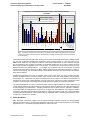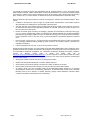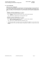
1
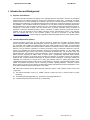
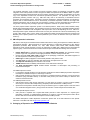
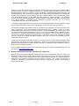
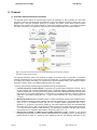
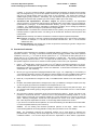
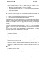
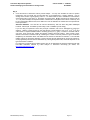
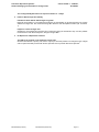
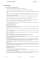
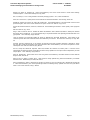
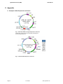
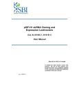
Lentivector Expression Systems:
Guide to Packaging and Transduction of
Target Cells
Cat. #s LV100A-1, LV201B-1,
LV500/510A-1, LV600-606A-1,
LV601B-1, LV900A-1
User Manual
See PAC or kit label for
storage temperature
(ver. 080707)
A limited-use label license covers this
product. By use of this product, you
accept the terms and conditions outlined
in the Licensing and Warranty Statement
contained in this user manual.
Lentivector Expression Systems:
Guide to Packaging and Transduction of Target Cells
Cat. #s LV100A-1 – LV601B-1,
& LV900A-1
Contents
I.
Introduction and Background
A.
B.
C.
D.
E.
F.
G.
H.
Purpose of this Manual
Lentiviral Expression Systems
SBI’s Expression Lentivectors
Packaging of Expression Constructs into Pseudoviral Particles
Delivery of Packaged Lentivector Constructs into Target Cells
Safety Guidelines
List of Components
Additional Required Materials
2
2
3
3
4
5
7
8
II. Protocol
A. Procedure Outline and General Comments
B. Pseudovirus Production
C. Pseudoviral Titer Estimation
D. Transduction of the Packaged Lentivector Expression Construct
9
10
11
13
III. Troubleshooting
A. Low Viral Titer (<105 ifu/ml)
B. Inefficient Transduction of Packaged Constructs
IV. References
15
15
17
V. Appendix
A.
B.
C.
D.
E.
F.
G.
Examples of SBI’s Expression Lentivectors
Functional Maps of Packaging Plasmids for FIV-based System
Functional Maps of Packaging Plasmids for HIV-based System
Percentage of Transduced Cells with Increasing MOI
Properties of copGFP Fluorescent Protein
Related Products
Technical Support
VI. Licensing and Warranty Statement
888-266-5066 (Toll Free)
650-968-2200 (outside US)
19
20
21
21
26
27
27
28
Page 1
System Biosciences (SBI)
User Manual
I. Introduction and Background
A. Purpose of this Manual
This manual provides information describing how to package lentivector expression constructs in pseudoviral
particles and use of packaged expression constructs for transduction of target cells. Specifically, it provides
critical instructions on how to package an FIV-based or HIV-based Lentivector Expression construct in VSV-G
pseudotyped viral particles by co-transfecting 293TN Producer Cells with a Lentivector Expression construct and
the pPACKF1™ (for FIV-based constructs) or pPACKH1™ (for HIV-based constructs) Packaging Plasmid Mix.
Recommendations are also provided for selection and use of FIV-based and HIV-based lentivector systems for
transducing a wide range of target cells. This manual does not include information about construction of
expression constructs in lentiviral expression vectors. Information on making constructs using these vectors is
available in the user manuals for each of SBI’s Lentivector Cloning and Expression Vectors. User manuals,
which are provided with each of the Lentivector products, can also be accessed on the SBI website
(http://www.systembio.com). Before using the reagents and material supplied with this product, please read the
entire user manual.
B. Lentiviral Expression Systems
Lentiviral expression vectors are the most effective vehicles for transducing and stably expressing different
effector molecules (siRNA, cDNA, DNA fragments, antisense, ribozymes, etc.) or reporter constructs in almost
any mammalian cell—including non-dividing cells and whole model organisms (Cann, 2000). As with standard
plasmid vectors, it is possible to introduce lentiviral constructs in plasmid form into the cells with low-to-medium
efficiency and get transient expression of effectors (reporters) using conventional transfection protocols.
However, by packaging the lentiviral expression construct into pseudoviral particles, you can obtain highly
efficient transduction (up to 100%)—even with the most difficult to transfect cells, such as primary, stem, and
differentiated cells. The expression construct transduced in cells is integrated into genomic DNA and provides
stable, long-term expression of siRNA, cDNA or reporter gene. Endogenously expressed siRNA effectors
provide long-term silencing of the target gene and allow the researcher to generate cell lines and transgenic
organisms with a stable knockdown phenotype for functional studies. Expression of full-length cDNAs from
integrated viral constructs is a unique tool to study gain-of-function effect for cellular phenotypes. Stably
integrated transcriptional reporter constructs are a novel approach to the study of transcriptional regulation in the
natural chromosomal environment and the monitoring of specific signaling pathways. Moreover, lentiviral
delivery does not produce the non-specific cell responses typically associated with chemical transfections or use
of an adenoviral delivery system (Gould, 2003, Cann, 2000).
SBI offers both FIV-based and HIV-based lentiviral expression systems. Both systems consist of three main
components:
(1) The lentiviral expression vector (e.g., shRNA construct in pSIF1-H1-Puro™ or cDNA construct in pCDF
lentivector)
(2) The lentiviral packaging plasmids (e.g., pPACKH1™ Packaging Plasmid mix )
(3) A pseudoviral particle producer cell line (e.g., 293TN cells)
Page 2
ver. 080707
www.systembio.com
Lentivector Expression Systems:
Guide to Packaging and Transduction of Target Cells
Cat. #s LV100A-1 – LV601B-1,
& LV900A-1
The lentiviral expression vector contains the genetic elements required for packaging, transduction, stable
integration of the viral expression construct into genomic DNA, and expression of the siRNA, cDNA, or reporter.
The lentiviral packaging plasmids provide all of the proteins essential for transcription and packaging of an RNA
copy of the expression construct into recombinant pseudoviral particles. For production of a high titer of
pseudoviral particles, producer cells (e.g., HEK 293 cells) need to be transiently co-transfected with the
expression and packaging vectors. Expression constructs packaged in pseudoviral particles are secreted by
producer cells in culture media and can be used directly to transduce expression constructs into target cells.
Following transduction into the target cells, this expression construct is reverse transcribed and integrated into
the genome of the target cell, providing a high level of expression of effector or reporter molecules.
The most popular lentiviral expression system is HIV based (Federico, 2003; Heiser, 2004; Machida, 2003).
Despite improved biosafety features, third generation HIV cloning vectors still pose a potential biohazard risk due
to the possible recombination with endogenous viral sequences to form a self-replicating HIV virus. SBI’s FIVbased Expression system addresses these issues since they are derived from a feline immunodeficiency virus
(Curran, 2002; Sauter, 2001; Loewen, 2003). Both of SBI’s HIV-based and FIV-based lentivector systems meet
Biosafety Level 2 (BSL-2) based on the criteria published by the Centers for Disease Control (for details see
section F).
C. SBI’s Expression Lentivectors
SBI offers a wide range of FIV-based and HIV-based lentivectors for cloning and expression of siRNA, cDNA and
transcriptional reporters. SBI’s lentivectors are a third generation of lentivectors developed for gene therapy
applications (Poeschla, 2003; Sodroski, J.G., 1997, 1999; Federico, 2003; Heiser, 2004; Machida, 2003).
Appendix A shows several examples of vectors for expression of siRNA (pSI), cDNA (pCD) and transcriptional
reporters (pTR). These lentivectors have similar functional maps and include the following common features:
•
•
•
•
•
•
•
Hybrid CMV-5’LTR (for FIV-based vectors) and hybrid RSV-5’LTR promoter (for HIV-based vectors)
provide a high level of expression of full-length pseudoviral constructs in 293 producer cells.
Genetic elements (cPPT, GAG, LTRs) necessary for the packaging, transduction, and stable integration of
the viral expression construct into genomic DNA.
SV40 origin for stable propagation of the lentiviral plasmid in 293 producer cells.
The pUC origin for high copy replication and maintenance of the plasmid in E.coli cells.
The ampicillin resistance gene for selection in E.coli cells.
WPRE element enhances stability and translation of the CMV-driven transcripts.
The SV40 polyadenylation signal enables efficient termination of transcription and processing of
recombinant transcripts.
For siRNA Expression vectors:
•
H1 expression cassette located in 3’ΔLTR provides constitutive and efficient RNA polymerase III-dependent
transcription of shRNA transcript in a wide range of cell lines.
•
CMV promoter promotes a high level of expression of copGFP (fluorescent reporter), puromycin-N-acetyl
transferase (drug selectable marker) or truncated H2Kk protein (cell surface marker) for detection and
selection of transduced cells.
For cDNA Expression vectors:
•
•
CMV promoter drives a high level of expression, in a wide variety of cell lines, of a gene of interest (cDNA)
which may be cloned into the multiple cloning site (MCS) located downstream of CMV promoter.
Optional second expression cassette with puromycin resistance gene or copGFP reporter under the control
of a constitutive elongation factor 1 (EF1) promoter for selection or FACS analysis of transduced cells.
For Transcriptional Reporter vectors:
•
CMV promoter is replaced with a hybrid TRE-mCMV promoter to drive expression of a reporter gene
(dscopGFP, luciferase or β-galactosidase) depending on presence of specific transcriptional factor which
binds to TRE element and activates transcription from the mCMV promoter.
For detailed descriptions of SBI’s Expression Lentivectors, please refer to the User Manual for each specific
lentivector or visit SBI’s web site at http://www.systembio.com.
D. Packaging of Expression Constructs into Pseudoviral Particles
888-266-5066 (Toll Free)
650-968-2200 (outside US)
Page 3
System Biosciences (SBI)
User Manual
Currently, the most efficient technology for producing a high titer of infectious, replication-incompetent lentiviral
particles is based on transient, coordinated expression of a lentiviral expression construct and all necessary
packaging proteins delivered into producer cells by simultaneous transfection with lentiviral expression and
packaging plasmids. When expressed in packaging cells, the highly-efficient hybrid CMV/5’LTR (or RSV/5’LTR)
promoter of the expression construct produces large numbers of the expression construct transcript that contains
all of the functional elements (i.e., Psi, RRE, and cPPT) required for efficient packaging. The expression
construct transcript is efficiently packaged into VSV-G pseudotyped viral particles with helper proteins produced
by the pPACK plasmids. Pseudoviral particles generated by producer cells within 48 – 72 hr can be
concentrated, frozen, and used in later experiments.
To facilitate packaging, SBI offers a 293TN producer cell line that was optimized for effective production of a high
titer of pseudoviral particles by introduction of the SV40 large T antigen and neomycin gene product.
The pPACKF1 Packaging Plasmid Mix consists of a combination of two plasmids—pFIV-34N and pVSV-G (see
maps in Appendix B). The pFIV-34N plasmid contains the structural (gag), regulatory (vif, gp4, rev, nef) and
replication (pol) genes which code for the proteins required to produce the lentivirus. The viral env gene, which
encodes the envelope protein that defines the tropism (i.e., the range of infectable cells), is deleted in the pFIV34N construct. The pVSV-G plasmid expresses the envelope glycoprotein of vesicular stomatitis virus (VSV-G)
from the CMV promoter, thus replacing lentiviral env gene. Viral particles, VSV-G protein pseudotyped, mediate
viral entry through lipid binding and plasma membrane fusion and can infect both mammalian and nonmammalian cells (Burns, 1993).
The pPACKH1 Packaging Plasmid Mix consists of an optimized mixture of three plasmids–pPACKH1-GAG,
pPACKH1-REV and pVSV-G (see maps in Appendix C). Similar to the FIV-based Packaging Plasmid Mix, the
pPACKH1 plasmids provide all necessary structural, replication, and integration proteins and helper functions to
produce VSV-G pseudotyped particles.
For more detailed descriptions and sequence information regarding our cloning and packaging lentivectors, visit
our web site at http://www.systembio.com.
E. Delivery of Packaged Lentivector Constructs into Target Cells
Pantropic VSV-G pseudotyped viral particles containing the RNA copy of the lentivector expression construct can
be efficiently used to deliver and stably express effector and reporter sequences in a wide range of mammalian
target cells. In order to provide guidelines for the use of lentivector delivery systems, we compared transduction
efficiencies of FIV-based and HIV-based vectors in different cell types. Fig. 1 shows a comparison of
transduction efficiencies of FIV-based and HIV-based lentivector systems for 27 different cell lines, including
primary and stem cells.
Page 4
ver. 080707
www.systembio.com
Lentivector Expression Systems:
Guide to Packaging and Transduction of Target Cells
Cat. #s LV100A-1 – LV601B-1,
& LV900A-1
Comparison of Transduction Efficiencies of FIV vs. HIV in different cell lines
at low MOI
12.8 6.9
1.4
1.3
FIV-based pSIF1-copGFP
HIV-based pSIH1-copGFP
1.2
1.1
1.0
ratio to H1299
0.9
0.8
0.7
0.6
0.5
0.4
0.3
0.2
0.1
HUVEC
(3 passage)
bone marrow,
mesenchymal
adipose tissue,
mesencymal
mouse Lin- ckit+
bone marrow
PBMC
CHO
cat
AML-5
CrFK
C6
rat
RAT-1
P388
NIH3T3
NB41
mouse
hamster
human
P19
THP-1
RAW 264.7
MOLT-4
K562
HL60
OVCAR-3
MCF-7
HepG2
BT-474
HeLa S3
293-T-BM
H1299
UMUC-3
0.0
primary/stem
Fig. 1. Comparison of transduction efficiencies for FIV-based and HIV-based packaged positive transduction
controls for 27 different cell lines based on FACS analysis percentage of GFP-positive cells at low Multiplicity
of Infection (MOI).
These data clearly demonstrate that unlike commonly used cancer cell lines (like H1299, HeLa, HeK295, HepG2,
etc.) that can be effectively transduced by lentivectors, some cell types (mouse Lin- ckit+ bone marrow, P19,
PBMC, HL60, P388) are more resistant to infection. More efficient transduction of more “resistant” cell types
may be possible by using a higher concentration of pseudoviral particles per cell in order to achieve the same
MOI, as demonstrated in Appendix C, but not in all cases. It is important to mention that FIV-based and HIVbased lentivectors have different tropism. For example, the FIV-based system is more effective at infecting
several of the tested mouse cell lines (P19, NB41, NIH3T3, P38) and some of the blood cells (MOLT-4, K562, Tcells from AML patient). The HIV-based system is more effective at infecting stem and primary cells (HUVEC,
bone marrow, adipose).
Pseudotyped lentiviruses have been successfully used to infect many other cell types, including neuronal,
dendritic, endothelial, retinal, pancreatic, hepatic, aortic smooth muscle cells, airway epithelia, skin fibroblasts,
macrophages, etc. Lentivectors have been successfully used also for direct in vivo delivery and expression of
transgenes in muscle, brain, airway epithelium, liver, pancreas, retina, and skin. For a more complete list of cells
or tissues, which have been successfully transduced with lentivectors, please see the Reference Section.
The pseudoviral particles can infect (or transduce) target cells and express effector or reporter molecules but
cannot replicate within target cells because the viral structural genes are absent and the LTRs are designed to
be self-inactivating upon transduction. Following transduction into the target cells, the expression cassette is
reverse transcribed and integrated into the genome of the target cell. After integration, the expression cassette
continuously and stably produces high levels of effector or reporter molecules in target cells. Target cells stably
expressing the effector molecule can be isolated using the selectable marker contained in the expression vector
construct (e.g., puromycin or copGFP).
F. Safety Guidelines
SBI’s Expression lentivectors together with the pPACK packaging plasmids comprise the third-generation
lentiviral expression system. The HIV-based lentivectors are based on the vectors developed for gene therapy
applications by Dr. J. G. Sodroski (US patent #5,665,577 and # 5,981,276).
888-266-5066 (Toll Free)
650-968-2200 (outside US)
Page 5
System Biosciences (SBI)
User Manual
The original FIV expression system was developed by Eric M. Poeschla, David J. Looney, and Flossie WongStaal at UCSD (Poeschla, 1998; Poeschla 2003). The feline immunodeficiency virus (FIV) was originally isolated
from cat blood. Despite common close exposure of humans to FIV through contact with domestic cats (including
bites, scratches, etc.), no human infection or disease has ever been associated with FIV (Poeschla, 2003).
Both FIV-based and HIV-based lentivector systems are designed to maximize their biosafety features, which
include:
•
A deletion in the enhancer of the U3 region of 3’ΔLTR ensures self-inactivation of the lentiviral construct
after transduction and integration into genomic DNA of the target cells.
•
The CMV promoter (in FIV-based vectors) and RSV promoter (in HIV-based vectors) upstream of 5’LTR in
the lentivector allow efficient Tat-independent production of viral RNA, reducing the number of genes from
HIV-1 that are used in this system.
•
Number of lentiviral genes necessary for packaging, replication and transduction is reduced to three (gag,
pol, rev), and the corresponding proteins are expressed from different plasmids (for HIV-based packaging
plasmids) lacking packaging signals and share no significant homology to any of the expression lentivectors,
pVSV-G expression vector, or any other vector, to prevent generation of recombinant replication-competent
virus.
•
None of the HIV-1 genes (gag, pol, rev) will be present in the packaged viral genome, as they are expressed
from packaging plasmids lacking packaging signal—therefore, the lentiviral particles generated are
replication-incompetent.
•
Pseudoviral particles will carry only a copy of your expression construct.
Despite the above safety features, use of HIV-based vectors falls within NIH Biosafety Level 2 criteria due to the
potential biohazard risk of possible recombination with endogenous viral sequences to form self-replicating virus,
or the possibility of insertional mutagenesis. For a description of laboratory biosafety level criteria, consult the
Centers
for
Disease
Control
Office
of
Health
and
Safety
Web
site
at
http://www.cdc.gov/od/ohs/biosfty/bmbl4/bmbl4s3.htm. It is also important to check with the health and safety
guidelines at your institution regarding the use of lentiviruses and always follow standard microbiological
practices, which include:
•
Wear gloves and lab coat all the time when conducting the procedure.
•
Always work with pseudoviral particles in a Class II laminar flow hood.
•
All procedures are performed carefully to minimize the creation of splashes or aerosols.
•
Work surfaces are decontaminated at least once a day and after any spill of viable material.
•
All cultures, stocks, and other regulated wastes are decontaminated before disposal by an approved
decontamination method such as autoclaving. Materials to be decontaminated outside of the immediate
laboratory area are to be placed in a durable, leakproof, properly marked (biohazard, infectious waste)
container and sealed for transportation from the laboratory.
Page 6
ver. 080707
www.systembio.com
Lentivector Expression Systems:
Guide to Packaging and Transduction of Target Cells
Cat. #s LV100A-1 – LV601B-1,
& LV900A-1
G. List of Components
pPACK Lentivector Packaging Kits
The pPACK Packaging Plasmid Mix is an optimized mixture of the packaging plasmids in an amount sufficient for
2
10 co-transfections with a lentivector expression construct in 10-cm tissue culture plates (or, alternatively, 75 cm
flasks). The positive control expression construct with copGFP reporter and shRNA sequence targeting Firefly
luciferase is provided in an amount sufficient for 6 control co-transfections with the pPACK packaging plasmid
mix.
pPACKF1™ Lentivector Packaging Kit (Cat. # LV100A-1)
for packaging FIV-based lentivector expression constructs
200 μl
20 μl
pPACKF1 Packaging Plasmid Mix: Mixture of pFIV-34N and pVSV-G plasmids
(0.5 μg/μl)
pSIF1-H1-siLuc-copGFP Positive Control Plasmid (0.5 μg/μl)
pPACKH1™ Lentivector Packaging Kit (Cat. # LV500A-1)
for packaging HIV-based lentivector expression constructs
200 μl
20 μl
•
pPACKH1 Packaging Plasmid Mix: Mixture of pPACKH1-GAG, pPACKH1REV, and pVSV-G plasmids (0.5 μg/μl)
pSIH1-H1-siLuc-copGFP Positive Control Plasmid (0.5 μg/μl)
The pPACK Plasmid Mixes and copGFP Positive Control Plasmid are shipped on dry ice or blue ice and
should be stored at -20°C upon receipt. Properly stored plasmids are stable for 12 months from the date
received.
888-266-5066 (Toll Free)
650-968-2200 (outside US)
Page 7
System Biosciences (SBI)
User Manual
Packaged Positive Transduction Controls
Packaged VSV-G pseudotyped Positive Transduction Controls are used to estimate and optimize transduction
efficiency of lentivector expression constructs and packaged GeneNet siRNA Libraries. The packaged positive
controls with copGFP reporter are provided in an amount sufficient to infect ≥2 × 10⁵ cells at an MOI of 1. The
constructs contain an shRNA targeting Firefly Luciferase. Proceed with Section II.D for the transduction
control protocol.
•
Page 8
The Packaged Controls are shipped on dry ice and should be immediately stored at -70°C upon receipt.
Avoid thawing and refreezing of pseudoviral particles! Each freeze-thaw cycle causes reduction of the
titer by 20-30%. Properly stored pseudoviral particles are stable for 6 months from the date received.
ver. 080707
www.systembio.com
Lentivector Expression Systems:
Guide to Packaging and Transduction of Target Cells
Cat. #s LV100A-1 – LV601B-1,
& LV900A-1
293TN Producer Cell Line (Cat. # LV900A-1)
The 293TN Human Kidney cell line is optimized for effective production of a high titer of pseudoviral particles and
stably expresses the SV40 large T antigen and neomycin gene products.
0.5 – 2 ml
•
293TN Producer Cell Line, ≥2 × 10⁶ cells (may vary by lot)
The 293TN Cell Line is shipped on dry ice and should be stored at -80°C or liquid nitrogen (gas phase)
upon receipt. Properly stored 293TN cells are stable for 1-12 months from the date received, depending on
storage conditions.
H. Additional Required Materials
SBI products:
•
•
•
293TN Human Kidney Producer Cell Line (SBI, Cat. # LV900A-1; or ATCC, 293T/17, Cat # CRL-11268)
Peg-It Virus concentration solution (SBI, Cat. # LV810A-1)
Lentivector Expression Construct (see http://www.systembio.com for a full list)
Expression constructs should be purified with a QIAGEN Endotoxin-free Plasmid Purification Kit. The
following kit combinations can be used for Midi scale preparation of endotoxin-free DNA:
¾ QIAfilter Plasmid Midi Kit, Cat. # 12243, and EndoFree Plasmid Maxi Kit, Cat. # 12362
¾ QIAfilter Plasmid Midi Kit, Cat. # 12243, and EndoFree Plasmid Buffer Set, Cat. # 19048
Please visit the QIAGEN website to download the specialized protocol that is not contained in the user
manual:
¾
http://www1.qiagen.com/literature/protocols/pdf/QP15.pdf
Additional reagents:
•
•
•
•
•
•
•
•
•
•
•
•
Dulbecco’s Modified Eagle’s Medium (D-MEM)
(high glucose with sodium pyruvate and L-glutamine; Invitrogen, Cat. # 11995073)
Fetal Bovine Serum (Invitrogen, Cat. # 16000036)
Puromycin (Sigma, Cat. # P8833)
Penicillin/Streptomycin (Invitrogen, Cat. # 15070063)
Trypsin-EDTA (Sigma, Cat. # T3924)
®
Polybrene (hexadimethrine bromide; Sigma, Cat. # H9268)
Millex-HV 0.45 μm PVDF filters (Millipore, Cat. # SLHVR25LS)
Tissue Culture Plates and Related Tissue Culture Supplies
Lipofectamine™ Reagent (Invitrogen, Cat. # 18324-111)
Plus™ Reagent (Invitrogen, Cat. # 11514-015)
Sterile TE Buffer (10 mM Tris pH 8.0, 0.1 mM EDTA pH 8.0)
H1299 human lung carcinoma cell line (ATCC, Cat. # CRL-5803)
888-266-5066 (Toll Free)
650-968-2200 (outside US)
Page 9
System Biosciences (SBI)
User Manual
II. Protocol
A. Procedure Outline and General Comments
The flowchart below outlines the general steps required for packaging of both FIV-based and HIV-based
expression constructs and transduction and expression of the viral expression construct in target cells. In the
diagram below, using the HIV-based system as an example, the positive control pSIH1-H1-siLuc-copGFP
plasmid, which expresses the copGFP reporter, is used as the expression construct. To construct a lentiviral
expression construct for your experiment, refer to the user manual provided with each specific lentivector.
Fig. 2. Procedure for transient production of pseudoviral particles and transduction of effector
expression construct into target cells.
The lentiviral expression system was designed to simplify all necessary steps in production of pseudoviral
particles and transduction of an expression construct into target cells. For general information and background
on working with lentiviral technology, we recommend the General Reviews listed in the Reference Section,
particularly, Federico, 2003, Cann (2000) and Buchschacher et al. (2000).
To ensure optimal results, follow these general guidelines during your experiments:
•
Lentiviral expression construct quality: To generate your specific lentiviral expression construct, refer to
the protocol in the user manual provided with the vector. Transfection efficiency significantly depends on the
quality of plasmid DNA. We recommend purifying plasmid DNA with a QIAGEN Endotoxin-free Plasmid Kit
(see Section I.F) or by CsCl centrifugation. You will need 2 μg of lentiviral expression construct in sterile TE
buffer with a concentration ranging from 0.2 – 2 μg/μl for each transfection in a 10-cm culture plate (or 75
cm2 flask).
•
Maintaining 293TN cell line: The 293TN cell line is a highly transfectable derivative of the HEK293 cell line
with constitutive expression of SV40 T-antigen and neomycin resistance gene. The 293TN cells should be
grown at 37°C in a humidified chamber with 5% CO2 in D-MEM medium supplemented with 4 mM Lglutamine, 1.5 g/l glucose, 100 units/ml penicillin G, 100 μg/ml streptomycin (90%), and heat-inactivated
fetal bovine serum (10%). With a doubling time of less than 24 hours, the 293TN cells should be split every
1 – 2 days when they reach 70 – 80% confluency. For subculturing, detach the cells with 0.25% trypsin,
0.03% EDTA at 37°C, add fresh culture medium, and split at a ratio of 1:3 – 1:5.
•
pSIF1-H1-siLuc-copGFP or pSIH1-H1-siLuc-copGFP Expression Vector: Included in the pPACK
Packaging Kit as a positive control to optimize and troubleshoot your packaging protocol. Specifically, this
plasmid should be used as a reference when quantifying viral titer and assaying expression of your
Page 10
ver. 080707
www.systembio.com
Lentivector Expression Systems:
Guide to Packaging and Transduction of Target Cells
Cat. #s LV100A-1 – LV601B-1,
& LV900A-1
construct. If you use a Lentivector without a copGFP reporter and expression of copGFP will not interfere
with your biological assay, you can mix the copGFP construct with your expression construct at a 1:100 ratio
and use it as internal positive control at every stage of your experiment. The vectors also express an
shRNA targeting Firefly Luciferase. Alternatively, you may determine titer by RT-PCR using one of SBI’s
UltraRapid Lentiviral Titer Kits (LV960A-1 [for human cells]; LV960B-1 [for mouse cells]).
•
Transfection with Lipofectamine™ and Plus™ reagent: The protocol provided in the experimental
section is based on our experience with lentivectors and 293TN cells, and is similar to the one provided for
Lipofectamine™ (Invitrogen). To maximize transfection efficiency, you may need to optimize transfection
conditions. For a detailed protocol, optimization of transfection conditions, and for scaling-up pseudoviral
production, please refer to Invitrogen’s Lipofectamine™ Reagent Manual.
•
Pseudoviral titer: The relative titer is measured based on flow cytometry or by fluorescent microscopy and
is always specific to a particular cell line. For titering, we use H1299 cells.. Below are some key terms used
in the protocol:
ifu/ml (infectious units/ml)—the relative concentration of infection-competent pseudoviral particles;
MOI (multiplicity of infection)—the ratio of infectious pseudoviral particles (ifu) to the number of cells being
6
5
infected. For example, if 1 × 10 cells are to be infected at an MOI of 0.1, then 1 × 10 ifu should be
added to the cells;
Transduction Efficiency—the average copy number of expression constructs per genome of target cell in
the infected (transduced) population.
B. Pseudovirus Production
The following protocol describes the generation of pseudoviral particles containing a copy of your lentivector
expression construct. The yield of recombinant lentiviral particles typically produced under these optimized
6
6
conditions is 10 ml of 1 – 3 × 10 ifu/ml for the FIV system and 2 – 5 × 10 ifu/ml for the HIV system per 10-cm
2
culture plate (or 75 cm flask) when measured by transduction of H1299 cells. This amount of pseudoviral
6
particles is generally sufficient to infect 5 – 10 × 10 target cells at an MOI equal to 1. We recommend including
the copGFP expression construct as an external or internal positive control in all of your experiments.
1.
6
2
Plate 3 × 10 293TN cells in a fresh 10-cm plate (or 75 cm flask) the day before transfection in 9 ml of DMEM medium, supplemented with glutamine and heat-inactivated serum (10% FBS) without antibiotics.
Disperse the cells by thorough pipetting to ensure even distribution. At the moment of transfection, the cells
should reach 50 – 70% confluency.
2.
Mix 20 μl (10 μg) of the pPACK Packaging Plasmid Mix with 2 μg (2 – 25 μl depending on concentration) of
your lentivector expression construct, then dilute the plasmid mixture into 400 μl D-MEM medium without
serum and antibiotics. Add 20 μl of Plus™ Reagent, mix, and incubate at room temperature for 15 min.
3.
Dilute 30 μl of Lipofectamine™ Reagent into 400 μl of D-MEM medium without serum and antibiotics. Mix
gently.
4.
Dropwise, add diluted Lipofectamine™ Reagent (from step 3) to DNA/Plus™ Reagent complex (from step
2), then mix gently by inversion and incubate at room temperature for 15 min.
5.
Add the DNA/ Plus™ Reagent/ Lipofectamine™ Reagent complex (from step 4) to the plate from step 1, and
mix complexes with medium gently by tilting the plates a few times and incubate at 37°C in a CO2 incubator
for 48 hours.
6.
We recommend collecting supernatants at 48 hours post-transfection (Step 5). Usually, the peak of virus
production is achieved at 48 hours. Collect all 10 ml of the pseudovirus-containing medium in a 15-ml
sterile, capped conical tube, and centrifuge at 3000 rpm at room temperature for 5 minutes to pellet cell
debris. Following centrifugation, transfer the viral supernatant into a fresh 15-ml sterile, capped conical
tube. Then aliquot the supernatant into sterile 1.5-ml microfuge tubes and store them at -80°C.
If you choose to concentrate the viral particles, we recommend following the User Manual of the SBI’s PEGit™ Virus Precipitation Solution (Cat. # LV810A-1).
Caution: You are working with infectious pseudoviral particles at this stage. Please follow the
recommended guidelines for working with BSL-2 safety class (see Section I.G for more details).
Note: The supernatant containing the pseudoviral particles can be used directly to determine the
pseudoviral titer, and directly used to infect target cells in vitro as long as the target cells can survive in
conditioned medium. You can also aliquot and store the supernatant at –80°C without cryoprotectant.
888-266-5066 (Toll Free)
650-968-2200 (outside US)
Page 11
System Biosciences (SBI)
User Manual
Freezing and thawing may result in 20 – 30% loss of viral titer with each cycle. Alternatively, to obtain a
higher titer, you may concentrate pseudovirus by the any of the following suggested methods:
•
SBI’s PEG-it™ Virus Precipitation Solution (Cat. # LV810A-1) (recommended)
•
Centrifuge the supernatant at 50,000 x g for 90 min at +4°C, then resuspend the pseudoviral-containing
precipitate in a smaller volume of D-MEM
•
Sucrose cushion centrifugation.
C. Pseudoviral Titer Estimation
We recommend that you estimate the titer of the pseudovirus-containing supernatant before proceeding with
transduction experiments for the following reasons:
•
To ensure that pseudoviral stock is viable
•
To determine the percentage of target cells which can be transduced with pseudoviral stock
•
To control the number of copies of integrated viral constructs per target cell
The protocol for measuring of relative titers assumes that the positive control expression plasmid—the control
copGFP plasmid—was mixed with your expression construct as an internal control at a ratio of 1:100 and is
packaged into pseudoviral particles. In an alternative approach, the copGFP control plasmid can be packaged
separately but in parallel with your construct, as an external control. In this scenario, the control plasmid would
be used to check and optimize the transfection/packaging steps (see Section B). If you are not using the
copGFP plasmid as an internal control, refer to the Alternative Methods note immediately following the titering
procedure.
To determine the relative pseudoviral titer, you will need to transduce the packaged lentivector expression
construct into the H1299 cell line in the presence of Polybrene (4 – 8 μg/ml) for 3 – 8 hours, allow 72 hours for
expression to start, and then count the number of cells expressing copGFP by fluorescent microscopy or by
FACS.
To check your titer, you can also choose a cell line appropriate for your experimental system. The lentiviral
constructs should effectively transduce most of the commonly used mammalian cell lines (see Section E).
Relative titers may vary up to 10 to 20-fold depending on the chosen cell line.
Day 1.
1.
5
For each viral stock, plate H1299 cells in a 24-well plate at a density of 0.6 – 1 × 10 cells per well 24 hours
prior to viral infection. Add 1 ml of complete D-MEM medium (with serum and antibiotics) and incubate cells
at 37°C with 5% CO2 overnight.
Day 2.
2.
Prepare complete D-MEM medium plus 10% FBS with Polybrene to a final concentration of 5 μg/ml.
Prepare an extra well as a control.
®
Note: Polybrene is a polycation that neutralizes charge interactions to increase binding between the
pseudoviral capsid and the cellular membrane. The optimal concentration of Polybrene depends on cell type
and may need to be empirically determined (usually in the range of 2 – 10 μg/ml). Excessive exposure to
Polybrene (>12 hr) can be toxic to some cells.
3.
Remove culture medium and replace with 0.5 ml of complete D-MEM medium with 10% serum and
Polybrene (from Step 2). For each pseudoviral stock, use three wells. Infect H1299 cells by adding 1 μl of
viral stock into the first well (dilution factor of 500), 10 μl of viral stock into the second well (dilution factor of
50), and 100 μl of viral stock into the third well (dilution factor of 5). For one additional well (mock well
control) add 0.5 ml of D-MEM medium with Polybrene (from Step 2). Incubate cells at 37°C with 5% CO2
overnight.
Day 3.
4.
Remove culture medium and replace with 1 ml of complete D-MEM medium (without Polybrene). Incubate
the cells at 37°C with 5% CO2 overnight.
Day 4.
5.
Page 12
Split the cells 1:3 to 1:5 if necessary, depending on the type of cells, and incubate in complete D-MEM for an
additional 24 hours.
ver. 080707
www.systembio.com
Lentivector Expression Systems:
Guide to Packaging and Transduction of Target Cells
Cat. #s LV100A-1 – LV601B-1,
& LV900A-1
Day 5.
6.
Count the fraction of fluorescent cells by FACS analysis. You may also visualize the cells for copGFP
fluorescence, but the results may be inaccurate due to inconsistencies in counting methods. Use an
average of the fraction of green-glowing cells in 5 – 10 random fields of view to estimate the overall fraction
of fluorescing cells on the plate (i.e., the fraction of infected cells). Multiply the fraction of infected cells by
1.5 × 10⁵ (in this example, the expected number of H1299 cells on the plate at the moment of infection), and
by the corresponding dilution factor, then divide into 0.5 ml to determine the relative titer of the pseudovirus
in the supernatant.
Alternative Methods: The viral titer can also be estimated by real time PCR using SBI’s UltraRapid
Lentiviral Titer Kit (Cat. # LV960A-1 [human cells] or Cat. # LV960B-1 [mouse cells]).
If you are using an expression vector with puromycin resistance, titer can be determined by puromycin
selection. Split the counted number of cells into aliquots in serial dilutions (1/10; 1/102; 1/103; 1/104) and
culture them in complete D-MEM medium containing puromycin (1 μg/ml). Continue to incubate cells for
one week. Every second day, replace the selection medium with fresh medium. After at least a week,
determine the fraction of infected cells by calculating the number of colonies present at a given cell dilution
multiplied by the cell dilution factor and divided into the number of cells taken for selection. Then calculate
relative viral titer as previously described. Please note that the titer determined by puromycin selection is
usually significantly less than the titer determined by counting GFP-positive cells, and it also depends on cell
type and selection conditions.
For Lentivector constructs with the H2Kk reporter, titer can be determined by staining transduced cells with
FITC-H2Kk antibodies, followed by FACS analysis or fluorescent microscopy (for details, see
http://www.miltenyibiotech.com).
888-266-5066 (Toll Free)
650-968-2200 (outside US)
Page 13
System Biosciences (SBI)
User Manual
D. Transduction of the Packaged Lentivector Expression Construct
General considerations:
•
The transduction efficiency of the expression construct varies significantly for different cells and
experimental conditions, including virus concentration, exposure time to virus, and growth area of cells. In
order to optimize transduction conditions, we recommend that you use either a titered pseudoviral stock
containing the positive control copGFP construct or your packaged expression construct mixed with the
copGFP construct (for details, see Section A). To determine the concentration of pseudoviral particles
required to provide the desired multiplicity of infection (MOI) for your target cells, you should do several
transductions with different concentrations of packaged pseudoviral particles containing the control plasmid.
Results of these test transductions should be used to determine an optimal concentration that yields the
optimal percentage of infected cells based on copGFP fluorescence. Note that some cell types, e.g. some
suspension cultures, may be rather resistant to infection (see Section I.E).
•
Expression of the lentiviral construct can be measured directly at about 48 – 72 hours after transduction
(“transient transduction”). Selecting stably transduced cells requires additional time after transduction. For
example, puromycin selection usually requires an additional two weeks. The decision to use “transiently
transduced” cells or selected cells depends on the nature of your target cells, biological assay, etc. Some
infected, actively dividing cells (e.g., 293, HT1080, HeLa, etc) may express the construct in 100% of cells at
an MOI of 1. For these “easy-to-transduce” cells, most biological assays can be performed at 48 – 72 hours
after transduction. However, some cells may only express the construct in 10 – 50% of cells, even when
transduced with a high concentration of infection-competent pseudoviral particles. For these “difficult-totransduce” cells, it is probably desirable to select the clones stably expressing the Lentivector construct for
experimental assays.
•
SBI’s Expression Lentivectors contain a deletion in the 3’LTR which leads to self-inactivation of the
lentiviral vector after reverse transcription and integration into genomic DNA. Although more than one copy
of a lentiviral construct may be integrated into the genome of a single cell, the lentiviral construct cannot
produce infectious viral particles. However, in spite of these safety features, please remember that you are
working with transducible pseudoviral particles. Although the particles are replication-incompetent, they are
infection-competent, so the expression cassette which they carry will infect, integrate, and express in any
mammalian cells. Please follow the recommended guidelines for working with BSL-2 safety class (see
Section I.F for more details).
The following protocol provides a general recommendation for transduction of your lentivector expression
construct packaged in pseudotyped viral particles into target cells. Use these guidelines as a starting point for
determining optimal conditions for your cells and experiments.
Day 1.
1.
5
Plate target cells in a 24-well plate at a density of 0.5 × 10 cells per well 24 hours prior to viral infection.
Add 0.5 ml of complete optimal medium (with serum and antibiotics) and incubate cells at 37°C with 5% CO2
overnight.
Note: It is possible to use other plate formats for transduction as well. In this case, the amount of cells
should be adjusted depending on the growth area of the well or plate.
Day 2.
2.
Prepare a mixture of complete medium with Polybrene at a final concentration of 5 μg/ml. Remove media
from plate wells and replace with 0.5 ml of this Polybrene/media mixture per well (for 24-well plate).
Note: Note: Mix the virus with the medium gently by rotation or inversion. Do not vortex.
Note: Polybrene® is a polycation that neutralizes charge interactions to increase binding between the
pseudoviral capsid and the cellular membrane. The optimal concentration of Polybrene depends on cell type
and may need to be empirically determined (usually in the range of 2 – 10 μg/ml). Excessive exposure to
Polybrene (>12 hr) can be toxic to some cells.
Page 14
ver. 080707
www.systembio.com
Lentivector Expression Systems:
Guide to Packaging and Transduction of Target Cells
3.
Cat. #s LV100A-1 – LV601B-1,
& LV900A-1
Infect target cells by adding the prepared virus. We suggest using several amounts of pseudoviral stock
(example: 1μl, 5μl and 10μl of virus). In addition, we recommend including a transduction well with a
copGFP positive control construct virus and other appropriate positive and negative control construct virus
preparations. Incubate cells at 37°C with 5% CO2 overnight. For extremely fast-growing and metabolizing
cell lines, such as 293T, use 3% FBS in the medium.
Day 3.
4.
Remove the culture medium and replace with 1 ml of complete medium (without Polybrene). Incubate the
cells at 37°C with 5% CO2 overnight.
Day 4.
5.
By day 4, the culture will be confluent (depending on cell type). Split it 1:3 to 1:5, depending on the type of
cells, and continue incubating for 48 hours in complete D-MEM.
Day 6.
6.
The infected target cells can be analyzed for transient expression of the expression construct using an
appropriate biological assay. If you have used an internal copGFP control, determine the percentage of
infected cells by counting fluorescing cells by flow cytometry. Alternatively, the infected cells can be
identified by selecting and counting based on selection marker genes contained in the expression construct
(e.g., puromycin), but the titer determined by puromycin selection is usually less than the titer determined by
copGFP selection.
888-266-5066 (Toll Free)
650-968-2200 (outside US)
Page 15
System Biosciences (SBI)
User Manual
III. Troubleshooting
A. Low Viral Titer (<105 ifu/ml)
1.
Poor Transfection Efficiency
293TN Cells have too high or too low density
Plate fewer or more cells in order to have about 50 – 70% confluency at transfection stage.
Lentivector expression construct DNA preparation is of poor quality
Purify plasmid DNA using a QIAGEN Endotoxin-free Plasmid Purification Kit or by phenol/chloroform
extraction followed by a CsCl gradient centrifugation.
Plasmid DNA/Lipofectamine™/Plus Reagent ratios are incorrect
Optimize the ratios using the guidelines provided in the Lipofectamine™ protocol.
2.
Inefficient Production of the pseudovirus
293TN Cells are of poor quality
Optimize growth conditions, check growth medium, and don’t grow 293TN cells for more than 20 passages.
Check for mycoplasma contamination. Do not overgrow the cells (do not allow the cells to reach more than
90% confluency in order to keep the culture continuously in logarithmic growth phase).
Pseudoviral supernatant harvested too early or too late
Harvest supernatant every 12 hours starting 24 hours after transfection for 2 – 3 days (24, 36, 48, 60, 72
hours), then titer each batch.
Lentiviral expression construct is too large
The packaging limit for the lentiviral system is 8.5 kb from 5’ LTR to 3’ dLTR. However, the efficiency of
packaging drops significantly at greater than 2 kb of cDNA insert length. For a 3 kb insert, the titers could be
10-fold lower than for a 1 kb insert.
Truncated viral RNA transcript
Re-check the lentivector construct sequence to confirm the absence of a polyadenylation (ATAAA) site
between the LTR elements.
B. Inefficient Transduction of Packaged Lentivector Expression Constructs
1.
Poor Infection Efficiency
Your stock contains low titer of virus
Optimize infection protocol by using standard pre-packaged pseudoviral stocks of copGFP positive control
which can be purchased from SBI (see Appendix F, Related Products).
Volume of infecting supernatant is too high
Keep volume as low as possible to achieve maximal adsorption of viral particles to the cells.
The assay is performed too early
Normally, the maximal expression of integrated provirus is expected to develop by 72 hours after infection.
However, some cells display delayed expression. Try the assay at a later time, such as 96 hours.
CMV promoter is not functional in target cells
Replace the CMV promoter with the elongation factor 1 (EF1) promoter in the expression construct.
Target cell line may be difficult to transduce
Check titer with 293TN or H1299 cells. Optimize the transduction protocol. Use a higher MOI.
Concentration of Polybrene added during titration is too high
Add and optimize Polybrene concentration in the range of 4 – 10 μg/ml.
Loss of pseudoviral titer during storage
Aliquot and store pseudoviral stock at –80°C. Each freeze-thaw cycle drops the titer about 30%. Use a
fresh aliquot for transduction.
Page 16
ver. 080707
www.systembio.com
Lentivector Expression Systems:
Guide to Packaging and Transduction of Target Cells
Cat. #s LV100A-1 – LV601B-1,
& LV900A-1
The cell might methylate some toxic sequences within 10 – 14 days
2.
Infection Affects Target Cell Viability
Pseudoviral stock medium affects target cell growth
Dilute the stock medium or concentrate the pseudovirus by centrifugation to minimize the amount of medium
added to the target cells. We recommend using SBI’s PEG-it™ Virus Precipitation Solution (Cat. # LV810A1).
Polybrene is toxic for target cells
Optimize the concentration and exposure time to Polybrene during the transduction step. It is also possible
to transduce the cells without Polybrene, though at a higher MOI.
3.
No Expression of Expression Construct
The CMV or H1 promoter is not functional in target cells
We have observed this in primary cells, but the only way to solve this problem is to change the type of target
cells or replace the CMV promoter with the EF1 promoter and H1 promoter with the U6 promoter.
888-266-5066 (Toll Free)
650-968-2200 (outside US)
Page 17
System Biosciences (SBI)
User Manual
IV. References
FIV and HIV Lentivector System Reviews:
Cann, A.J.(ed). (2000) RNA Viruses. A Practical Approach. Oxford Univ. Press.
Curran MA, Nolan GP. Nonprimate lentiviral vectors. Curr Top Microbiol Immunol. 2002; 261: 75-105.
Curran MA, Nolan GP. Recombinant feline immunodeficiency virus vectors. Preparation and use. Methods Mol Med. 2002; 69:
335-50
Federico, M. Methods in Molecular Biology. Volume 229. Lentivirus gene engineering protocols. (2003), Humana Press.
Heiser, W.C. (ed). Methods in Molecular Biology. Volume 246. Gene delivery to mammalian cells. Volume 2: Viral Gene
transfer techniques. (2004), Humana Press.
Loewen N, Barraza R, Whitwam T, Saenz DT, Kemler I, Poeschla EM. FIV Vectors. Methods Mol Biol. 2003; 229: 251-71.
Machida, C.A. (ed). Viral vectors for gene therapy. Methods and Protocols. (2003), Humana Press.
Naldini L. Lentiviruses as gene transfer agents for delivery to non-dividing cells. Curr Opin Biotechnol. 1998 Oct; 9(5): 457-63.
Sauter SL, Gasmi M. FIV vector systems. Somat Cell Mol Genet. 2001 Nov; 26(1-6): 99-129.
Sauter SL, Gasmi M, Dubensky TW Jr. A highly efficient gene delivery system derived from feline immunodeficiency virus
(FIV). Methods Mol Med. 2003; 76: 405-32.
Prototypes of SBI’s Lentivectors:
Poeschla EM, Wong-Staal F, Looney DJ. Efficient transduction of nondividing human cells by feline immunodeficiency virus
lentiviral vectors. Nat Med. 1998 Mar; 4(3): 354-7.
Poeschla, E.M., Looney, D.J., and Wong-Staal, F. (2003) Lentiviral nucleic acids and uses thereof. US Patent NO. 6,555,107
B2
Sodroski, J.G. Vector containing HIV packaging sequences, packging defective HIV vectors, and uses thereof. US patent
#5,665,577. (1997) September 9.
Sodroski, J.G. Vectors containing HIV packaging sequences, packaging defective HIV vectors, and uses thereof. US patent #
5,981,276. (1999) November 9.
Delivery of Lentiviral Expression Constructs with Lentivector Systems into Target Cells:
Alisky JM, Hughes SM, Sauter SL, Jolly D, Dubensky TW Jr, Staber PD,
Chiorini JA, Davidson BL. Transduction of murine cerebellar neurons with recombinant FIV and AAV5 vectors. Neuroreport.
2000 Aug 21; 11(12): 2669-73.
Brooks AI, Stein CS, Hughes SM, Heth J, McCray PM Jr, Sauter SL, Johnston JC, Cory-Slechta DA, Federoff HJ, Davidson
BL. Functional correction of established central nervous system deficits in an animal model of lysosomal storage disease with
feline immunodeficiency virus-based vectors. Proc Natl Acad Sci U S A. 2002 Apr 30; 99(9): 6216-21.
Buchschacher, G.L., and Wong-Staal, F. (2000) Development of lentiviral vectors for gene theraphy for human diseases.
Blood. 95:2499-2504.
Burns, J.C., Friedmann, T., Driever, W., Burrascano, M., and Yee, J.K. (1993) Vesicular stomatitis virus G glycoprotein
pseudotyped retroviral vectors: concentration to a very high titer and efficient gene transfer into mammalian and nonmammalian cells. Proc. Natl. Acad. Sci. USA. 90:8033-8034.
Curran MA, Kaiser SM, Achacoso PL, Nolan GP. Efficient transduction of nondividing cells by optimized feline
immunodeficiency virus vectors.
Mol Ther. 2000 Jan; 1(1): 31-8.
Curran MA, Ochoa MS, Molano RD, Pileggi A, Inverardi L, Kenyon NS, Nolan
GP, Ricordi C, Fenjves ES. Efficient transduction of pancreatic islets by feline immunodeficiency virus vectors 1.
Transplantation. 2002 Aug 15; 74(3): 299-306.
DePolo NJ, Reed JD, Sheridan PL, Townsend K, Sauter SL, Jolly DJ, Dubensky TW Jr. VSV-G pseudotyped lentiviral vector
particles produced in human cells are inactivated by human serum. Mol Ther. 2000 Sep; 2(3): 218-22.
Page 18
ver. 080707
www.systembio.com
Lentivector Expression Systems:
Guide to Packaging and Transduction of Target Cells
Cat. #s LV100A-1 – LV601B-1,
& LV900A-1
Derksen TA, Sauter SL, Davidson BL. Feline immunodeficiency virus vectors. Gene transfer to mouse retina following
intravitreal injection. J Gene Med. 2002 Sep-Oct; 4(5): 463-9.
Dull, T., Zufferey, R., et al. A third generation of lentivirus packaging system. J. Virol., 1988. 92: 8468-8471.
Gould, D.J. and Favorov, P. (2003) Vectors for the treatment of autoimmune diseases. Gene Therapy 10:912-927.
Haskell RE, Hughes SM, Chiorini JA, Alisky JM, Davidson BL. Viral-mediated delivery of the late-infantile neuronal ceroid
lipofuscinosis gene, TPP-I to the mouse central nervous system. Gene Ther. 2003 Jan; 10(1): 34-42.
Hughes SM, Moussavi-Harami F, Sauter SL, Davidson BL. Viral-mediated gene transfer to mouse primary neural progenitor
cells.
Mol Ther. 2002 Jan; 5(1): 16-24.
Kang Y, Stein CS, Heth JA, Sinn PL, Penisten AK, Staber PD, Ratliff KL, Shen H, Barker CK, Martins I, Sharkey CM, Sanders
DA, McCray PB Jr, Davidson BL. In vivo gene transfer using a nonprimate lentiviral vector pseudotyped with Ross River Virus
glycoproteins. J Virol. 2002 Sep; 76(18): 9378-88.
Lotery AJ, Derksen TA, Russell SR, Mullins RF, Sauter S, Affatigato LM, Stone EM, Davidson BL. Gene transfer to the
nonhuman primate retina with recombinant feline immunodeficiency virus vectors. Hum Gene Ther. 2002 Apr 10; 13(6): 68996.
Price MA, Case SS, Carbonaro DA, Yu XJ, Petersen D, Sabo KM, Curran MA, Engel BC, Margarian H, Abkowitz JL, Nolan
GP, Kohn DB, Crooks GM. Expression from second-generation feline immunodeficiency virus vectors is impaired in human
hematopoietic cells. Mol Ther. 2002 Nov; 6(5): 645-52.
Sinn PL, Hickey MA, Staber PD, Dylla DE, Jeffers SA, Davidson BL, Sanders, DA, McCray PB Jr. Lentivirus vectors
pseudotyped with filoviral envelope glycoproteins transduce airway epithelia from the apical surface independently of folate
receptor alpha. J Virol. 2003 May; 77(10): 5902-10.
Stein CS, Davidson BL. Gene transfer to the brain using feline immunodeficiency virus-based lentivirus vectors. Methods
Enzymol. 2002; 346: 433-54.
Wang G, Sinn PL, Zabner J, McCray PB Jr. Gene transfer to airway epithelia using feline immunodeficiency virus-based
lentivirus vectors. Methods Enzymol. 2002; 346: 500-14.
Wang G, Slepushkin V, Zabner J, Keshavjee S, Johnston JC, Sauter SL, Jolly, DJ, Dubensky TW Jr, Davidson BL, McCray PB
Jr. Feline immunodeficiency virus vectors persistently transduce nondividing airway epithelia and correct the cystic fibrosis
defect. J Clin Invest. 1999 Dec; 104(11): R55-62.
888-266-5066 (Toll Free)
650-968-2200 (outside US)
Page 19
System Biosciences (SBI)
User Manual
V. Appendix
A. Examples of SBI’s Expression Lentivectors
Fig. 3. HIV-based shRNA Cloning and Expression Lentivector.
Included as a positive control in the pPACKH1 packaging kit to
monitor packaging efficiency.
Fig. 4. FIV-based cDNA Expression Lentivector.
Page 20
ver. 080707
www.systembio.com
Lentivector Expression Systems:
Guide to Packaging and Transduction of Target Cells
Cat. #s LV100A-1 – LV601B-1,
& LV900A-1
Fig. 5. HIV-based PathNet™ Transcriptional Reporter Lentivector.
B. Functional Maps of Packaging Plasmids for FIV-based Lentivector System
Fig. 6. Functional maps of plasmids in pPACKF1 Packaging Plasmid
Mix—pFIV-34N packaging plasmid (left) and pVSV-G pseudotyping plasmid
(right).
888-266-5066 (Toll Free)
650-968-2200 (outside US)
Page 21
System Biosciences (SBI)
User Manual
C. Functional Maps of Packaging Plasmids for HIV-based Lentivector System
Fig. 7. Functional maps of plasmids in pPACKH1 Packaging Plasmid
Mix—pPACKH1-GAG (top left) and pPACKH1-REV (top right) packaging
plasmids, and pVSV-G pseudotyping plasmid (bottom).
D. Transduction Efficiencies of Different Cell Lines with Increasing Relative Concentration of
Viral Particles for FIV-based and HIV-based Lentivectors
Human Cell Lines
Page 22
ver. 080707
www.systembio.com
Lentivector Expression Systems:
Guide to Packaging and Transduction of Target Cells
100%
Cat. #s LV100A-1 – LV601B-1,
& LV900A-1
H1299
(human non-small cell lung carcinoma)
100%
80%
% infected cells
% infected cells
80%
60%
40%
FIV-based pSIF1-copGFP
20%
HIV-based pSIH1-copGFP
0%
60%
40%
FIV-based pSIF1-copGFP
20%
HIV-based pSIH1-copGFP
0%
0
1
2
100%
3
4
5
6
7
8
0
9 10 11 12 13 14 15
1
2
4
5
6
7
8
Viral Titer (arbitrary units)
293-T-BM
(human embryonic kidney)
HeLa S3
(human cervix carcinoma)
100%
9
10
11
% infected cells
80%
60%
40%
FIV-based pSIF1-copGFP
20%
HIV-based pSIH1-copGFP
0%
60%
40%
FIV-based pSIF1-copGFP
20%
HIV-based pSIH1-copGFP
0%
0
1
2
3
4
5
6
7
8
9 10 11 12 13 14 15
0
1
2
3
4
Viral Titer (arbitrary units)
80%
80%
% infected cells
100%
100%
60%
40%
FIV-based pSIF1-copGFP
20%
5
6
7
8
9 10 11 12 13 14 15 16 17
Viral Titer (arbitrary units)
HEPG2
(human hepatocellular liver carcinoma)
% infected cells
3
Viral Titer (arbitrary units)
80%
% infected cells
UMUC-3
(human bladder carcinoma)
BT-474
(human breast ductal carcinoma)
60%
40%
FIV-based pSIF1-copGFP
20%
HIV-based pSIH1-copGFP
HIV-based pSIH1-copGFP
0%
0%
0
1
2
3
4
5
6
7
8
9
10
11
12
Viral Titer (arbitrary units)
888-266-5066 (Toll Free)
650-968-2200 (outside US)
0
1
2
3
4
5
6
7
8
9
10
Viral Titer (arbitrary units)
Page 23
System Biosciences (SBI)
User Manual
Human Cell Lines (cont’d)
MCF-7
(human breast adenocarcinoma)
100%
FIV-based pSIF1-copGFP
80%
% infected cells
80%
% infected cells
OVCAR-3
(human ovarian adenocarcinoma)
100%
60%
40%
FIV-based pSIF1-copGFP
20%
HIV-based pSIH1-copGFP
60%
40%
20%
HIV-based pSIH1-copGFP
0%
0%
0
1
2
3
4
5
6
7
8
9
10
11
12
0
1
2
100%
K562
(human chronic myelogenous leukemia)
4
6
7
8
9
10
11
12
11
12
FIV-based pSIF1-copGFP
% infected cells
80%
60%
40%
FIV-based pSIF1-copGFP
20%
5
HL60
(human acute myeloid leukemia)
100%
80%
% infected cells
3
Viral Titer (arbitrary units)
Viral Titer (arbitrary units)
HIV-based pSIH1-copGFP
60%
40%
20%
HIV-based pSIH1-copGFP
0%
0%
0
1
2
3
4
5
6
7
8
9
10
0
1
2
100%
MOLT-4
(human acute lymphoblastic leukemia)
% infected cells
% infected cells
5
6
7
8
9
10
FIV-based pSIF1-copGFP
80%
60%
40%
FIV-based pSIF1-copGFP
HIV-based pSIH1-copGFP
0%
HIV-based pSIH1-copGFP
60%
40%
20%
0%
0
1
2
3
4
5
6
7
8
9
10
11
12
Viral Titer (arbitrary units)
Page 24
4
THP-1
(human acute monocytic leukemia)
100%
80%
20%
3
Viral Titer (arbitrary units)
Viral Titer (arbitrary units)
ver. 080707
0
1
2
3
4
5
6
7
8
9
10
Viral Titer (arbitrary units)
www.systembio.com
Lentivector Expression Systems:
Guide to Packaging and Transduction of Target Cells
Cat. #s LV100A-1 – LV601B-1,
& LV900A-1
Human Primary/Stem Cell Lines
PBMC (donor)
(peripheral blood mononuclear cells)
100%
FIV-based pSIF1-copGFP
80%
80%
HIV-based pSIH1-copGFP
% infected cells
% infected cells
100%
HUVEC (3 passages) (donor)
(human umbilical vein endothelial cells)
60%
40%
20%
60%
40%
FIV-based pSIF1-copGFP
20%
HIV-based pSIH1-copGFP
0%
0%
0
1
2
3
4
5
6
7
8
9
10
0
1
2
3
bone marrow human mesenchymal
stem cells (donor)
100%
5
6
7
8
9
10 11 12 13 14 15
AML (donor)
(acute myelogenous leukemia)
100%
80%
80%
% infected cells
% infected cells
4
Viral Titer (arbitrary units)
Viral Titer (arbitrary units)
60%
40%
FIV-based pSIF1-copGFP
20%
HIV-based pSIH1-copGFP
0%
60%
40%
FIV-based pSIF1-copGFP
HIV-based pSIH1-copGFP
20%
0%
0
5
10
15
20
25
30
35
40
45
50
0
5
10
Viral Titer (arbitrary units)
15
20
25
30
35
40
45
50
55
Viral Titer (arbitrary units)
adipose tissue human mesenchymal
stem cells (donor)
100%
% infected cells
80%
60%
40%
FIV-based pSIF1-copGFP
20%
HIV-based pSIH1-copGFP
0%
0
2
4
6
8
10
12
14
16
18
20
22
24
Viral Titer (arbitrary units)
888-266-5066 (Toll Free)
650-968-2200 (outside US)
Page 25
System Biosciences (SBI)
User Manual
Mouse Cell Lines
RAW 264.7
(mouse leukaemic monocyte macrophage)
100%
100%
80%
80%
P19
(mouse embryo teratocarcinoma)
% infected cells
% infected cells
FIV-based pSIF1-copGFP
60%
40%
FIV-based pSIF1-copGFP
20%
HIV-based pSIH1-copGFP
60%
40%
20%
HIV-based pSIH1-copGFP
0%
0%
0
1
2
3
4
5
6
7
8
9
10
0
1
2
3
NB41
(mouse neuroblastoma)
100%
5
6
7
8
9
10 11 12 13 14 15
NIH3T3
(mouse embryonic fibroblast)
100%
80%
% infected cells
80%
% infected cells
4
Viral Titer (arbitrary units)
Viral Titer (arbitrary units)
60%
40%
FIV-based pSIF1-copGFP
20%
60%
40%
FIV-based pSIF1-copGFP
20%
HIV-based pSIH1-copGFP
HIV-based pSIH1-copGFP
0%
0%
0
1
2
3
4
5
6
7
8
0
9 10 11 12 13 14 15 16
1
2
P388
(mouse lymphocytic leukemia)
100%
3
4
5
6
7
8
9
10
50
55
Viral Titer (arbitrary units)
Viral Titer (arbitrary units)
mouse Lin- ckit+ bone marrow
stem cells
100%
FIV-based pSIF1-copGFP
HIV-based pSIH1-copGFP
% infected cells
% infected cells
80%
FIV-based pSIF1-copGFP
80%
60%
40%
20%
0%
60%
40%
20%
0%
0
1
2
3
4
5
6
7
8
9
10
Viral Titer (arbitrary units)
Page 26
HIV-based pSIH1-copGFP
ver. 080707
0
5
10
15
20
25
30
35
40
45
Viral Titer (arbitrary units)
www.systembio.com
Lentivector Expression Systems:
Guide to Packaging and Transduction of Target Cells
Cat. #s LV100A-1 – LV601B-1,
& LV900A-1
Rat Cell Lines
C6
(rat glioma cell line)
100%
80%
80%
% infected cells
% infected cells
RAT-1
(rat embryonic fibroblast)
100%
60%
40%
FIV-based pSIF1-copGFP
20%
60%
40%
FIV-based pSIF1-copGFP
HIV-based pSIH1-copGFP
20%
HIV-based pSIH1-copGFP
0%
0%
0
1
2
3
4
5
6
7
8
9
10
0
1
2
Viral Titer (arbitrary units)
CrFK
(cat, domestic, kidney cells)
100%
100%
80%
80%
% infected cells
% infected cells
4
5
6
7
8
9
10
Hamster Cell Line
Feline (Cat) Cell Line
60%
40%
FIV-based pSIF1-copGFP
HIV-based pSIH1-copGFP
20%
3
Viral Titer (arbitrary units)
CHO
(Chinese hamster ovary)
60%
40%
FIV-based pSIF1-copGFP
HIV-based pSIH1-copGFP
20%
0%
0%
0
1
2
3
4
5
6
7
8
9
10
Viral Titer (arbitrary units)
0
1
2
3
4
5
6
7
8
9
10
Viral Titer (arbitrary units)
E. Properties of the CopGFP Fluorescent Protein
The pSIF1-H1-siLuc-copGFP, pSIH1-copGFP, and pSIH1-H1-siLuc-copGFP Vectors contain the full-length
copGFP gene with optimized human codons for high level of expression of the fluorescent protein from the CMV
promoter in mammalian cells. The copGFP marker is a novel natural green monomeric GFP-like protein from
copepod (Pontellina sp.). The copGFP protein is a non-toxic, non-aggregating protein with fast protein
maturation, high stability at a wide range of pH (pH 4 – 12), that does not require any additional cofactors or
substrates. The copGFP protein has very bright fluorescence that exceeds at least 1.3 times the brightness of
EGFP, the widely used Aequorea victoria GFP mutant. The copGFP protein emits green fluorescence with the
following characteristics:
emission wavelength max – 502 nm
quantum yield – 0.6
excitation wavelength max – 482 nm
extinction coefficient – 70,000 M-1 cm-1
Due to its exceptional properties, copGFP is an excellent fluorescent marker which can be used instead of EGFP
for monitoring delivery of expression constructs into cells.
888-266-5066 (Toll Free)
650-968-2200 (outside US)
Page 27
System Biosciences (SBI)
User Manual
F. Related Products
•
UltraRapid Lentiviral Titer Kit (Cat. # LV960A-1 [human cells], LV960B-1 [mouse cells])
Allows you to measure copy number (MOI) of integrated lentiviral constructs in genomic DNA of target cells
after transduction with constructs made in any of SBI’s FIV or HIV-based Lentivectors or GeneNet™ siRNA
Libraries.
•
NF-κB/293/GFP Transcriptional Reporter Cell Line (Cat. # TR800A-1)
This human embryonic kidney (HEK)-293-based cell line with a 300-fold NF-κB-dependent activation of a
copGFP reporter gene—3 times more sensitive than competitor cell lines—makes analysis of Nuclear Factor
kappa B (NF-κB) pathway activation more sensitive and reliable.
•
LentiMag™ Magnetotransduction Kit (Cat. # LV800A-1)
A novel, simple, and highly effective approach to increase transduction efficiency with SBI’s lentiviral vectors
compared to the standard method of Polybrene®-aided transduction.
•
PEG-it™ Virus Precipitation Solution (Cat. # LV810A-1)
Simple and highly effective means to concentrate lentiviral vector inocula produced with SBI’s pPACK
Lentivector packaging systems.
•
Cloning and Expression Lentivectors (many)
Choose from FIV and HIV-based shRNA, miRNA, cDNA, or TRE cloning and expression vectors. For a list
of currently available vectors, please visit our website at www.systembio.com.
G. Technical Support
For more information about SBI products and to download manuals in PDF format, please visit our web site:
http://www.systembio.com
For additional information or technical assistance, please call or email us at:
System Biosciences (SBI)
1616 North Shoreline Blvd.
Mountain View, CA 94043
Phone: (650) 968-2200
(888) 266-5066 (Toll Free)
Fax:
(650) 968-2277
E-mail:
General Information: [email protected]
Technical Support: [email protected]
Ordering Information: [email protected]
Page 28
ver. 080707
www.systembio.com
Lentivector Expression Systems:
Guide to Packaging and Transduction of Target Cells
Cat. #s LV100A-1 – LV601B-1,
& LV900A-1
VI. Licensing and Warranty Statement
Limited Use License
Use of the pPACK Lentivector Packaging Kit, Packaged Positive Transduction Control, or 293TN Producer Cell Line (i.e., the
“Product”) is subject to the following terms and conditions. If the terms and conditions are not acceptable, return all components
of the Product to System Biosciences (SBI) within 7 calendar days. Purchase and use of any part of the Product constitutes
acceptance of the above terms.
The purchaser of the Product is granted a limited license to use the Product under the following terms and conditions:
The Product shall be used by the purchaser for internal research purposes only. The Product is expressly not designed,
intended, or warranted for use in humans or for therapeutic or diagnostic use.
The Product may not be resold, modified for resale, or used to manufacture commercial products without prior written consent
of SBI.
This Product should be used in accordance with the NIH guidelines developed for recombinant DNA and genetic research.
FIV Vector System
This Product is for non-clinical research use only. Use of this Product to produce products for sale or for any diagnostic,
therapeutic, clinical (including pre-clinical), veterinary or high throughput drug discovery purpose (the screening of more than
10,000 compounds per day) is prohibited. In order to obtain a license to use this product for these commercial purposes,
contact The Regents of the University of California. This Product or the use of this Product is covered by U.S. Patent No.
6,555,107 owned by The Regents of the University of California.
HIV Vector System
This product is for non-clinical research use only. Use of this Product to produce products for resale or for any diagnostic,
therapeutic, clinical, veterinary, or food purpose is prohibited. In order to obtain a license to use this Product for these
commercial purposes, contact the Office of Research and Technology Ventures at the Dana-Farber Cancer Institute, Inc. in
Boston, Massachusetts, USA. This Product or the use of this Product is covered by U.S. Patents Nos. 5,665,577 and
5,981,276 (and foreign equivalents) owned by the Dana-Farber Cancer Institute, Inc.
WPRE Technology
SBI has a license to sell the Product containing WPRE, under the terms described below. Any use of the WPRE outside of
SBI’s Product or the Products’ intended use requires a license as detailed below. Before using the Product containing WPRE,
please read the following license agreement. If you do not agree to be bound by its terms, contact SBI within 10 days for
authorization to return the unused Product containing WPRE and to receive a full credit.
The WPRE technology is covered by patents issued to The Salk Institute for Biological Studies.
SBI grants you a non-exclusive license to use the enclosed Product containing WPRE in its entirety for its intended use. The
Product containing WPRE is being transferred to you in furtherance of, and reliance on, such license. Any use of WPRE
outside of SBI’s Product or the Product’s intended use requires a license from the Salk Institute for Biological Studies.
This license agreement is effective until terminated. You may terminate it at any time by destroying all Products containing
WPRE in your control. It will also terminate automatically if you fail to comply with the terms and conditions of the license
agreement. You shall, upon termination of the license agreement, destroy all Products containing WPRE in you control, and so
notify SBI in writing.
This License shall be governed in its interpretation and enforcement by the laws of California.
Contact for WPRE Licensing: The Salk Institute for Biological Studies, 10010 North Torrey Pines Road, La Jolla, CA 92037;
Attn: Office for Technology Management; Phone: (858) 435-4100 extension 1275; Fax: (858) 450-0509.
CMV Promoter
The CMV promoter is covered under U.S. Patents 5,168,062 and 5,385,839 and its use is permitted for research purposes
only. Any other use of the CMV promoter requires a license from the University of Iowa Research Foundation, 214 Technology
Innovation Center, Iowa City, IA 52242
CopGFP Control
This product contains a proprietary nucleic acid coding for a proprietary fluorescent protein(s) intended to be used for research
purposes only. Any use of the proprietary nucleic acids other than for research use is strictly prohibited. USE IN ANY OTHER
APPLICATION REQUIRES A LICENSE FROM EVROGEN. To obtain such a license, please contact Evrogen at
[email protected].
SBI has pending patent applications on various features and components of the Product. For information concerning licenses for
commercial use, contact SBI.
Purchase of the product does not grant any rights or license for use other than those explicitly listed in this Licensing and
Warranty Statement. Use of the Product for any use other than described expressly herein may be covered by patents or subject
888-266-5066 (Toll Free)
650-968-2200 (outside US)
Page 29
System Biosciences (SBI)
User Manual
to rights other than those mentioned. SBI disclaims any and all responsibility for injury or damage which may be caused by the
failure of the buyer or any other person to use the Product in accordance with the terms and conditions outlined herein.
Limited Warranty
SBI warrants that the Product meets the specifications described in the accompanying Product Analysis Certificate. If it is proven
to the satisfaction of SBI that the Product fails to meet these specifications, SBI will replace the Product or provide the purchaser
with a refund. This limited warranty shall not extend to anyone other than the original purchaser of the Product. Notice of
nonconforming products must be made to SBI within 30 days of receipt of the Product.
SBI’s liability is expressly limited to replacement of Product or a refund limited to the actual purchase price. SBI’s liability does
not extend to any damages arising from use or improper use of the Product, or losses associated with the use of additional
materials or reagents. This limited warranty is the sole and exclusive warranty. SBI does not provide any other warranties of any
kind, expressed or implied, including the merchantability or fitness of the Product for a particular purpose.
SBI is committed to providing our customers with high-quality products. If you should have any questions or concerns about any
SBI products, please contact us at (888) 266-5066.
© 2008, System Biosciences (SBI).
Page 30
ver. 080707
www.systembio.com