1
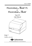
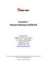
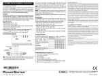
Product no. 03-96
Protein A ELISA
Instruction Manual
for samples with or without IgG
IMMUNSYSTEM I.M.S. AB
www.immunsystem.com
Edition
Edition090811M_draft
KI-090921M
All
Copyright©©IMMUNSYSTEM
IMMUNSYSTEMI.M.S.
I.M.S.
AB
Allrights
rights reserved.
reserved. Copyright
AB
Instruction manual for Protein A ELISA kit
IMMUNSYSTEM, Product No 03-96
Table of content
1.
2.
3.
4.
5.
6.
7.
8.
9.
10.
11.
12.
13.
14.
15.
16.
1.
Background ...................................................................... 2
Assay principle ................................................................. 4
Kit contents....................................................................... 5
Materials not provided ...................................................... 6
Precautions ....................................................................... 6
Reagent preparation .......................................................... 7
Sample and standard preparation ....................................... 8
Test procedure ................................................................ 10
Analysis of results ........................................................... 12
Performance .................................................................... 13
Tips and hints ................................................................. 16
Related products ............................................................. 16
References ...................................................................... 17
Quick start guide, samples without IgG ........................... 18
Quick start guide, samples with IgG ................................ 19
Contact and order information......................................... 20
Background
1.1 Staphylococcal Protein A (SpA)
SpA is an immunoglobulin (IgG)-binding
protein found in the bacterial cell wall of
Staphylococcus aureus (ref 1). SpA binds to
most mammalian IgG and can be used for
detection or purification of such antibodies (ref
2). Affinity chromatography on SpA columns is
widely used for the purification of monoclonal
and polyclonal antibodies. SpA may sometimes
leak from the column and contaminate the
preparation which may give false results in
immunological assays. In the in vivo setting,
SpA can react with IgG causing anaphylactic
reactions (ref 1). Thus, it is important that antibody preparations are free
from SpA before being used.
Page 2 of 20
Instruction manual for Protein A ELISA kit
IMMUNSYSTEM, Product No 03-96
This kit has been developed and is primarily tested with recombinant SpA
from GE Healthcare, which is also the source of the included SpA reference.
Native SpA and other sources of recombinant SpA generally give good
recoveries, but in some cases it may be necessary to use the same SpA
source for the standard as for the samples being analysed.
1.2 Non-specific interactions between SpA and IgG
Analysing for the presence of SpA in samples containing IgG involves two
major problems which must be overcome in order to obtain reliable results.
Fc binding. SpA reacts with the Fc region of IgG from most animal
species which makes its specific detection by immunoassay difficult. Antibodies specific for SpA will bind specifically as well as non-specifically due to
the Fc reactivity of SpA. In this kit, the problem is solved by using chicken
anti-SpA IgY. Chicken antibody is one of the few immunoglobulins that does
not bind SpA in the Fc region.
IgG
Y
IgY
Y
IgG binds SpA nonspecifically in the Fc region.
This problem is overcome using chicken IgY
since it is not Fc reactive to SpA.
Other non-specific interactions. In samples containing
mammalian IgG, immunologically active epitopes of SpA can be blocked by
non-specifically bound IgG. To overcome this problem, SpA can be analysed
at low pH (ref 4) since this should cause SpA and IgG to dissociate.
However, high affinity antibodies (e.g. human IgG and certain mouse
monoclonals) bind to SpA even below pH 3. Unfortunately, the specific
immunological detection of SpA is very difficult at such low pH and an assay
at this pH would likely have an unsatisfactory analytical performance.
Instead, we have chosen to establish a flexible assay system that can be
tailored to each customer’s situation. The standard references are made with
known amounts of IgG present. The IgG solution, which is added to the
standard references, should be as similar as possible to the IgG found in the
Page 3 of 20
Instruction manual for Protein A ELISA kit
IMMUNSYSTEM, Product No 03-96
samples. To dissociate SpA and IgG, the samples and standard references
are boiled for 4 minutes. In other words, the assay is standardised with the
same IgG that is present in the samples. The possible error from the
blocking of antigenic epitopes is therefore eliminated.
2.
Assay principle
This ELISA kit is designed to detect SpA in IgG-containing solutions (e.g.
monoclonal antibody preparations), in acid eluates from SpA columns, or in
other liquid preparations. It is a sandwich ELISA based on microtitre strips
precoated with the primary antibody, affinity-purified chicken anti-SpA. SpA
from the sample is bound to the microwell and detected by biotinylated
chicken anti-SpA antibody. Next, a streptavidin horseradish peroxidase
conjugate is bound to the biotin conjugate. Finally, the substrate is added
and reacts with the horseradish peroxidase to produce a colour change.
Colour development indicates a positive result, and the colour can be read
visually or by a microplate spectrophotometer at 450 nm. Samples to be
tested with this assay are often of acidic pH. In order for the test to work
properly, such samples should be neutralised. If a sample contains
mammalian IgG, SpA may interact with the IgG and be partially blocked,
falsely lowering the signal. To avoid this problem, samples containing IgG
are treated to denature the IgG and expose SpA epitopes. A schematic
outline of the assay is shown.
Page 4 of 20
Instruction manual for Protein A ELISA kit
IMMUNSYSTEM, Product No 03-96
1
YY
SpA from the sample is bound to wells precoated
with the primary antibody, chicken anti-SpA IgY
Incubate 1 hr.
at room temperature
2
B
Y
YY
Biotinylated chicken anti-SpA IgY is
added as secondary antibody
Incubate 1 hr.
at room temperature
Strepavidin HRP is added and binds
to the biotin conjugate
Incubate 30 min.
at room temperature
3
B
Y
XX
YY
4
Substrate
B
Y
XX
YY
Incubate 10 min. at
room temp. (dark)
Coloured product
Substrate is added and and reacts with HRP to produce a coloured product.
Finally, stop solution is added to stop the chromogenic reaction.
3.
Kit contents
• Microtitre strips 8x12, precoated
with chicken anti-SpA antibody.
• Biotinylated anti-SpA IgY, 200 µL.
100x solution. Contains preservative.
• PBS-Tween tablets, 3 tablets.
Sufficient for 3x0.5 L Phosphate
Buffered Saline with 0.1% Tween.
• HRP Conjugate, 200 µL. Streptavidin
horseradish peroxidase conjugate.
100x solution. Contains preservative.
• Protein A reference, 200 µL. 0.5
mg/mL SpA solution (recombinant
origin). Contains preservative.
• Substrate solution, 25 mL.
TMB/H2O2 solution.
• Tween 20 concentrate, 2 mL.
• 2 M Tris Buffer, 25 mL.
• Stop solution, 20 mL. 1 M HCl.
(Caution: Strong Acid)
Page 5 of 20
Instruction manual for Protein A ELISA kit
IMMUNSYSTEM, Product No 03-96
4.
Materials not provided
• Pipettes: 10 to 1,000 µL
• Eppendorf tubes
• Microplate spectrophotometer
• 10-mL tubes
• Disposable pipette tips
• Ultra pure water
Additional materials needed to assay samples with IgG:
• SpA-free IgG solution
• Centrifuge (2000g)
• Water bath
• Vial rack or floating aid
5.
Precautions
• Carefully read and follow all
instructions.
• Use a separate pipette tip for each
sample.
• Store the kit and all reagents at +2 to
+8°C (35 to 45°F).
• Do not pipette by mouth.
• All reagents should equilibrate to room
temperature +18 to +25°C (64 to 77°F)
before use.
• Standard references must be used
for each new test series.
• Unused microtitre wells should be
stored sealed at +2 to +8° C.
• Use only ultra pure water for
preparation of reagents.
• Do not mix components or instruction
booklets from kits with different
expiration dates (different batches).
• The Substrate solution and 2 M Tris
buffer are irritating to eyes,
respiratory system and skin. Avoid
contact with skin and eyes.
• Be careful to prevent contamination of
kit components.
• The Stop solution contains HCl. This
is a strong acid that can cause burns.
Handle with care.
• Do not use the kit beyond expiration
date.
• This kit is for research use only.
• Do not eat, drink, or smoke where
specimens or kit reagents are
handled.
Page 6 of 20
Instruction manual for Protein A ELISA kit
IMMUNSYSTEM, Product No 03-96
6.
Reagent preparation
The examples below calculate the approximate amount needed to assay 96
microtitre wells (i.e. 12 strips). The amount of Neutralisation and Dilution
buffer needed depends on the pH and concentration of the samples and
therefore varies with each test.
• Microtitre wells, Substrate solution and Stop solution are ready for
use.
• Protein A reference. Should be diluted according to step 7.4.
• Tween solution. Prepare Tween solution by diluting the concentrate 1:5
in ultra pure water. Example: Mix 1 mL Tween concentrate with 4 ml
water. Vortex.
• Neutralisation buffer. Prepare by diluting Tween solution 1:5 in Tris
Buffer. Example: Mix 1 mL Tween solution with 4 mL Tris buffer. Vortex.
• Dilution buffer. Prepare by diluting Neutralisation buffer 1:10 in your
acid elution buffer (i.e. sample matrix buffer). Example: Mix 1 mL
Neutralisation buffer with 9 mL elution buffer. Vortex.
• PBS-Tween (PBS-T) buffer. Dissolve 1 tablet in 500 mL ultra pure
water. Store prepared PBS-T buffer at + 4°C but not for more than 3
days.
• Biotinylated anti-SpA IgY Concentrate. Dilute 1:100 in PBS-T buffer.
The solution is not stable for more than 24 hours. Example: Mix 120 µL
Biotinylated anti-SpA IgY Concentrate with 12 mL PBS-T buffer. Vortex.
• HRP conjugate Concentrate. Dilute 1:100 in PBS-T buffer. The solution
is not stable for more than 24 hours. Example: Mix 120 µL HRP
conjugate Concentrate with 12 mL PBS-T buffer. Vortex.
Page 7 of 20
Instruction manual for Protein A ELISA kit
IMMUNSYSTEM, Product No 03-96
7.
Sample and standard preparation
After reagent preparation, the samples and standard references are
prepared before the assay is performed. The steps for sample preparation
and test procedure are also summarised in the Quick Start Guides on pages
18 and 19.
7.1 Preparing samples (samples with and without IgG):
Neutral samples: SpA easily binds to glass and plastic material. However,
in the presence of 0.05% Tween 20 this binding is inhibited. In order to avoid
this, add Tween solution, 1:400, to the sample tubes before adding sample.
Next, add sample and mix well. Example: For 2 mL sample volume, add 5
µL Tween solution.
Acidic samples: Samples should be neutralised to improve the specific
immunological detection of SpA. Determine the elution volume of your acid
eluted samples. Add 1/10 volume of Neutralisation buffer to each sample
(dilution factor: 0.9). Example: Add 100 µL Neutralisation buffer to 0.9 mL
sample volume. Measure the pH and add Neutralisation buffer until neutral
pH is reached. Note the volume of buffer added in order to calculate dilution
factor.
7.2 Preparing samples (only for samples with IgG):
7.2.1 Determine the IgG concentration in your samples by measuring the
optical density at 280 nm (OD280). Divide the OD280 value with 1.36 to
get the concentration.
7.2.2 Dilute the samples with Dilution buffer so that all samples contain the
same IgG concentration (note the volume of buffer added in order to
calculate dilution factor).
7.2.3 Transfer 500 µL of each sample with adjusted IgG concentration to a
labelled Eppendorf tube (not provided).
Page 8 of 20
Instruction manual for Protein A ELISA kit
IMMUNSYSTEM, Product No 03-96
7.3 Preparing Solution G (only for samples with IgG):
7.3.1 Determine the concentration of a SpA-free IgG solution (see 7.2.1).
7.3.2 Add the IgG solution to Dilution buffer in a 10-mL tube (not provided).
Example: Add the IgG solution to 5 mL Dilution buffer (approximately
4 mL is needed to prepare standards I, II and III). Adjust the IgG
concentration in Solution G to the same as in the samples (see 7.2.2).
7.4 Preparing standard references:
7.4.1 Label four Eppendorf tubes from I to IV and prepare the standards as
indicated below.
Std
I
II
III
IV
Source solution
40 µL Protein A ref.
40 µL Std I
200 µL Std II
100µL Std III
Samples w/o
IgG
PBS-Tween
Samples with
IgG
Solution G
760 µL
960 µL
800 µL
900 µL
760 µL
960 µL
800 µL
See step 7.5
SpA conc.
(ng/mL)
25000
1000
200
20
7.5 Removal of IgG (only for samples with IgG):
7.5.1 Make a hole in the cap of the Eppendorf tubes. Place your samples
and Standard III (200 ng/mL) in a rack or floating aid and boil in a
water bath for 4 minutes. Remove from the water bath and let cool for
5-10 minutes at room temperature.
7.5.2 Centrifuge the boiled tubes at 2000g for 60 seconds.
7.5.3 Add 100µL of the supernatant of your boiled samples and Standard III
into 900µL of Dilution Buffer. This gives Standard IV at 20 ng/mL.
Note: This dilution of samples and standards after boiling can be omitted. For
more information see section 11, Tips and Hints.
Page 9 of 20
Instruction manual for Protein A ELISA kit
IMMUNSYSTEM, Product No 03-96
8.
Test procedure
8.1 Performing the assay:
8.1.1 Add 100µL of PBS-Tween for samples WITHOUT IgG or Dilution
Buffer for samples WITH IgG into row B to H for wells allocated for
the standard references.
8.1.2 Add 200 µL of Standard IV to wells in row A. It is recommended to
test standard references in duplicate. Dilute the standard in the ELISA
plate according to figure 1.
Figure 1. 1:2 dilutions of Standard IV in the ELISA plate
A
B
C
D
E
F
G
H
Strip 1 2
Std IV: Add 200µL of Std IV into row A. [20ng/mL]
Std V:
Transfer 100µL of Std IV into row B. Mix well. [10ng/mL]
Std VI: Transfer 100µL of Std V into row C. Mix well. [5ng/mL]
Std VII: Transfer 100µL of Std VI into row D. Mix well. [2.5ng/mL]
Std VIII: Transfer 100µL of Std VII into row E. Mix well. [1.25ng/mL]
Std IX: Transfer 100µL of Std VIII into row F. Mix well. [0.625ng/mL]
Std X:
Transfer 100µL of Std IX into row G. Mix well. [0.313ng/mL]
Blank:
Discard 100µL of Std X from row G.
100µL PBS-Tween or Dilution Buffer to row H.
8.1.3 Add 100 µL of samples to appropriate wells. It is recommended to test
samples in duplicate.
8.1.4 Cover with plastic wrap or tape and incubate the plate for 1 hour at
room temperature.
8.1.5 Wash the strips by emptying the wells and then rinsing 4 times with
PBS-T buffer, filling all wells to the top for each rinse. Empty the wells
by tapping the plate upside down onto a paper towel to remove the
last traces of fluid.
8.1.6 Add 100 µL Biotinylated anti-SpA IgY (diluted) to each well. Cover
with plastic wrap or tape and incubate the plate for 1 hour at room
temperature.
Page 10 of 20
Instruction manual for Protein A ELISA kit
IMMUNSYSTEM, Product No 03-96
8.1.7 Wash the strips (see step 8.1.5). Add 100 µL HRP conjugate (diluted)
to each well. Cover with plastic wrap or tape and incubate the plate for
30 minutes at room temperature.
8.1.8 Wash the strips (see step 8.1.5). Add 100 µL Substrate solution to
each well and incubate the plate 10 minutes at room temperature in
the dark. Begin timing after the first well is filled.
8.1.9 Stop the reaction by adding 100 µL Stop solution to each well. Add the
Stop solution in the same order as the Substrate solution was added.
8.1.10 Measure the absorbance of the samples and standard references at
450 nm within 30 minutes after adding the Stop solution.
Page 11 of 20
Instruction manual for Protein A ELISA kit
IMMUNSYSTEM, Product No 03-96
9.
Analysis of results
To determine the exact SpA concentrations in your samples, use data
reduction software to automatically generate an appropriate standard curve
such as a four-parameter curve fit. Most ELISA reader software programs
can correct for background automatically, so consult the user’s manual for
your specific software.
9.1 Manually estimating SpA sample concentration
Standard curve: Calculate the mean for the standard reference duplicates
and correct for background by subtracting the mean absorbance value for
the blank. Plot Corrected OD450 vs. Concentration on a semi-log paper.
OD450 (Standard IV to X) - OD450 (PBS-Tween or Dilution Buffer)
= Corrected OD450
Samples: Calculate the mean absorbance value for the sample duplicates
and correct for background by subtracting the mean absorbance value for
the blank.
From the corrected OD values, approximate the sample concentration (CS)
from the standard curve. To calculate the final SpA concentration (CF) in
each of the samples, divide CS by the final dilution factor (DF) for each
sample. When a sample has been diluted several times, i.e. with
neutralisation buffer and then dilution buffer, multiply the dilution factors to
get the final dilution factor
Final SpA concentration (ng/mL): CF = CS /DF
Page 12 of 20
Instruction manual for Protein A ELISA kit
IMMUNSYSTEM, Product No 03-96
10. Performance
The kit has been validated according to general guidelines. An excerpt of
results is shown below. The performance information should only be taken
as guidance. For quality control purposes, the assay should be validated inhouse with respect to common parameters. In the shown results we have
used recombinant SpA from GE Healthcare and human IgG (mix of
subclasses, primarily IgG1 och IgG2).
10.1 Validity of assay
For samples without IgG, Standard IX (0.625 ng/mL) should have a
corrected absorbance value greater than 0.1. For samples with IgG,
Standard VIII (prepared as 1.25 ppm) should have a corrected absorbance
value greater than 0.1.
Note: These plots should not be used
for quantification of samples
10.2 Sensitivity:
The functional sensitivity is 0.15 ng/mL (0.15 ppm1). This is set as the
concentration where the intra assay CV<10% and the OD-signal is higher
than ODBLANK + 3*SDBLANK.
1
The unit parts per million, ppm, is the concentration of Protein A relative to the
concentration of IgG, e.g. 1 ng/mL Protein A in 1mg/mL of IgG equals 1 ppm.
Page 13 of 20
Instruction manual for Protein A ELISA kit
IMMUNSYSTEM, Product No 03-96
10.3 Linearity:
The linear range is 0.15 – 33 ng/mL (0.15 – 33 ppm). This is set as the
concentration where recovery of standards is 80-120% and a plot of
2
Quantified vs. Theoretical concentration gives R >0.990.
10.4 Specificity
Recovery has been tested with different SpA sources, both native and
recombinant. Recoveries were in the range 75-150%.
Page 14 of 20
Instruction manual for Protein A ELISA kit
IMMUNSYSTEM, Product No 03-96
10.5 Precision:
Coefficients of variation (CV) for duplicate measurements are typically
<10%. The average between-day CV was <15% when tested on 6 different
occasions for three samples. The samples were tested in a concentration
range 0.5 – 150 ng/mL (ppm).
10.6 Recovery:
Samples at three different concentrations were spiked with three different
amounts. The recovery was within ±15% for all samples and amounts
spiked.
10.7 Matrix effect:
The binding affinity of SpA differs between IgG from different species but
also between isotypes of IgG from the same species. Thus, the accuracy of
the results will depend on the IgG that is used for the standards. Even with
the same IgG, the graph shows the importance of normalizing the IgGconcentration in samples and standards.
10.8 Antigen excess:
No antigen excess effect was seen for standards tested up 1600 ppm.
Page 15 of 20
Instruction manual for Protein A ELISA kit
IMMUNSYSTEM, Product No 03-96
11. Tips and hints
Dilution of standards and samples for samples with IgG
The 1:10 dilution of standards and samples that is performed after boiling of
samples can be omitted. This may be desired if a ten-fold dilution of samples
makes them too diluted. In this case, Standard III in the table (section 7.4)
should be further diluted 1:10 in solution G, giving Standard IV at 20 ng/mL.
Do not mix these two procedures! It is important to keep the same exact
procedure for samples and standards.
12. Related products
MouSelect
Product number: 09-04
The MouSelect kit contains the basic components needed to set up a
sandwich ELISA for the assessment of Protein A binding. The components
are designed to aid in an early selection of polyclonal or monoclonal
antibodies, to a preferential purification on Protein A (from Staphylococcus
aureus). The kit can also be used to optimise the Protein A binding of
antibodies.
anti-Protein A
Product number: 01-008
IgY fraction. After purification, the antibodies can be used in various
immunological applications to detect Protein A (from Staphylococcus
aureus).
Page 16 of 20
Instruction manual for Protein A ELISA kit
IMMUNSYSTEM, Product No 03-96
13. References
•
Forsgren A. et al., SpA and its exploitation. In: Staphylococci and
Staphylococcal infections 2 (Eds. C.S.F. Easmon and C. Adlam), pp.
429-480, Academic Press Inc., London (1983).
•
Lindmark R. et al., Binding of immunoglobulins to SpA and
immunoglobulin levels in mammalian sera. J. Immunol. Methods, 62, 113 (1983).
•
Langone J.J. et al., Radioimmunoassays for SpA of Staphylococcus
aureus. J. Immunol. Methods, 63, 145-157 (1983).
•
Berglund A. et al., G01N 33/563 G01N 33/569; Ways to determine Fcbinding bacterial polypeptides. The Swedish Patent Administration,
Edition number, 448 190 (1987).
•
Larsson A. et al., A microELISA useful for determination of SpA-binding
monoclonal antibodies. Hybridoma, 9, 289-294 (1990).
Page 17 of 20
Instruction manual for Protein A ELISA kit
IMMUNSYSTEM, Product No 03-96
14. Quick start guide, samples without IgG
Add Tween or neutralise samples if
necessary.
Step 7.1, page 8
Prepare standards in PBS-Tween.
Step 7.4, page 9
Add standards and samples to wells.
Step 8.1.1-8.1.4, page 10
- 20 to 0.313 ng/mL.
- 100 µL/well
- Incubate 60 min in RT
Wash x4 and add 1:100 diluted
Biotinylated anti-SpA IgY.
Step 8.1.5-8.1.6, page 10
- 100 µL/well
- Incubate 60 min in RT
Wash x4 and add 1:100 diluted HRP
conjugate.
Step 8.1.7, page 11
- 100 µL/well
- Incubate 30 min in RT
Wash x4 and add substrate solution.
Step 8.1.8, page 11
- 100 µL/well
- Incubate 10 min, dark
Add stop solution and read plate at
450 nm.
Step 8.1.9-8.1.10, page 11
- 100 µL/well
Page 18 of 20
Instruction manual for Protein A ELISA kit
IMMUNSYSTEM, Product No 03-96
15. Quick start guide, samples with IgG
Add Tween or neutralise samples if necessary.
Step 7.1, page 8
Determine and normalise IgG concentration of
samples.
Step 7.2, page 8
- [IgG] = OD280/1.36
Prepare standards in IgG-solution, same
concentration and isotype as samples.
Step 7.3-7.4, page 9
Boil standard III and samples. Centrifuge and
dilute supernatant 10x in Dilution buffer.
Step 7.5, page 9
- 4 min. boil
- 2000g for 60 sec
- 10x dilution
Add standards and samples to wells, take from
supernatant.
Step 8.1.1-8.1.4, page 10
- 20 to 0.3125 ng/mL
- 100 µL/well
- Incubate 60 min in RT
Wash x4 and add 1:100 diluted Biotinylated
anti-SpA IgY.
Step 8.1.5-8.1.6, page 10
- 100 µL/well
- Incubate 60 min in RT
Wash x4 and add 1:100 diluted HRP
conjugate.
Step 8.1.7, page 11
- 100 µL/well
- Incubate 30 min in RT
Wash x4 and add substrate solution.
Step 8.1.8, page 11
- 100 µL/well
- Incubate 10 min, dark
Add stop solution and read plate at 450 nm.
Step 8.1.9-8.1.10, page 11
- 100 µL/well
Page 19 of 20
Instruction manual for Protein A ELISA kit
IMMUNSYSTEM, Product No 03-96
16. Contact and order information
IMMUNSYSTEM I.M.S. AB
Dag Hammarskjölds väg 26
S-751 83 Uppsala, Sweden
Phone +46 18 53 89 09
Fax +46 18 53 89 97
www.immunsystem.com
Page 20 of 20