1
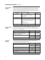
pcDNA™4/TO/myc-His A, B, and C Expression vectors with C-terminal tags designed for use with the T-REx™ System Catalog no. V1030-20 Revision date: 13 November 2010 Manual part no. 25-0287 MAN0000117 Corporate Headquarters Invitrogen Corporation 1600 Faraday Avenue Carlsbad, CA 92008 T: 1 760 603 7200 F: 1 760 602 6500 E: [email protected] For country-specific contact information visit our web site at www.invitrogen.com User Manual ii Table of Contents Contents and Storage . ......................................................................................................................................... iv Accessory Products. .............................................................................................................................................. v Introduction . .................................................................................................................. 1 System Overview . ..................................................................................................................................................1 Methods . ........................................................................................................................ 4 Cloning into pcDNA™4/TO/myc-His A, B, and C . ..........................................................................................4 Transfection and Analysis . ...................................................................................................................................9 Creating Stable Cell Lines. ..................................................................................................................................12 Appendix. ..................................................................................................................... 15 Recipes....................................................................................................................................................................15 Zeocin™ ...................................................................................................................................................................16 pcDNA™4/TO/myc-His Vector . ........................................................................................................................18 pcDNA™4/TO/myc-His/lacZ Vector . ..............................................................................................................20 Technical Support . ...............................................................................................................................................21 Purchaser Notification . .......................................................................................................................................22 References . ............................................................................................................................................................23 iii Contents and Storage Contents 20 μg (40 μL at 0.5 μg/μL) each of pcDNA™4/TO/myc-His A, B, and C in TE buffer*, pH 8.0 20 μg (40 μL at 0.5 μg/μL) pcDNA™4/TO/myc-His/lacZ™ TE buffer, pH 8.0 *TE Buffer, pH 8.0: 10 mM Tris-HCl, 1 mM EDTA, pH 8.0 Shipping/Storage iv The plasmids are shipped on wet ice. Upon receipt, store the plasmids at –20°C. Accessory Products Introduction The products listed below are intended for use with the pcDNA™4/TO/myc-His vectors. T-REx™ System The pcDNA™4/TO/myc-His vectors are designed for use with the T-REx™ System for tetracycline-regulated expression of your gene of interest in mammalian cells. The Core System includes the inducible expression vector of choice, the regulatory vector, and primers for sequencing. The Complete System includes the Core System plus inducing and selection agents. See below for a detailed description of the contents of each T-REx™ Kit. For more information on the T-REx™ System, refer to our website (www.invitrogen.com) or contact Technical Support (see page 21). T-REx™ Kit Inducible Expression Vector Complete System T-REx™ Cell Lines K1030-02 pcDNA 4/TO/myc-His For your convenience, Invitrogen has available three mammalian cell lines that stably express the Tet repressor. T-REx™-293 cells and T-REx™-HeLa cells express the Tet repressor from pcDNA™6/TR and should be maintained in medium containing blasticidin. T-REx™-U2OS cells express the Tet repressor from pCEP4/tetR as described in Yao et al., 1998 and should be maintained in medium containing hygromycin. Note that the pCEP4/tetR plasmid is episomally maintained in the T-REx™-U2OS cell line, but is stable under hygromycin selection. Expression of your gene of interest from pcDNA4/TO/myc-His™ may be assayed by transfection of your pcDNA™4/TO/myc-His construct into any of the T-REx™ cell lines and induction with tetracycline. Ordering information is provided below. Cell Line Source Catalog no. ™ Human embryonic kidney R710-07 ™ Human cervical adenocarcinoma R714-07 ™ Human osteosarcoma R712-07 ™ Human lymphocyte R722-07 T-REx -293 T-REx -HeLa T-REx -U2OS T-REx -Jurkat T-REx™ System Components K1030-01 ™ pcDNA 4/TO/myc-His Core System Catalog no. ™ Many of the reagents used in the T-REx™ System are available separately from Invitrogen. See the table below for ordering information. Item Amount Catalog no. pcDNA 6/TR 20 μg V1025-20 Blasticidin S HCl, powder 50 mg R210-01 Blasticidin S HCl, liquid 20 mL A11139-02 1g R250-01 ™ Zeocin™ Continued on next page v Accessory Products, Continued Detecting Fusion Proteins A number of antibodies are available from Invitrogen that can be used to detect expression of your fusion protein from pcDNA™4/TO/myc-His. Horseradish peroxidase (HRP)-conjugated antibodies allow one-step detection in western blots using colorimetric or chemiluminescent detection methods. The amount of antibody supplied is sufficient for 25 westerns. Antibody Anti-myc Epitope Catalog no. Detects 10 amino acid epitope derived from c-myc (Evans et al., 1985): R950-25 EQKLISEEDL Anti-myc-HRP Same as above R951-25 Anti-His(C-term) Detects the C-terminal polyhistidine (6His) tag (requires the free carboxyl group for detection) (Lindner et al., 1997): R930-25 HHHHHH-COOH Anti-His(C-term)-HRP Purifying Fusion Proteins Same as above The polyhistidine (6His) tag can be used to purify the recombinant fusion protein with a metal-chelating resin such as ProBond™. Ordering information for ProBond™ resin is provided below: Item Quantity Catalog no. ProBond Purification System 6 purifications K850-01 ProBond ™ Purification System with Anti-myc-HRP Antibody 1 Kit K852-01 ProBond™ Resin 50 mL R801-01 150 mL R801-15 50 columns R640-50 ™ Purification Columns Additional Products The table below list additional products that can be used with the T-REx™ System and the pcDNA™4/TO/myc-His vectors. Item Phosphate-Buffered Saline (PBS) pH 7.4 (1X) ® One Shot TOP10F´ (chemically competent) ™ Electrocomp TOP10F´ ™ PureLink Quick Plasmid Miniprep Kit -Gal Assay Kit -Gal Staining Kit vi R931-25 Quantity Catalog no. 500 mL 10010-023 1000 mL 10010-031 21 50 L C3030-03 5 80 L C665-55 50 preps K2100-10 80 mL K1455-01 1 kit K1465-01 Introduction System Overview pcDNA™4/TO/ myc-His A, B, and C Vectors pcDNA™4/TO/myc-His A, B, and C are 5.1 kb expression vectors designed for use with the T-REx™ System available from Invitrogen (see page v for ordering information). The vectors allow tetracycline-regulated expression of the gene of interest in mammalian host cells cotransfected with the pcDNA™6/TR vector (see page vi). Features of the pcDNA™4/TO/myc-His vectors allow purification and detection of expressed proteins. The vectors contain the following elements: Hybrid promoter consisting of the human cytomegalovirus immediate-early (CMV) promoter and tetracycline operator 2 (TetO2) sites for high-level tetracycline-regulated expression in a wide range of mammalian cells (see below) Three reading frames to facilitate in-frame cloning with a C-terminal peptide encoding the c-myc epitope and a polyhistidine (6His) tag Zeocin™ resistance gene for selection of stable cell lines (Mulsant et al., 1988) (see page 15 for more information) The control plasmid, pcDNA™4/TO/myc-His/lacZ, is included for use as a positive control for transfection and tetracycline-regulated expression in the cell line of choice. For more information about pcDNA™6/TR and the T-REx™ System, refer to the T-REx™ System manual or our website (www.invitrogen.com), or contact Technical Support (see page 21). For ordering information, see page v. A Note About pcDNA™4/TO/ myc-His The pcDNA™4/TO/myc-His vectors contain two tetracycline operator 2 (TetO2) sites within the human CMV promoter for tetracycline-regulated expression of your gene of interest (Yao et al., 1998). The TetO2 sequences serve as binding sites for 4 Tet repressor molecules (comprising two Tet repressor homodimers) and confer tetracycline-responsiveness to your gene of interest. The Tet repressor is expressed from the pcDNA™6/TR plasmid. For more details about the TetO2 sequences, see the next page. For more information about the pcDNA™6/TR plasmid and the Tet repressor, refer to the T-REx™ System manual. The T-REx™ System manual is available for downloading from our website (www.invitrogen.com) or from Technical Support (see page 21). In the absence of tetracycline, expression of your gene of interest is repressed by the binding of Tet repressor homodimers to the TetO2 sequences. Addition of tetracycline to the cells derepresses the hybrid CMV/TetO2 promoter in pcDNA™4/TO/myc-His and allows expression of your gene of interest. Continued on next page 1 Overview, Continued Tet Operator Sequences The promoters of bacterial tet genes contain two types of operator sequences, O1 and O2, that serve as high affinity binding sites for the Tet repressor (Hillen and Berens, 1994; Hillen et al., 1983). Each O1 and O2 site binds to one Tet repressor homodimer. While Tet repressor homodimers bind to both tet operators with high affinity, studies have shown that the affinity of the Tet repressor homodimer for O2 is three- to five-fold higher than for O1 (Hillen and Berens, 1994). Tet operators have been incorporated into heterologous eukaryotic promoters to allow tetracycline-regulated gene expression in mammalian cells (Gossen and Bujard, 1992; Yao et al., 1998). In the T-REx™ System, two copies of the O2 operator sequence (TetO2) were inserted into the strong CMV promoter of pcDNA™4/TO/myc-His to allow regulated expression of your gene of interest by tetracycline. We use the TetO2 operator sequence in pcDNA™4/TO/myc-His to maximize repression of basal gene expression. For more detailed information about tet operators, refer to Hillen and Berens (1994). Yao et al. (1998) have demonstrated that the location of tet operator sequences in relation to the TATA box of a heterologous promoter is critical to the function of the tet operator. Regulation by tetracycline is only conferred upon a heterologous promoter by proper spacing of the TetO2 sequences from the TATA box (Yao et al., 1998). For this reason, the first nucleotide of the TetO2 operator sequence has been placed 10 nucleotides after the last nucleotide of the TATA element in the CMV promoter in pcDNA™4/TO/myc-His. Refer to the diagrams on pages 5–7 for the sequence and placement of the TetO2 sequences in relation to the TATA box. In other tetracycline-regulated systems, the TetO2 sequences are located upstream of the TATA element in the promoter of the inducible expression vector (Gossen and Bujard, 1992). These systems differ substantially from the T-REx™ System in that they use regulatory molecules composed of the Tet repressor fused to a viral transactivation domain. The presence of viral transactivation domains appears to overcome the requirement for specific positioning of the TetO2 sequences in relation to the TATA box of the heterologous promoter. However, the presence of viral transactivation domains has been found to have deleterious effects in some mammalian cell lines. Continued on next page 2 Overview, Continued Experimental Outline Use the following outline to clone and express your gene of interest in the pcDNA™4/TO/myc-His vector. Step Action 1 Consult the multiple cloning sites diagrammed on pages 5–7 to determine which vector (A, B, or C) should be used to clone your gene in frame with the C-terminal c-myc epitope and the polyhistidine tag. 2 Ligate your insert into pcDNA™4/TO/myc-His and transform into E. coli. Select transformants on 50 to 100 μg/mL ampicillin or 25 to 50 mg/mL Zeocin™ in Low Salt LB. (see page 15 for recipe). 3 Analyze your transformants for the presence and orientation of the insert by restriction digestion. 4 Select a transformant with the correct restriction pattern and use sequencing to confirm that your gene is cloned in frame with the C-terminal peptide. 5 Cotransfect your pcDNA™4/TO/myc-His construct and pcDNA™6/TR into the cell line of choice using your own method of transfection, and induce expression of your gene of interest with tetracycline. Generate a double stable cell line, if desired. For more information about pcDNA™6/TR, refer to the T-REx™ System manual. 6 Test for expression of your recombinant gene by western blot analysis or functional assay. For antibodies to the c-myc epitope or the C-terminal polyhistidine tag, see page vi. 7 To purify your recombinant protein, you may use metal-chelating resin such as ProBond™. ProBond™ resin is available separately from Invitrogen (see page vi for ordering information). 3 Methods Cloning into pcDNA™4/TO/myc-His A, B, and C Maintaining pcDNA™4/TO/ myc-His Vectors Many E. coli strains are suitable for the propagation and maintenance of this vector including TOP10F´, DH5F´, JM109, and INVF´. We recommend that you propagate vectors containing inserts in E. coli strains that are recombination deficient (recA) and endonuclease A deficient (endA). To propagate and maintain pcDNA™4/TO/myc-His vectors, use 10 ng of each vector to transform a recA, endA E. coli strain like like TOP10F´, DH5, JM109, or equivalent. Select transformants on LB agar plates containing 50 to 100 μg/mL ampicillin or 25 to 50 μg/mL Zeocin™ in Low Salt LB (see page 15 for recipe). Be sure to prepare a glycerol stock of each plasmid for long-term storage (see page 8). Cloning Considerations Your insert should contain a Kozak translation initiation sequence with an ATG start codon for proper initiation of translation (Kozak, 1987; Kozak, 1991; Kozak, 1990). An example of a Kozak consensus sequence is provided below. Other sequences are possible, but the G or A at position –3 and the G at position +4 (shown in bold) illustrates the most commonly occurring sequence with strong consensus. Replacing one of the two bases at these positions provides moderate consensus, while having neither results in weak consensus. The ATG initiation codon is shown underlined. (G/A)NNATGG To express your gene as a recombinant fusion protein, you must clone your gene in frame with the C-terminal peptide. The vector is supplied in three reading frames to facilitate cloning. See pages 5–7 to develop a cloning strategy. If you wish to express your protein without the C-terminal peptide, be sure to include a stop codon. Continued on next page 4 Cloning into pcDNA™4/TO/myc-His A, B, and C, Continued Multiple Cloning Site of Version A Below is the multiple cloning site for pcDNA™4/TO/myc-His A. Restriction sites are labeled to indicate the cleavage site. Potential stop codons are underlined. The boxed nucleotides indicate the variable region. The multiple cloning site has been confirmed by sequencing and functional testing. The vector sequence of pcDNA™4/TO/myc-His A is available for downloading from our website (www.invitrogen.com) or from Technical Support (see page 21). For a map and a description of the features of pcDNA4/TO/myc-His™ A, refer to the Appendix, pages 18–19. CMV Forward priming site 721 AAAATCAACG GGACTTTCCA AAATGTCGTA ACAACTCCGC CCCATTGACG CAAATGGGCG 781 GTAGGCGTGT ACGGTGGGAG GTCTATATAA GCAGAGCTCT CCCTATCAGT GATAGAGATC Tetracycline operator (TetO2) TATA box Tetracycline operator (TetO2) 841 TCCCTATCAG TGATAGAGAT CGTCGACGAG CTCGTTTAGT GAACCGTCAG ATCGCCTGGA 901 GACGCCATCC ACGCTGTTTT GACCTCCATA GAAGACACCG GGACCGATCC AGCCTCCGGA Pme I* Afl II Hind III 961 BstX I* Xho I Xba I Apa I Age I Polyhistidine (6xHis) region AAA CTC ATC TCA GAA GAG GAT CTG AAT ATG CAT ACC GGT CAT CAT CAC Lys Leu Ile Ser Glu Glu Asp Leu Asn Met His Thr Glu His His His Pme I* 1127 Not I ATTCTGCAGA TATCCAGCAC AGTGGCGGCC GCTCGAGTCT AGAGGGCCCT TC GAA CAA Glu Gln c-myc epitope 1079 BstX I* EcoR I BamH I CTCTAGCGTT TAAACTTAAG CTTGGTACCG AGCTCGGATC CACTAGTCCA GTGTGGTGGA Pst I EcoR V 1021 Asp718 I Kpn I BGH Reverse priming site CAT CAC CAT TGA GT TTAAACCCGC TGATCAGCCT CGACTGTGCC TTCTAGTTGC His His His *** *Note that there are two Pme I sites and two BstX I sites in the polylinker. Continued on next page 5 Cloning into pcDNA™4/TO/myc-His A, B, and C, Continued Multiple Cloning Site of Version B Below is the multiple cloning site for pcDNA™4/TO/myc-His B. Restriction sites are labeled to indicate the cleavage site. Potential stop codons are underlined. The boxed nucleotides indicate the variable region. The multiple cloning site has been confirmed by sequencing and functional testing. The vector sequence of pcDNA™4/TO/myc-His B is available for downloading from our website (www.invitrogen.com) or from Technical Support (see page 21). For a map and a description of the features of pcDNA™4/TO/myc-His B, refer to the Appendix, pages 18–19. CMV Forward priming site 721 AAAATCAACG GGACTTTCCA AAATGTCGTA ACAACTCCGC CCCATTGACG CAAATGGGCG Tetracycline operator (TetO2) TATA box 781 GTAGGCGTGT ACGGTGGGAG GTCTATATAA GCAGAGCTCT CCCTATCAGT GATAGAGATC 841 TCCCTATCAG TGATAGAGAT CGTCGACGAG CTCGTTTAGT GAACCGTCAG ATCGCCTGGA 901 GACGCCATCC ACGCTGTTTT GACCTCCATA GAAGACACCG GGACCGATCC AGCCTCCGGA 961 CTCTAGCGTT TAAACTTAAG CTTGGTACCG AGCTCGGATC CACTAGTCCA GTGTGGTGGA Tetracycline operator (TetO2) Pme I* Afl II Hind III Pst I EcoR V 1021 Asp718 I Kpn I BstX I* Xba I Apa I Sac II Age I CAA AAA CTC ATC TCA GAA GAG GAT CTG AAT ATG CAT ACC GGT CAT CAT Gln Lys Leu Ile Ser Glu Glu Asp Leu Asn Met His Thr Glu His His Polyhistidine (6xHis) region 1128 Xho I ATTCTGCAGA TATCCAGCAC AGTGGCGGCC GCTCGAGTCT AGAGGGCCCG CGGTTC GAA Glu c-myc epitope 1080 Not I BstX I* EcoR I BamH I Pme I* BGH Reverse priming site CAC CAT CAC CAT TGA GTTTAAAC CCGCTGATCA GCCTCGACTG TGCCTTCTAG His His His His *** *Note that there are two Pme I sites and two BstX I sites in the polylinker. Continued on next page 6 Cloning into pcDNA™4/TO/myc-His A, B, and C, Continued Multiple Cloning Site of Version C Below is the multiple cloning site for pcDNA™4/TO/myc-His C. Restriction sites are labeled to indicate the cleavage site. Potential stop codons are underlined. The boxed nucleotides indicate the variable region. The multiple cloning site has been confirmed by sequencing and functional testing. The vector sequence of pcDNA™4/TO/myc-His C is available for downloading from our website (www.invitrogen.com) or from Technical Support (see page 21). For a map and a description of the features of pcDNA™4/TO/myc-His C, refer to the Appendix, pages 18–19. CMV Forward priming site 721 AAAATCAACG GGACTTTCCA AAATGTCGTA ACAACTCCGC CCCATTGACG CAAATGGGCG Tetracycline operator (TetO2) TATA box 781 GTAGGCGTGT ACGGTGGGAG GTCTATATAA GCAGAGCTCT CCCTATCAGT GATAGAGATC 841 TCCCTATCAG TGATAGAGAT CGTCGACGAG CTCGTTTAGT GAACCGTCAG ATCGCCTGGA 901 GACGCCATCC ACGCTGTTTT GACCTCCATA GAAGACACCG GGACCGATCC AGCCTCCGGA 961 CTCTAGCGTT TAAACTTAAG CTTGGTACCG AGCTCGGATC CACTAGTCCA GTGTGGTGGA Tetracycline operator (TetO2) Pme I* Afl II Hind III Pst I EcoR V 1021 Not I Xho I BstE II Age I Polyhistidine (6xHis) region CTC ATC TCA GAA GAG GAT CTG AAT ATG CAT ACC GGT CAT CAT CAC CAT Leu Ile Ser Glu Glu Asp Leu Asn Met His Thr Glu His His His His Pme I* 1126 BstX I* BstX I* EcoR I BamH I ATTCTGCAGA TATCCAGCAC AGTGGCGGCC GCTCGAGGTC ACCCATTC GAA CAA AAA Glu Gln Lys c-myc epitope 1078 Asp718 I Kpn I BGH Reverse priming site CAC CAT TGA GTTTAA ACCCGCTGAT CAGCCTCGAC TGTGCCTTCT AGTTGCCAGC His His *** *Note that there are two Pme I sites and two BstX I sites in the polylinker. Continued on next page 7 Cloning into pcDNA™4/TO/myc-His A, B, and C, Continued Transformation Method You may use any method of your choice for transformation. Chemical transformation is the most convenient method for many researchers. Electroporation is the most efficient and the method of choice for large plasmids. E. coli Transformation Transform your ligation mixtures into a competent recA, endA E. coli strain (e.g., TOP10F´, DH5) and select on LB agar plates containing 50–100 μg/mL ampicillin or 25–50 μg/mL Zeocin™ in Low Salt LB (see page 15 for recipe). Select 10–20 clones and analyze for the presence and orientation of your insert. MEND ION AT RECOM Important Preparing a Glycerol Stock 8 Any E. coli strain that contains the complete Tn5 transposable element (i.e., DH5F´IQ, SURE, SURE2) encodes the ble (bleomycin resistance gene). These strains will confer resistance to Zeocin™. For the most efficient selection, we recommend that you choose an E. coli strain that does not contain the Tn5 gene (i.e., TOP10F´). We recommend that you sequence your construct with the CMV Forward and BGH Reverse primers (not included in the kit) to confirm that your gene contains an ATG start codon and is cloned in frame with the C-terminal peptide. Refer to the diagrams on pages 5–7 for the sequences and location of the priming sites in each vector. For Invitrogen’s custom primer synthesis services, refer to our website (www.invitrogen.com) or contact Technical Support (see page 21). Once you have identified the correct clone, purify the colony and make a glycerol stock for long-term storage. Keep a DNA stock of your plasmid at –20°C. 1. Streak the original colony out on an LB plate containing 50 μg/mL ampicillin, or 25 μg/mL Zeocin™ in Low Salt LB (see page 15 for recipe). Incubate the plate at 37°C overnight. 2. Isolate a single colony and inoculate into 1–2 mL of LB containing 50 μg/mL ampicillin, or 25 μg/mL Zeocin™ in Low Salt LB. 3. Grow the culture to mid-log phase (OD600 = 0.5–0.7). 4. Mix 0.85 mL of culture with 0.15 mL of sterile glycerol and transfer to a cryovial. 5. Store at –80°C. Transfection and Analysis Introduction Once you have cloned your gene of interest into pcDNA™4/TO/myc-His and have prepared clean plasmid preparations of your pcDNA™4/TO/myc-His construct and pcDNA™6/TR, you are ready to cotransfect the plasmids into the mammalian cell line of choice. We recommend that you include the positive control vector (see below) and a mock transfection to evaluate your results. General guidelines are provided on the next page for cotransfection and induction. Refer to the T-REx™ System manual for more detailed information on pcDNA™6/TR, transfection, and induction of expression using tetracycline. Plasmid Preparation Plasmid DNA for transfection into eukaryotic cells must be clean and free of phenol and sodium chloride. Contaminants will kill the cells, and salt will interfere with lipid complexing, decreasing transfection efficiency. We recommend isolating plasmid DNA using the PureLink™ HQ Mini Plasmid Purification Kit (page vi). Other methods of obtaining high quality plasmid DNA may be suitable. Positive Control pcDNA™4/TO/myc-His /lacZ™ is provided as a positive control vector for mammalian cell transfection and expression (see page 20) and may be used to optimize transfection conditions for your cell line. Cotransfection of the positive control vector and pcDNA™6/TR results in the induction of -galactosidase expression upon addition of tetracycline. A successful cotransfection will result in -galactosidase expression that can be easily assayed by staining with X-gal (see below). Assay for -galactosidase Activity You may assay for -galactosidase expression by activity assay using cell-free lysates (Miller, 1972) or by staining the cells for activity. Invitrogen offers the -Gal Assay Kit and the -Gal Staining Kit for fast and easy detection of -galactosidase expression (see page vi for ordering information). Continued on next page 9 Transfection and Analysis, Continued Important Because tetracycline-regulated expression in the T-REx™ System is based on a repression/derepression mechanism, the amount of Tet repressor that is expressed in the host cell line from pcDNA™6/TR will determine the level of transcriptional repression of the Tet operator sequences in your pcDNA™4/TO/myc-His construct. Tet repressor levels should be sufficiently high to suitably repress basal level transcription. We have varied the ratio of pcDNA™6/TR and pcDNA™4/TO/myc-His plasmid that we transiently cotransfect into mammalian cells to optimize repression and inducibility of the hybrid CMV/TetO2 promoter in pcDNA™4/TO/myc-His. We recommend that you cotransfect your mammalian host cell line with a ratio of at least 6:1 (w/w) pcDNA™6/TR: pcDNA™4/TO/myc-His plasmid DNA, but you may want to try varying ratios of pcDNA™6/TR: pcDNA™4/TO/myc-His plasmid to optimize repression and expression for your particular cell line and your gene of interest. General guidelines are provided below to cotransfect your pcDNA™4/TO/myc-His Cotransfection and Induction with construct (or the control plasmid) and pcDNA™6/TR into your cell line of interest and to induce expression of your protein of interest with tetracycline. Refer to the Tetracycline T-REx™ System manual for more information on transfection and the preparation and handling of tetracycline. Use cells that are approximately 60% confluent for transfection. Cotransfect pcDNA™6/TR and your pcDNA™4/TO/myc-His construct at a ratio of 6:1 (w:w) into the cell line of choice using your preferred method. Absolute amounts of plasmid will vary depending on the method of transfection and the cell line used. After transfection, add fresh medium and allow the cells to recover for 24 hours before induction. Remove medium and add fresh medium containing the appropriate concentration of tetracycline to the cells. In general, we recommend that you add tetracycline to a final concentration of 1 μg/mL (5 μL of a 1 mg/mL stock solution per 5 mL of medium) to the cells and incubate the cells for 24 hours at 37°C to obtain maximal induction of your protein of interest. Harvest the cells and assay for expression of your gene of interest. Continued on next page 10 Transfection and Analysis, Continued Detecting Recombinant Fusion Proteins If you have cloned your gene in frame with the C-terminal peptide, you may use the Anti-myc antibodies or the Anti-His(C-term) antibodies to detect expression of your recombinant fusion protein from pcDNA™4/TO/myc-His (see page vi for ordering information). To detect fusion protein by western blot, you will need to prepare a cell lysate from transfected cells. We recommend that you perform a time course to optimize expression of the fusion protein (e.g., 12, 24, 36, 48 hours etc. after tetracycline induction). Use the protocol below to lyse cells. Other protocols are also suitable. 1. Wash cell monolayers (~5 105 to 1 106 cells) once with phosphate-buffered saline solution (see page vi). 2. Scrape cells into 1 mL PBS and pellet the cells at 1,500 g for 5 minutes. 3. Resuspend in 50 μL Cell Lysis Buffer (see page 15 for recipe). Other cell lysis buffers are also suitable. Vortex. 4. Incubate cell suspension at 37°C for 10 minutes to lyse the cells. Note: You may prefer to lyse the cells at room temperature or on ice if degradation of your protein is a potential problem. 5. Centrifuge the cell lysate at 10,000 g for 10 minutes to pellet nuclei and transfer the supernatant to a fresh tube. Assay the lysate for protein concentration. Note: Do not use protein assays utilizing Coomassie Blue or other dyes. NP-40 interferes with the binding of the dye with the protein. Purification 6. Add SDS-PAGE sample buffer to a final concentration of 1X and boil the sample for 5 minutes. 7. Load 20 μg of lysate onto an SDS-PAGE gel and electrophorese. Use the appropriate percentage of acrylamide to resolve your fusion protein. The C-terminal peptide containing the c-myc epitope and the polyhistidine (6His) tag will add approximately 3 kDa to the size of your protein. You will need 5 106 to 1 107 transfected cells for purification of your protein using ProBond™ resin (or another metal-chelating resin). Refer to the manufacturer’s instructions before attempting to purify your fusion protein. To prepare cells for lysis, refer to the protocol on page 14. 11 Creating Stable Cell Lines Introduction Once you have established that you can induce the expression of your construct, you may create a stable cell line that inducibly expresses your gene of interest. pcDNA™4/TO/myc-His contains the Zeocin™ resistance gene to allow selection of stable lines using Zeocin™. Note that your gene of interest will be constitutively expressed if you transfect your pcDNA™4/TO/myc-His construct into mammalian host cells prior to transfecting the pcDNA™6/TR plasmid. For more information on selection of stable cell lines using pcDNA™6/TR and blasticidin, refer to the T-REx™ System manual. Reminder: When generating a stable cell line expressing the Tet repressor (from pcDNA™6/TR), select for clones that express the highest levels of Tet repressor to use as hosts for your pcDNA™4/TO/myc-His expression plasmid. Those clones which express the highest levels of Tet repressor should exhibit the most complete repression of basal transcription of your gene of interest. Determining Antibiotic Sensitivity To generate a stable cell line expressing your protein of interest, you need to determine the minimum concentration of Zeocin™ required to kill your untransfected host cell line. Typically, concentrations between 50 and 1000 μg/mL Zeocin™ are sufficient to kill the untransfected host cell line. Test a range of concentrations (see below) to ensure that you determine the minimum concentration necessary for your cell line. For instructions on how to prepare and store Zeocin™, see pages 16–17. Note: Before transfecting your host cell line with pcDNA™6/TR, perform a similar experiment to determine the minimum concentration of blasticidin required to kill the untransfected cell line. Refer to the T-REx™ System manual for information about blasticidin. 1. Plate or split a confluent plate so the cells will be approximately 25% confluent. Prepare a set of 7 plates. 2. The next day, substitute culture medium with medium containing varying concentrations of Zeocin™ (e.g., 0, 50, 125, 250, 500, 750, and 1,000 μg/mL). 3. Replenish the selective medium every 3–4 days, and observe the percentage of surviving cells. 4. Count the number of viable cells at regular intervals to determine the appropriate concentration of Zeocin™ that prevents growth within 2 weeks after addition of Zeocin™. Continued on next page 12 Creating Stable Cell Lines, Continued Effect of Zeocin™ on Sensitive and Resistant Cells Zeocin™'s method of killing is quite different from blasticidin, neomycin, and hygromycin. Cells do not round up and detach from the plate. Sensitive cells may exhibit the following morphological changes upon exposure to Zeocin™: Vast increase in size (similar to the effects of cytomegalovirus infecting permissive cells) Abnormal cell shape Presence of large empty vesicles in the cytoplasm (breakdown of the endoplasmic reticulum and golgi apparatus or scaffolding proteins) Breakdown of plasma and nuclear membrane (appearance of many holes in these membranes) Eventually, these "cells" will completely break down and only "strings" of protein will remain. Zeocin™-resistant cells should continue to divide at regular intervals to form distinct colonies. There should not be any distinct morphological changes in Zeocin™-resistant cells when compared to cells not under selection with Zeocin™. For more information about Zeocin™, see page 16. Possible Sites for Linearization To obtain stable transfectants, you may choose to linearize your pcDNA™4/TO/myc-His construct before transfection. While linearizing your vector may not improve the efficiency of transfection, it increases the chances that the vector does not integrate in a way that disrupts either the gene of interest or other elements important for mammalian expression. The table below lists unique sites that may be used to linearize your construct prior to transfection. Other restriction sites are also possible. Be sure that your insert does not contain the restriction enzyme site you wish to use to linearize your vector. Enzyme Restriction Site (bp) Location (A,B,C) Mun I 161 Upstream of CMV promoter Nru I 208 Upstream of CMV promoter Sap I 3219 (A), 3223 (B), 3215 (C) Backbone Eam1105 I 4228 (A), 4232 (B), 4224 (C) Ampicillin gene Fsp I 4450 (A), 4454 (B), 4446 (C) Ampicillin gene Pvu I 4598 (A), 4602 (B), 4594 (C) Ampicillin gene Sca I 4708 (A), 4712 (B), 4704 (C) Ampicillin gene Ssp I 5032 (A), 5036 (B), 5028 (C) Backbone Continued on next page 13 Creating Stable Cell Lines, Continued Selecting Stable Integrants Once you have determined the appropriate Zeocin™ concentration to use for selection, you can generate a stable cell line expressing pcDNA™6/TR and your pcDNA™4/TO/myc-His construct. First generate a stable cell line expressing pcDNA™6/TR, and then use this cell line as the host for your pcDNA™4/TO/myc-His construct. Use Zeocin™ to select for double stable clones. Remember to maintain your cells in medium containing blasticidin as well. Dual Selection of Stable Integrants 1. Transfect your cell line of choice with your pcDNA™4/TO/myc-His using the desired protocol. Include a sample of untransfected cells as a negative control. 2. 24 hours after transfection, wash the cells and add fresh medium to the cells. 3. 48 hours after transfection, split the cells into fresh medium containing Zeocin™ at the appropriate concentration for your cell line. Split the cells such that they are no more than 25% confluent. If the cells are too dense, the antibiotic will not kill the untransfected cells. 4. Replenish medium every 3–4 days until Zeocin™-resistant colonies are detected. 5. Pick at least 20 foci and expand them to test for tetracycline-inducible gene expression. If you wish to select for stable cell lines by dual selection, you may cotransfect your pcDNA™4/TO/myc-His expression plasmid and pcDNA™6/TR into your cell line of choice, and select with Zeocin™ and blasticidin. Pick and expand at least 40 foci to screen for tetracycline-regulated expression of your gene of interest. Preparing Cells for Use the procedure below to prepare cells for lysis prior to purification of your protein using ProBond™. You will need 5 106 to 1 107 stably transfected cells for Lysis purification of your protein using ProBond™ (see the ProBond™ System manual). Lysing Cells 14 1. Seed cells in either five T-75 flasks or 2 to 3 T-175 flasks. 2. Grow the cells in selective medium until they are 50% confluent. 3. Add the appropriate concentration of tetracycline and induce expression of your protein of interest to the desired level. 4. Harvest the cells by treating with trypsin-EDTA for 2 to 5 minutes or by scraping the cells in PBS. 5. Inactivate the trypsin by diluting with fresh medium (if necessary) and transfer the cells to a sterile microcentrifuge tube. 6. Centrifuge the cells at 1,500 rpm for 5 minutes. Resuspend cell pellet in PBS. 7. Centrifuge the cells at 1,500 rpm for 5 minutes. You may lyse the cells immediately or freeze in liquid nitrogen and store at –70°C until needed. If you are using ProBond™ resin, refer to the ProBond™ Purification manual for details about sample preparation. If you are using another metal-chelating resin, refer to the manufacturer’s instructions. Appendix Recipes Low Salt LB Medium with Zeocin™ For Zeocin™ to be active, the salt concentration of the bacterial medium must remain low (< 90 mM) and the pH must be 7.5. For selection in E. coli, it is imperative that you prepare LB broth and plates using the following recipe. Note the lower salt content of this medium. Failure to use lower the salt content of your LB medium will result in nonselection due to inactivation of the drug. Low Salt LB Medium: 10 g Tryptone 5 g NaCl 5 g Yeast Extract Cell Lysis Buffer 1. Combine the dry reagents above and add deionized, distilled water to 950 mL. Adjust pH to 7.5 with 5 M NaOH. Bring the volume up to 1 liter. For plates, add 15 g/L agar before autoclaving. 2. Autoclave on liquid cycle at 15 lbs/sq. in. and 121°C for 20 minutes. 3. Thaw Zeocin™ on ice and vortex before removing an aliquot. 4. Allow the medium to cool to at least 55°C before adding the Zeocin™ to a 25 g/mL final concentration. 5. Store plates at 4°C in the dark. Plates containing Zeocin™ are stable for 1–2 weeks. 50 mM Tris, pH 7.8 150 mM NaCl 1% Nonidet P-40 1. This solution can be prepared from the following common stock solutions. For 100 mL, combine: 1 M Tris base 5 M NaCl Nonidet P-40 5 mL 3 mL 1 mL 2. Bring the volume up to 90 mL with deionized water and adjust the pH to 7.8 with HCl. 3. Bring the volume up to 100 mL. Store at room temperature. Note: Protease inhibitors may be added at the following concentrations: 1 mM PMSF 1 μg/mL pepstatin 1 μg/mL leupeptin 15 Zeocin™ Zeocin™ Zeocin™ belongs to a family of structurally related bleomycin/phleomycin-type antibiotics isolated from Streptomyces. Antibiotics in this family are broad spectrum antibiotics that act as strong antibacterial and antitumor drugs. They show strong toxicity against bacteria, fungi (including yeast), plants, and mammalian cells (Baron et al., 1992; Drocourt et al., 1990; Mulsant et al., 1988; Perez et al., 1989). The Zeocin™ resistance protein has been isolated and characterized (Calmels et al., 1991; Drocourt et al., 1990). This protein, the product of the Sh ble gene (Streptoalloteichus hindustanus bleomycin gene), is a 13.7 kDa protein that binds Zeocin™ and inhibits its DNA strand cleavage activity. Expression of this protein in eukaryotic and prokaryotic hosts confers resistance to Zeocin™. Molecular Weight, Formula, and Structure The formula for Zeocin™ is C60H89N21O21S3 and and the molecular weight is 1,535. The diagram below shows the structure of Zeocin™. CONH2 H H2 N N H O H N CH3 HO N O ++ Cu N H N H N O O N O NH O N H2 N H N CH3 HO R S N S CH3 H OH O O CH3 R = NH2 N HN NH NH2 OH H2N O O HO O MW = 1,535 O HO Applications of Zeocin™ OH OH Zeocin™ is used for selection in mammalian cells (Mulsant et al., 1988); plants (Perez et al., 1989); yeast (Baron et al., 1992); and prokaryotes (Drocourt et al., 1990). Suggested concentrations of Zeocin™ for selection in mammalian cell lines and E. coli are listed below: Organism Zeocin™ Concentration and Selective Medium E. coli 25–50 g/mL in low salt LB medium* (see page 15 for recipe) Mammalian Cells 50–1000 g/mL (varies with cell line) * Efficient selection requires that the concentration of NaCl be no more than 5 g/liter (< 90 mM). Continued on next page 16 Zeocin™, Continued Handling Zeocin™ Preparing and Storing Zeocin™ High salt and acidity or basicity inactivate Zeocin™. Therefore, we recommend that you reduce the salt in bacterial medium and adjust the pH to 7.5 to keep the drug active (see Low Salt LB Medium with Zeocin™, page 15). Note that the pH and salt concentration do not need to be adjusted when preparing tissue culture medium containing Zeocin™. Store Zeocin™ at –20°C and thaw on ice before use. Zeocin™ is light sensitive. Store the drug, and plates or medium containing drug, in the dark at 4°C. You may store culture medium containing Zeocin™ at 4°C protected from exposure to light for up to 1 month. Wear gloves, a laboratory coat, and safety glasses or goggles when handling Zeocin™-containing solutions. Zeocin™ is toxic. Do not ingest or inhale solutions containing the drug. Zeocin™ is available from Invitrogen (see page v for ordering information). Prepare 1.25 mL aliquots of Zeocin™ at a concentration of 100 mg/mL in autoclaved, deionized water. The stability of Zeocin™ is guaranteed for six months, if stored at –20°C and protected from exposure to light. 17 pcDNA™4/TO/myc-His Vector BGH pA f1 EM-7 P pcDNA4/TO/ myc-His A, B, C 5.1 kb ci n n UC o Ze p 5151 nucleotides i ri 40 o SV A m p i c i l li Comments for pcDNA4/TO/myc-His A or 6xHis Pme I c-myc epitope CM V O2 Tet X 2 Age I The figure below summarizes the features of the pcDNA™4/TO/myc-His vectors. The vector sequences for pcDNA™4/TO/myc-His A, B, and C are available for downloading from our website (www.invitrogen.com) or from Technical Support (see page 21). Pme I Afl II Hind III Asp718 I Kpn I BamH I BstX I EcoR I Pst I EcoR V BstX I Not I Xho I Xba I* Apa I* Sac II** BstE II*** Map of pcDNA™4/TO/ myc-His o ri SV40 pA CMV promoter: bases 232-958 TATA box: bases 804-810 Tetracycline operator 2 (2X TetO2) sequences: bases 820-859 CMV Forward priming site: bases 769-789 Multiple cloning site: bases 968-1069 c-myc epitope: bases 1073-1102 Polyhistidine (6xHis) tag: bases 1118-1135 BGH reverse priming site: bases 1158-1175 BGH polyadenylation sequence: bases 1164-1388 f1 origin: bases 1434-1862 SV40 promoter and origin: bases 1867-2211 EM-7 promoter: bases 2253-2319 Zeocin resistance gene: bases 2320-2694 SV40 early polyadenylation sequence: bases 2824-2954 pUC origin: bases 3337-4010 (complementary strand) bla promoter: bases 5016-5114 (complementary strand) Ampicillin (bla) resistance gene: bases 4155-5015 (complementary strand) *Unique in versions A and B only ** Unique in version B only ***Unique in version C only Continued on next page 18 pcDNA™4/TO/myc-His Vector, Continued Features of pcDNA™4/TO/ myc-His pcDNA™4/TO/myc-His A (5151 bp), pcDNA™4/TO/myc-His B (5155 bp), and pcDNA™4/TO/myc-His C (5147 bp) contain the following elements. All features have been functionally tested. Feature Benefit Human cytomegalovirus (CMV) immediate early promoter Permits high-level expression of your gene of interest (Andersson et al., 1989; Boshart et al., 1985; Nelson et al., 1987). CMV Forward priming site Allows sequencing in the sense orientation. Tetracycline operator (O2) sequences Two tandem 19 nucleotide repeats which serve as binding sites for Tet repressor homodimers (Hillen and Berens, 1994). Multiple cloning site Allows insertion of your gene of interest c-myc epitope (Glu-Gln-Lys-Leu-Ile-SerGlu-GluAsp-Leu) Allows detection of your recombinant protein with the Anti-myc Antibody or Anti-myc-HRP Antibody (see page vi) (Evans et al., 1985). C-terminal polyhistidine (6His) tag Permits purification of your recombinant protein on metal-chelating resin such as ProBond™. In addition, the C-terminal polyhistidine tag is the epitope for the Anti-His(C-term) Antibody (Lindner et al., 1997) and the Anti-His(C-term)-HRP Antibody (see page vi). BGH Reverse priming site Permits sequencing of the non-coding strand. Bovine growth hormone (BGH) polyadenylation signal Allows efficient transcription termination and polyadenylation of mRNA (Goodwin and Rottman, 1992). f1 origin Allows rescue of single-stranded DNA. SV40 early promoter and origin Allows efficient, high-level expression of the Zeocin™ resistance gene in mammalian cells and episomal replication in cells expressing SV40 large T antigen. EM-7 promoter Synthetic prokaryotic promoter for expression of the Zeocin™ resistance gene in E. coli. Zeocin™ resistance (Sh ble ) gene (expressed from the SV40 early promoter or the EM-7 promoter) Selection of stable transfectants in mammalian cells (Drocourt et al., 1990; Mulsant et al., 1988) and transformants in E. coli. SV40 early polyadenylation signal Allows efficient transcription termination and polyadenylation of mRNA. pUC origin Permits high-copy number replication and maintenance in E. coli. bla promoter Allows expression of the ampicillin (bla) resistance gene. Ampicillin resistance gene (-lactamase) Selection of transformants in E. coli. 19 pcDNA™4/TO/myc-His/lacZ Vector lacZ BGH pA f1 or i P ri 40 o SV CM V O2 Tet X 2 c-myc epitope 8198 bp UC p 8198 nucleotides ci n n Comments for pcDNA4/TO/myc-His/lacZ o ri Ze SV40 pA CMV promoter: bases 232-958 TATA box: bases 804-810 Tetracycline operator 2 (2X TetO2) sequences: bases 820-859 CMV Forward priming site: bases 769-789 LacZ gene: bases 1039-4095 c-myc epitope: bases 4120-4149 Polyhistidine (6xHis) tag: bases 4165-4182 BGH reverse priming site: bases 4205-4222 BGH polyadenylation sequence: bases 4211-4435 f1 origin: bases 4481-4909 SV40 promoter and origin: bases 4914-5258 EM-7 promoter: bases 5300-5366 Zeocin resistance gene: bases 5367-5741 SV40 early polyadenylation sequence: bases 5871-6000 pUC origin: bases 6384-7057 (complementary strand) bla promoter: bases 8063-8161 (complementary strand) Ampicillin (bla) resistance gene: bases 7202-8062 (complementary strand) 20 o EM-7 A m p i c i l li pcDNA4/TO/ myc-His/lacZ 6xHis Pme I The figure below summarizes the features of the pcDNA™4/TO/myc-His/lacZ vector. The vector sequence for pcDNA™4/TO/myc-His/lacZ is available for downloading from our website (www.invitrogen.com) or from Technical Support (see the next page). Age I Map of pcDNA™4/TO/ myc-His/lacZ Not I Xho I BstE II BstB I pcDNA™4/TO/myc-His/lacZ is a 8198 bp control vector containing the gene for -galactosidase. It was constructed by ligating a 3.1 kb Pst I–Not I fragment containing the lacZ gene into the Pst I–Not I site pcDNA™4/TO/myc-His A. Pme I Afl II Hind III BamH I Pst I Description Technical Support Web Resources Contact Us Visit the Invitrogen website at www.invitrogen.com for: Technical resources, including manuals, vector maps and sequences, application notes, MSDSs, FAQs, formulations, citations, handbooks, etc. Complete technical support contact information Access to the Invitrogen Online Catalog Additional product information and special offers For more information or technical assistance, call, write, fax, or email. Additional international offices are listed on our website (www.invitrogen.com). Corporate Headquarters: 5791 Van Allen Way Carlsbad, CA 92008 USA Tel: 1 760 603 7200 Tel (Toll Free): 1 800 955 6288 Fax: 1 760 602 6500 E-mail: [email protected] Japanese Headquarters: LOOP-X Bldg. 6F 3-9-15, Kaigan Minato-ku, Tokyo 108-0022 Tel: 81 3 5730 6509 Fax: 81 3 5730 6519 E-mail: [email protected] European Headquarters: Inchinnan Business Park 3 Fountain Drive Paisley PA4 9RF, UK Tel: 44 (0) 141 814 6100 Tech Fax: 44 (0) 141 814 6117 E-mail: [email protected] MSDS Material Safety Data Sheets (MSDSs) are available at www.invitrogen.com/msds. Certificate of Analysis The Certificate of Analysis (CofA) provides detailed quality control information for each product. CofAs are available on our website. Go to www.invitrogen.com/support and search for the Certificate of Analysis by product lot number, which is printed on the box. Limited Warranty Invitrogen (a part of Life Technologies Corporation) is committed to providing our customers with high-quality goods and services. Our goal is to ensure that every customer is 100% satisfied with our products and our service. If you should have any questions or concerns about an Invitrogen product or service, contact our Technical Support Representatives. All Invitrogen products are warranted to perform according to specifications stated on the certificate of analysis. The Company will replace, free of charge, any product that does not meet those specifications. This warranty limits the Company’s liability to only the price of the product. No warranty is granted for products beyond their listed expiration date. No warranty is applicable unless all product components are stored in accordance with instructions. The Company reserves the right to select the method(s) used to analyze a product unless the Company agrees to a specified method in writing prior to acceptance of the order. Invitrogen makes every effort to ensure the accuracy of its publications, but realizes that the occasional typographical or other error is inevitable. Therefore the Company makes no warranty of any kind regarding the contents of any publications or documentation. If you discover an error in any of our publications, please report it to our Technical Support Representatives. Life Technologies Corporation shall have no responsibility or liability for any special, incidental, indirect or consequential loss or damage whatsoever. The above limited warranty is sole and exclusive. No other warranty is made, whether expressed or implied, including any warranty of merchantability or fitness for a particular purpose 21 Purchaser Notification Limited Use Label License No. 22: Vectors and Clones Encoding Histidine Hexamer This product is licensed under U.S. Patent Nos. 5,284,933 and 5,310,663 and foreign equivalents from Hoffmann-LaRoche, Inc., Nutley, NJ and/or HoffmannLaRoche Ltd., Basel, Switzerland and is provided only for use in research. Information about licenses for commercial use is available from QIAGEN GmbH, Max-Volmer-Str. 4, D-40724 Hilden, Germany. Limited Use Label License No. 358: Research Use Only The purchase of this product conveys to the purchaser the limited, nontransferable right to use the purchased amount of the product only to perform internal research for the sole benefit of the purchaser. No right to resell this product or any of its components is conveyed expressly, by implication, or by estoppel. This product is for internal research purposes only and is not for use in commercial applications of any kind, including, without limitation, quality control and commercial services such as reporting the results of purchaser’s activities for a fee or other form of consideration. For information on obtaining additional rights, please contact [email protected] or Out Licensing, Life Technologies, 5791 Van Allen Way, Carlsbad, California 92008. 22 References Andersson, S., Davis, D. L., Dahlbäck, H., Jörnvall, H., and Russell, D. W. (1989). Cloning, Structure, and Expression of the Mitochondrial Cytochrome P-450 Sterol 26-Hydroxylase, a Bile Acid Biosynthetic Enzyme. J. Biol. Chem. 264, 8222-8229. Ausubel, F. M., Brent, R., Kingston, R. E., Moore, D. D., Seidman, J. G., Smith, J. A., and Struhl, K. (1994). Current Protocols in Molecular Biology (New York: Greene Publishing Associates and Wiley-Interscience). Baron, M., Reynes, J. P., Stassi, D., and Tiraby, G. (1992). A Selectable Bifunctional b-Galactosidase: Phleomycinresistance Fusion Protein as a Potential Marker for Eukaryotic Cells. Gene 114, 239-243. Boshart, M., Weber, F., Jahn, G., Dorsch-Häsler, K., Fleckenstein, B., and Schaffner, W. (1985). A Very Strong Enhancer is Located Upstream of an Immediate Early Gene of Human Cytomegalovirus. Cell 41, 521-530. Calmels, T., Parriche, M., Burand, H., and Tiraby, G. (1991). High Efficiency Transformation of Tolypocladium geodes Conidiospores to Phleomycin Resistance. Curr. Genet. 20, 309-314. Drocourt, D., Calmels, T. P. G., Reynes, J. P., Baron, M., and Tiraby, G. (1990). Cassettes of the Streptoalloteichus hindustanus ble Gene for Transformation of Lower and Higher Eukaryotes to Phleomycin Resistance. Nuc. Acids Res. 18, 4009. Evans, G. I., Lewis, G. K., Ramsay, G., and Bishop, V. M. (1985). Isolation of Monoclonal Antibodies Specific for c-myc Proto-oncogene Product. Mol. Cell. Biol. 5, 3610-3616. Goodwin, E. C., and Rottman, F. M. (1992). The 3´-Flanking Sequence of the Bovine Growth Hormone Gene Contains Novel Elements Required for Efficient and Accurate Polyadenylation. J. Biol. Chem. 267, 16330-16334. Gossen, M., and Bujard, H. (1992). Tight Control of Gene Expression in Mammalian Cells by Tetracycline-Responsive Promoters. Proc. Natl. Acad. Sci. USA 89, 5547-5551. Hillen, W., and Berens, C. (1994). Mechanisms Underlying Expression of Tn10 Encoded Tetracycline Resistance. Annu. Rev. Microbiol. 48, 345-369. Hillen, W., Gatz, C., Altschmied, L., Schollmeier, K., and Meier, I. (1983). Control of Expression of the Tn10-encoded Tetracycline Resistance Genes: Equilibrium and Kinetic Investigations of the Regulatory Reactions. J. Mol. Biol. 169, 707-721. Kozak, M. (1987). An Analysis of 5´-Noncoding Sequences from 699 Vertebrate Messenger RNAs. Nuc. Acids Res. 15, 8125-8148. Kozak, M. (1991). An Analysis of Vertebrate mRNA Sequences: Intimations of Translational Control. J. Cell Biol. 115, 887-903. Kozak, M. (1990). Downstream Secondary Structure Facilitates Recognition of Initiator Codons by Eukaryotic Ribosomes. Proc. Natl. Acad. Sci. USA 87, 8301-8305. Lindner, P., Bauer, K., Krebber, A., Nieba, L., Kremmer, E., Krebber, C., Honegger, A., Klinger, B., Mocikat, R., and Pluckthun, A. (1997). Specific Detection of His-tagged Proteins With Recombinant Anti-His Tag scFvPhosphatase or scFv-Phage Fusions. BioTechniques 22, 140-149. Miller, J. H. (1972). Experiments in Molecular Genetics (Cold Spring Harbor, New York: Cold Spring Harbor Laboratory). Mulsant, P., Tiraby, G., Kallerhoff, J., and Perret, J. (1988). Phleomycin Resistance as a Dominant Selectable Marker in CHO Cells. Somat. Cell Mol. Genet. 14, 243-252. Nelson, J. A., Reynolds-Kohler, C., and Smith, B. A. (1987). Negative and Positive Regulation by a Short Segment in the 5´-Flanking Region of the Human Cytomegalovirus Major Immediate-Early Gene. Mol. Cell. Biol. 7, 41254129. Perez, P., Tiraby, G., Kallerhoff, J., and Perret, J. (1989). Phleomycin Resistance as a Dominant Selectable Marker for Plant Cell Transformation. Plant Mol. Biol. 13, 365-373. Sambrook, J., Fritsch, E. F., and Maniatis, T. (1989). Molecular Cloning: A Laboratory Manual, Second Edition (Plainview, New York: Cold Spring Harbor Laboratory Press). Yao, F., Svensjö, T., Winkler, T., Lu, M., Eriksson, C., and Eriksson, E. (1998). Tetracycline Repressor, tetR, Rather than the tetR-Mammalian Cell Transcription Factor Fusion Derivatives, Regulates Inducible Gene Expression in Mammalian Cells. Hum. Gene Ther. 9, 1939-1950. ©2009, 2010 Life Technologies Corporation. All rights reserved. For research use only. Not intended for any animal or human therapeutic or diagnostic use. 23 Corporate Headquarters Invitrogen Corporation 5791 Van Allen Way Carlsbad, CA 92008 T: 1 760 603 7200 F: 1 760 602 6500 E: [email protected] For country-specific contact information, visit our web site at www.invitrogen.com User Manual