1
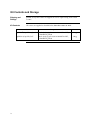
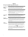
pcDNA™4/myc-His A, B, and C Catalog no. V863-20 Rev. Date: 27 October 2010 Manual part no. 25-0236 MAN0000078 Corporate Headquarters Invitrogen Corporation 1600 Faraday Avenue Carlsbad, CA 92008 T: 1 760 603 7200 F: 1 760 602 6500 E: [email protected] For country-specific contact information visit our web site at www.invitrogen.com User Manual ii Table of Contents Kit Contents and Storage..................................................................................................................................... iv Introduction ................................................................................................................... 1 Product Overview ..................................................................................................................................................1 Methods ......................................................................................................................... 2 Cloning into pcDNA™4/myc-His A, B, and C ....................................................................................................2 Transfection and Analysis.....................................................................................................................................6 Creating Stable Cell Lines .....................................................................................................................................8 Appendix...................................................................................................................... 11 pcDNA™4/myc-His Vector..................................................................................................................................11 pcDNA™4/myc-His/lacZ.....................................................................................................................................13 Zeocin™ ..................................................................................................................................................................14 Recipes ...................................................................................................................................................................16 Accessory Products ..............................................................................................................................................17 Technical Support.................................................................................................................................................19 Purchaser Notification .........................................................................................................................................20 References..............................................................................................................................................................21 iii Kit Contents and Storage Shipping and Storage pcDNA™4/myc-His vectors are shipped on wet ice. Upon receipt, store vectors at –20°C. Kit Contents All vectors are supplied as detailed below. Store the vectors at –20°C. Item Composition 20 g 40 L of 0.5 g/μL vector in 10 mM Tris-HCl, 1 mM EDTA, pH 8.0 20 g pcDNA™4/myc-His/lacZ iv Amount pcDNA 4/myc-His A, B, and C 40 L of 0.5 g/μL vector in 10 mM Tris-HCl, 1 mM EDTA, pH 8.0 ™ Introduction Product Overview Description of the System pcDNA™4/myc-His A, B, and C are 5.1 kb vectors designed for overproduction of recombinant proteins in mammalian cell lines. Features of the vectors allow purification and detection of expressed proteins (see pages 11-12 for more information). High-level stable and transient expression can be carried out in most mammalian cells. The vectors contain the following elements: Human cytomegalovirus immediate-early (CMV) promoter for high-level expression in a wide range of mammalian cells Three reading frames to facilitate in-frame cloning with a C-terminal peptide encoding the myc (c-myc) epitope and a polyhistidine (6xHis) metal-binding tag Zeocin™ resistance gene for selection of stable cell lines (Mulsant et al., 1988) (see page 14 for more information). Episomal replication in cell lines that are latently infected with SV40 or that express the SV40 large T antigen (e.g., COS7). The control plasmid, pcDNA™4/myc-His/lacZ is included for use as a positive control for transfection, expression, and detection in the cell line of choice. Experimental Outline Use the following outline to clone and express your gene of interest in pcDNA™4/myc-His: 1. Consult the multiple cloning sites described on pages 3-4 to determine which vector (A, B, or C) to use for cloning your gene in frame with the C-terminal myc epitope and the polyhistidine tag. 2. Ligate your insert into the appropriate vector and transform into E. coli. Select transformants on 50 to 100 μg/mL ampicillin or 25 to 50 g/mL Zeocin™ in Low Salt LB. For more information, see page 16. 3. Analyze your transformants for the presence of insert by restriction digestion. 4. Select a transformant with the correct restriction pattern and use sequencing to confirm that your gene is cloned in-frame with the C-terminal peptide. 5. Transfect your construct into the cell line of choice using your own method of transfection. Generate a stable cell line, if desired. 6. Test for expression of your recombinant gene by western blot analysis or functional assay. For antibodies to the myc epitope or the C-terminal polyhistidine tag, see page 18. 7. To purify your recombinant protein, you may use metal-chelating resin such as ProBond™. ProBond™ resin is available separately (see page 17). Methods Cloning into pcDNA™4/myc-His A, B, and C General Molecular Biology Techniques For help with DNA ligations, E. coli transformations, restriction enzyme analysis, purification of single-stranded DNA, DNA sequencing, and DNA biochemistry, refer to Molecular Cloning: A Laboratory Manual (Sambrook et al., 1989) or Current Protocols in Molecular Biology (Ausubel et al., 1994). E. coli Strain Many E. coli strains are suitable for the growth of this vector including TOP10F´, DH5F´, JM109, and INVF´. We recommend that you propagate vectors containing inserts in E. coli strains that are recombination deficient (recA) and endonuclease A deficient (endA). For your convenience, TOP10F´ is available from Invitrogen as chemically competent or electrocompetent cells (see page 17). Transformation Method You may use any method of your choice for transformation. Chemical transformation is the most convenient for most researchers. Electroporation is the most efficient and the method of choice for large plasmids. Maintaining pcDNA™4/myc-His To propagate and maintain the pcDNA™4/myc-His vectors, use a small amount of the supplied 0.5 g/μL stock solution in TE, pH 8.0 to transform a recA, endA E. coli strain like TOP10F´, DH5, JM109, or equivalent. Select transformants on LB plates containing 50 to 100 g/mL ampicillin or 25 to 50 g/mL Zeocin™ in Low Salt LB. Be sure to prepare a glycerol stock of each plasmid for long-term storage (see page 5). Cloning Considerations Your insert should contain a Kozak translation initiation sequence for proper initiation of translation (Kozak, 1987; Kozak, 1991; Kozak, 1990). An example of a Kozak consensus sequence is provided below. Note that other sequences are possible, but the A at position -3 and the G at position +4 are the most critical for function (shown in bold). The ATG initiation codon is shown underlined. ANNATGG To express your gene as a recombinant fusion protein, you must clone your gene in frame with the C-terminal peptide. The vector is supplied in three reading frames to facilitate cloning. See pages 3-4 to develop a cloning strategy. If you wish to express your protein WITHOUT the C-terminal peptide, be sure to include a stop codon. Continued on next page 2 Cloning into pcDNA™4/myc-His A, B, and C, Continued Multiple Cloning Site of Version A Below is the multiple cloning site for pcDNA™4/myc-His A. Restriction sites are labeled to indicate the cleavage site. The boxed nucleotides indicate the variable region. Note that there is a stop codon between the BamH I site and the BstX I site. The multiple cloning site has been confirmed by sequencing and functional testing. The vector sequence of pcDNA™4/myc-His A is available for downloading from our website (www.invitrogen.com) or from Technical Support (see page 19). T7 promoter/priming site 861 Hind III BstX I* 922 Pst I EcoR V EcoR I BstX I* Not I Xba I myc epitope Apa I BstB I CTC GAG TCT AGA GGG CCC TTC GAA CAA AAA CTC ATC TCA GAA GAG GAT CTG AAT Leu Glu Ser Arg Gly Pro Phe Glu Gln Lys Leu Ile Ser Glu Glu Asp Leu Asn Polyhistidine tag Age I 1030 BamH I ATC CAC TAG TCC AGT GTG GTG GAA TTC TGC AGA TAT CCA GCA CAG TGG CGG CCG Ile His *** Ser Ser Val Val Glu Phe Cys Arg Tyr Pro Ala Gln Trp Arg Pro Xho I 976 Kpn I Acc65 I ATTAATACGA CTCACTATAG GGAGACCCAA GCTGGCTAGT TAA GCT TGG TAC CGA GCT CGG Ala Trp Tyr Arg Ala Arg Pme I ATG CAT ACC GGT CAT CAT CAC CAT CAC CAT TGA GTTTAAACCC GCTGATCAGC Met His Thr Glu His His His His His His *** BGH Reverse priming site 1083 CTCGACTGTG CCTTCTAG *Note that there are two BstX I sites in the polylinker. Multiple Cloning Site of Version B Below is the multiple cloning site for pcDNA™4/myc-His B. Restriction sites are labeled to indicate the cleavage site. The boxed nucleotides indicate the variable region. The multiple cloning site has been confirmed by sequencing and functional testing. The vector sequence of pcDNA™4/myc-His B is available for downloading from our website (www.invitrogen.com) or from Technical Support (see page 19). T7 promoter/priming site Hind III Acc65 I Kpn I BamH I 861 ATTAATACGA CTCACTATAG GGAGACCCAA GCTGGCTAGT TAAG CTT GGT ACC GAG CTC GGA Leu Gly Thr Glu Leu Gly Pst I EcoR V BstX I* EcoR I BstX I* Not I 923 TCC ACT AGT CCA GTG TGG TGG AAT TCT GCA GAT ATC CAG CAC AGT GGC GGC CGC Ser Thr Ser Pro Val Trp Trp Asn Ser Ala Asp Ile Gln His Ser Gly Gly Arg Xho I Xba I Apa I Sac II BstB I myc epitope 977 TCG AGT CTA GAG GGC CCG CGG TTC GAA CAA AAA CTC ATC TCA GAA GAG GAT Ser Ser Leu Glu Gly Pro Arg Phe Glu Gln Lys Leu Ile Ser Glu Glu Asp Age I Polyhistidine tag Pme I 1028 CTG AAT ATG CAT ACC GGT CAT CAT CAC CAT CAC CAT TGA GTTT AAACCCGCTG Leu Asn Met His Thr Gly His His His His His His *** BGH Reverse priming site 1081 ATCAGCCTCG ACTGTGCCTT CTAGTTGCCA *Note that there are two BstX I sites in the polylinker. Continued on next page 3 Cloning into pcDNA™4/myc-His A, B, and C, Continued Multiple Cloning Site of Version C Below is the multiple cloning site for pcDNA™4/myc-His C. Restriction sites are labeled to indicate the cleavage site. The boxed nucleotides indicate the variable region. The multiple cloning site has been confirmed by sequencing and functional testing. The vector sequence of pcDNA™4/myc-His C is available for downloading from our website (www.invitrogen.com) or from Technical Support (see page 19). T7 promoter/priming site Hind III Acc65 I Kpn I 861 ATTAATACGA CTCACTATAG GGAGACCCAA GCTGGCTAGT TA AGC TTG GTA CCG AGC Ser Leu Val Pro Ser BamH I BstX I* EcoR I Pst I EcoR V BstX I* 918 TCG GAT CCA CTA GTC CAG TGT GGT GGA ATT CTG CAG ATA TCC AGC ACA GTG Ser Asp Pro Leu Val Gln Cys Gly Gly Ile Leu Gln Ile Ser Ser Thr Val Not I Xho I BstE II BstB I myc epitope 969 GCG GCC GCT CGA GGT CAC CCA TTC GAA CAA AAA CTC ATC TCA GAA GAG GAT Ala Ala Ala Arg Gly His Pro Phe Glu Gln Lys Leu Ile Ser Glu Glu Asp Age I Polyhistidine tag Pme I 1020 CTG AAT ATG CAT ACC GGT CAT CAT CAC CAT CAC CAT TGA GTTTAAACCC Leu Asn Met His Thr Gly His His His His His His *** BGH Reverse priming site 1069 GCTGATCAGC CTCGACTGTG CCTTCTAGTT GC *Note that there are two BstX I sites in the polylinker. E. coli Transformation Important Transform your ligation mixtures into a competent recA, endA E. coli strain (e.g., TOP10F´, DH5) and select on LB plates containing 50–100 μg/mL ampicillin or 25–50 g/mL Zeocin™ in Low Salt LB (see page 16). Select 10–20 clones and analyze for the presence and orientation of your insert. Any E. coli strain that contains the complete Tn5 transposable element (i.e., DH5F´IQ, SURE, SURE2) encodes the ble (bleomycin resistance gene). These strains will confer resistance to Zeocin™. For the most efficient selection, we recommend that you choose an E. coli strain that does not contain the Tn5 gene (i.e., TOP10, DH5, DH10, etc.). Continued on next page 4 MEND ION AT RECOM Cloning into pcDNA™4/myc-His A, B, and C, Continued Preparing a Glycerol Stock We recommend that you sequence your construct with the T7 Forward and BGH Reverse primers to confirm that your gene is fused in frame with the myc epitope and the C-terminal polyhistidine tag. For ordering primers, see page 17. Once you have identified the correct clone, be sure to purify the colony and make a glycerol stock for long-term storage. It is also a good idea to keep a DNA stock of your plasmid at –20°C. 1. Streak the original colony out on an LB plate containing 50 g/mL ampicillin or 25 g/mL Zeocin™ in Low Salt LB. Incubate the plate at 37°C overnight. 2. Isolate a single colony and inoculate into 1–2 mL of LB with 50 g/mL ampicillin. 3. Grow the culture to mid-log phase (OD600 = 0.5–0.7). 4. Mix 0.85 mL of culture with 0.15 mL of sterile glycerol and transfer to a cryovial. 5. Store at –80°C. 5 Transfection and Analysis Introduction Once you have confirmed that your construct is in the correct orientation and fused in frame with the C-terminal peptide, you are ready to transfect your cell line of choice. We recommend that you include the positive control vector and a mock transfection to evaluate your results. Plasmid Preparation Plasmid DNA for transfection into eukaryotic cells must be very clean and free from phenol and sodium chloride. Contaminants will kill the cells, and salt will interfere with lipids, decreasing transfection efficiency. We recommend isolating plasmid DNA using the PureLink™ HiPure Miniprep Kit or the PureLink™ HiPure Midiprep Kit (see page 17 for ordering information). Methods of Transfection For established cell lines (e.g. HeLa), consult original references or the supplier of your cell line for the optimal method of transfection. We recommend that you follow exactly the protocol for your cell line. Pay particular attention to medium requirements, when to pass the cells, and at what dilution to split the cells. Further information is provided in Current Protocols in Molecular Biology (see page 21). Methods for transfection include calcium phosphate (Chen and Okayama, 1987; Wigler et al., 1977), lipid-mediated (Felgner et al., 1989; Felgner and Ringold, 1989) and electroporation (Chu et al., 1987; Shigekawa and Dower, 1988). Invitrogen offers the Lipofectamine™ 2000 Reagent for mammalian transfection For more details, call Technical Support (see page 19) or visit our website at www.invitrogen.com. Positive Control pcDNA™4/myc-His/lacZ is provided as a positive control vector for mammalian cell transfection and expression (see page 13) and may be used to optimize transfection conditions for your cell line. The gene encoding -galactosidase is expressed in mammalian cells under the control of the CMV promoter. A successful transfection will result in -galactosidase expression that can be easily assayed (see below). Assay for -galactosidase Activity You may assay for -galactosidase expression by activity assay using cell-free lysates (Miller, 1972) or by staining the cells for activity. Invitrogen offers the -Gal Assay Kit and the -Gal Staining Kit for fast and easy detection of -galactosidase expression (see page 17). Continued on next page 6 Transfection and Analysis, Continued Detecting Fusion Proteins Several antibodies are available from Invitrogen to detect expression of your fusion protein from pcDNA™4/myc-His (see page 18). To detect fusion protein by western blot, you will need to prepare a cell lysate from transfected cells. We recommend that you perform a time course to optimize expression of the fusion protein (e.g. 24, 48, 72 hours, etc. after transfection). To lyse cells: 1. Wash cell monolayers (~106 cells) once with phosphate-buffered saline (PBS). 2. Scrape cells into 1 mL PBS and pellet the cells at 1,500 × g for 5 minutes. 3. Resuspend in 50 L Cell Lysis Buffer (see page 16). Other lysis buffers may be suitable. 4. Incubate cell suspension at 37°C for 10 minutes to lyse the cells. 5. Centrifuge the cell lysate at 10,000 × g for 10 minutes to pellet nuclei and transfer the supernatant to a fresh tube. Assay the lysate for protein concentration. Note: Do not use protein assays utilizing Coomassie® Blue or other dyes. NP-40 interferes with the binding of the dye with the protein. 6. Add SDS-PAGE sample buffer to a final concentration of 1X and boil the sample for 5 minutes. 7. Load 20 g of lysate onto an SDS-PAGE gel and electrophorese. Use the appropriate percentage of acrylamide to resolve your fusion protein. The C-terminal peptide containing the myc epitope and the polyhistidine tag will add approximately 3 kDa to the size of your protein. Purification You will need 5 × 10⁶ to 1 × 10⁷ transfected cells for purification of your protein on a 2 mL ProBond™ column (or other metal-chelating column). Refer to the manufacturer's instructions before attempting to purify your fusion protein. To prepare cells for lysis, refer to the protocol on page 10. 7 Creating Stable Cell Lines Introduction The pcDNA™4/myc-His vectors contain the Zeocin™ resistance gene for selection of stable cell lines using Zeocin™. We recommend that you test the sensitivity of your mammalian host cell to Zeocin™ as natural resistance varies among cell lines. General information and guidelines are provided below for your convenience. For more information about Zeocin™, refer to page 14. Effect of Zeocin™ on Sensitive and Resistant Cells The method of killing with Zeocin™ is quite different from neomycin and hygromycin. Cells do not round up and detach from the plate. Sensitive cells will exhibit the following morphological changes upon exposure to Zeocin™: • Vast increase in size • Abnormal cell shape • Presence of large empty vesicles in the cytoplasm (breakdown of the endoplasmic reticulum and golgi apparatus or scaffolding proteins) • Breakdown of plasma and nuclear membrane (appearance of many holes in these membranes). Eventually, these "cells" will completely break down and only "strings" of protein will remain. Zeocin™-resistant cells should continue to divide at regular intervals to form distinct colonies. There should not be any distinct morphological changes in Zeocin™-resistant cells when compared to cells not under selection with Zeocin™. Selection in Mammalian Cell Lines To generate a stable cell line expressing your protein, you need to determine the minimum concentration of Zeocin™ required to kill your untransfected host cell line. Typically, concentrations between 50 and 1,000 g/mL Zeocin™ are sufficient to kill the untransfected host cell line. Test a range of concentrations (see below) to ensure that you determine the minimum concentration necessary for your cell line. 1. Seed cells (2 × 10⁵ cells/60 mm plate) for each time point and allow cells to adhere overnight. 2. The next day, substitute culture medium with medium containing varying concentrations of Zeocin™ (e.g., 0, 50, 125, 250, 500, 750, and 1,000 g/mL). 3. Replenish the selective medium every 3–4 days, and observe the percentage of surviving cells. 4. Count the number of viable cells at regular intervals to determine the appropriate concentration of Zeocin™ that prevents growth. Continued on next page 8 Creating Stable Cell Lines, Continued To obtain stable transfectants, you may choose to linearize your vector before transfection. While linearizing your vector may not improve the efficiency of transfection, it increases the chances that the vector does not integrate in a way that disrupts the gene of interest. The table below lists unique sites that may be used to linearize your construct prior to transformation. Other restriction sites are possible. Note that the cleavage site is indicated for versions A, B, and C of pcDNA™4/myc-His. Be sure that your insert does not contain the restriction enzyme site you wish to use to linearize your vector. Possible Sites for Linearization Enzyme Restriction Site (bp) (A,B,C) Location Supplier Bgl II 13 Upstream of CMV promoter Many Mfe I 161 Upstream of CMV promoter New England Biolabs Nru I 209 Upstream of CMV promoter Many Mlu I 229 5´ end of CMV promoter Many Bst1107 I 2881 (A), 2885 (B), 2877 (C) End of SV40 poly A AGS*, Fermentas, Takara, Boehringer-Mannhiem Eam1105 I 4153 (A), 4157 (B), 4149 (C) Ampicillin gene AGS*, Fermentas, Takara Fsp I 4375 (A), 4379 (B), 4371 (C) Ampicillin gene Many Pvu I 4523 (A), 4527 (B), 4519 (C) Ampicillin gene Many Sca I 4633 (A), 4637 (B), 4629 (C) Ampicillin gene Many Ssp I 4957 (A), 4961 (B), 4953 (C) Ampicillin gene Many *Angewandte Gentechnologie Systeme Selecting Stable Integrants Once the appropriate Zeocin™ concentration is determined, you can generate a stable cell line with your construct. 1. Transfect your cells using the appropriate protocol for your cell line. Include a sample of untransfected cells as a negative control. 2. After transfection, wash the cells once with 1X PBS and add fresh medium to the cells. 3. 48 hours after transfection, split the cells into fresh medium containing Zeocin™ at the appropriate concentration for your cell line. Split the cells such that they are no more than 25% confluent. 4. Replenish selective medium every 3–4 days until Zeocin™-resistant colonies are detected. 5. Pick and expand colonies. Continued on next page 9 Creating Stable Cell Lines, Continued Preparing Cells for Use the procedure below to prepare cells for lysis prior to purification of your protein on ProBond™. You will need 5 × 10⁶ to 1 × 10⁷ cells for purification of Lysis your protein on a 2 mL ProBond™ column (see ProBond™ Purification System manual). Lysis of Cells 1. Seed cells in five T-75 flasks or 2 to 3 T-175 flasks. 2. Grow the cells in selective medium until they are 80–90% confluent. 3. Harvest the cells by treating with trypsin-EDTA for 2 to 5 minutes or by scraping the cells in PBS. 4. Inactivate the trypsin by diluting with fresh medium (if necessary) and transfer the cells to a sterile microcentrifuge tube. 5. Centrifuge the cells at 240 × g for 5 minutes. Resuspend the cell pellet in PBS. 6. Centrifuge the cells at 240 × g for 5 minutes. You may lyse the cells immediately or freeze in liquid nitrogen and store at –80°C until needed. If you are using ProBond™ resin, refer to the Probond™ Purification System manual for details about sample preparation for chromatography. If you are using other metal-chelating resin, refer to the manufacturer's instruction for recommendations on sample preparation. 10 Appendix pcDNA™4/myc-His Vector The figure below summarizes the features of the pcDNA™4/myc-His vectors. The vector sequences for pcDNA™4/myc-His A, B, and C are available for downloading from our website (www.invitrogen.com) or from Technical Support (see page 19). Age I V CM P f1 Term ri 40 o SV EM-7 n Ze oc A m p i ci l l i 5.1 kb in C 6xHis or i pcDNA4/ myc-His A, B, C pU Comments for pcDNA4/Myc-His 5075 nucleotides BGH pA SV40 pA T7 Hind III Acc65 I Kpn I BamH I BstX I EcoR I Pst I EcoR V BstX I Not I Xho I BstE II* Xba I* Apa I* Sac II** BstB I myc epitope Pme I Map of pcDNA™4/myc-His *There is a unique BstE II site, but no Xba I or Apa I sites in version C. **There is a unique Sac II site between the Apa I site and the BstB I site in version B only. CMV promoter: bases 209-863 T7 promoter/priming site: bases 863-882 Multiple cloning site: bases 902-999 myc epitope: bases 997-1026 Polyhistidine tag: bases 1042-1059 BGH reverse priming site: bases 1082-1099 BGH polyadenylation signal: bases 1085-1312 f1 origin: bases 1358-1786 SV40 promoter and origin: bases 1814-2122 EM-7 promoter: bases 2170-2225 Zeocin resistance gene: bases 2244-2618 SV40 polyadenylation signal: bases 2748-2878 pUC origin: bases 3261-3934 Ampicillin resistance gene: bases 4079-4939 Continued on next page 11 pcDNA™4/myc-His Vector, Continued Features of pcDNA™4/myc-His pcDNA™4/myc-His A (5075 bp), pcDNA™4/myc-His B (5079 bp), and pcDNA™4/myc-His C (5071 bp) contain the following elements. All features have been functionally tested. Feature Benefit Human cytomegalovirus (CMV) immediate-early promoter/enhancer Permits efficient, high-level expression of your recombinant protein (Andersson et al., 1989; Boshart et al., 1985; Nelson et al., 1987). T7 promoter/priming site Allows for in vitro transcription in the sense orientation and sequencing through the insert. Multiple cloning site in three reading frames Allows insertion of your gene and facilitates cloning in frame with the myc epitope and polyhistidine C-terminal tag. myc epitope (Glu-Gln-Lys-Leu-Ile-Ser-Glu-Glu-AspLeu) Allows detection of your recombinant protein with the Antimyc Antibody the Anti-myc-HRP Antibody, or the Anti-myc-AP Antibody (Evans et al., 1985) (see page 18 for ordering). C-terminal polyhistidine (6xHis) tag Permits purification of your recombinant protein on metalchelating resin such as ProBond™. In addition, the C-terminal polyhistidine tag is the epitope for the Anti-His(C-term) Antibody, the Anti-His (C-term)-HRP Antibody and the Anti-His(C-term)-AP (Lindner et al., 1997) (see page 18). BGH reverse priming site Permits sequencing through the insert. Bovine growth hormone (BGH) polyadenylation signal Efficient transcription termination and polyadenylation of mRNA (Goodwin and Rottman, 1992). f1 origin Allows rescue of single-stranded DNA. SV40 early promoter and origin Allows efficient, high-level expression of the Zeocin™ resistance gene and episomal replication in cells expressing the SV40 large T antigen. EM-7 promoter Synthetic promoter based on the bacteriophage T7 promoter for expression of the Zeocin™ resistance gene in E. coli. Zeocin™ resistance gene Selection of transformants in E. coli and stable transfectants in mammalian cells (Drocourt et al., 1990; Mulsant et al., 1988). SV40 polyadenylation signal Efficient transcription termination and polyadenylation of mRNA. pUC origin High-copy number replication and growth in E. coli. Ampicillin resistance gene (-lactamase) Selection of transformants in E. coli. 12 pcDNA™4/myc-His/lacZ pcDNA™4/myc-His/lacZ is a 8120 bp control vector containing the gene for galactosidase. This vector was constructed by ligating a 3,880 bp BamH I-Stu I fragment containing the CMV promoter and the Zeocin™ resistance gene from pcDNA™4/myc-His B to a 4,240 bp BamH I-Stu I fragment containing the lacZ gene, myc epitope, and polyhistidine tag from pcDNA™3.1/myc-His/lacZ. The figure below summarizes the features of the pcDNA™4/myc-His/lacZ vector. The vector sequence for pcDNA™4/myc-His/lacZ is available for downloading from our website (www.invitrogen.com) or by contacting Technical Support (see page 19). Age I V CM P lacZ BGH pA Not I Xho I BstE II BstB I T7 Hind III BamH I Pst I myc epitope f1 or i SV40 pA C in pU Ze oc 8.1 kb EM-7 pcDNA4/ myc-His/lacZ n CMV promoter: bases 209-863 T7 promoter/priming site: bases 863-882 LacZ ORF: bases 963-4019 myc epitope: bases 4044-4073 Polyhistidine tag: bases 4089-4106 BGH reverse priming site: bases 4129-4146 BGH polyadenylation signal: bases 4132-4359 f1 origin: bases 4405-4833 SV40 promoter and origin: bases 4861-5169 EM-7 promoter: bases 5217-5272 Zeocin resistance gene: bases 5291-5665 SV40 polyadenylation signal: bases 5795-5925 pUC origin: bases 6308-6981 Ampicillin resistance gene: bases 7126-7986 Term ri 40 o SV Ampicilli Comments for pcDNA4/Myc-His/lacZ 8120 nucleotides 6xHis Pme I Map of Control Vector 13 Zeocin™ The pcDNA™4/myc-His vectors contain the Zeocin™ resistance gene for selection of stable cell lines using Zeocin™. We recommend that you test the sensitivity of your mammalian host cell to Zeocin™ as natural resistance varies among cell lines. General information and guidelines are provided in this section for your convenience. Introduction Zeocin™ is a member of the bleomycin/phleomycin family of antibiotics isolated from Streptomyces. Antibiotics in this family are broad spectrum antibiotics that act as strong anti-bacterial and anti-tumor drugs. They show strong toxicity against bacteria, fungi (including yeast), plants, and mammalian cells. Zeocin™ The Zeocin™ resistance protein has been isolated and characterized (Calmels et al., 1991; Drocourt et al., 1990). This protein, the product of the Sh ble gene (Streptoalloteichus hindustanus bleomycin gene), is a 13.7 kDa protein that binds Zeocin™ in a stoichiometric manner to inhibit its DNA strand cleavage activity. Expression of this protein in eukaryotic and prokaryotic hosts confers resistance to Zeocin™. Molecular Weight, Formula, and Structure The formula for Zeocin™ is C55H86O21N20S2Cu-HCl and the molecular weight is 1,527.5 daltons. Zeocin™ is an HCl salt. The diagram below shows the structure of Zeocin™. CONH2 H H2 N N H O H N CH3 HO N Cu NH O N H N N H N O O N O O ++ H 2N H N CH3 HO R S N S CH3 H OH O O CH3 R = HN NH2 N NH NH2 OH H2N O O HO O HO OH OH O Continued on next page 14 Zeocin™, Continued Applications of Zeocin™ Zeocin™ is used for selection in mammalian cells (Mulsant et al., 1988); plants (Perez et al., 1989); yeast (Baron et al., 1992); and prokaryotes (Drocourt et al., 1990). Suggested concentrations of Zeocin™ for selection in mammalian cell lines and E. coli are listed below: Organism Zeocin™ Concentration and Selective Medium E. coli 25–50 g/mL in low salt LB medium* (see page 16 for recipe) Mammalian Cells 50–1,000 g/mL (varies with cell line) *Efficient selection requires that the concentration of NaCl be no more than 5 g/liter (< 90 mM). Handling Zeocin™ • High salt and acidity or basicity inactivates Zeocin™. Therefore, we recommend that you reduce the salt in bacterial medium and adjust the pH to 7.5 to keep the drug active (see page 16). • Store Zeocin™ at –20°C and thaw on ice before use. • Zeocin™ is light sensitive. Store drug, plates, and medium containing drug in the dark. • Wear gloves, a laboratory coat, and safety glasses or goggles when handling solutions containing Zeocin™. Zeocin™ is toxic. Do not ingest or inhale solutions containing the drug. 15 Recipes Low Salt LB Medium with Zeocin™ For Zeocin™ to be active, the salt concentration of the medium must remain low (<90 mM) and the pH must be 7.5. For selection in E. coli, it is imperative that you prepare LB broth and plates using the following recipe. Note the lower salt content of this medium. Failure to use low salt LB medium will result in non-selection due to inactivation of the drug. Low Salt LB Medium: 10 g Tryptone 5 g NaCl 5 g Yeast Extract Cell Lysis Buffer 1. Combine the dry reagents above and add deionized, distilled water to 950 mL. Adjust pH to 7.5 with 5 M NaOH. Bring the volume up to 1 liter. For plates, add 15 g/L agar before autoclaving. 2. Autoclave on liquid cycle at 15 lbs/sq. in. and 121°C for 20 minutes. 3. Thaw Zeocin™ on ice and vortex before removing an aliquot. 4. Allow the medium to cool to at least 55°C before adding the Zeocin™ to 25 g/mL final concentration. 5. Store plates at 4°C in the dark. Plates containing Zeocin™ are stable for 1-2 weeks. 50 mM Tris, pH 7.8 150 mM NaCl 1% Nonidet P-40 1. This solution can be prepared from the following common stock solutions. For 100 mL, combine: 1 M Tris base 5 mL 5 M NaCl 3 mL Nonidet P-40 1 mL 2. Bring the volume up to 90 mL with deionized water and adjust the pH to 7.8 with HCl. 3. Bring the volume up to 100 mL. Store at room temperature. Note: Protease inhibitors may be added at the following concentrations: 1 mM PMSF 1 g/mL pepstatin 1 g/mL leupeptin 16 Accessory Products Introduction The following products may be used with the pcDNA™4/myc-His vectors. For details, visit www.invitrogen.com or contact Technical Support (page 19). Item ProBond™ Purification System Catalog no. Amount 6 × 2 mL precharged, prepacked ProBond™ resin columns and buffers for native and denaturing purification K850-01 50 mL R801-01 150 mL R801-15 5 × 80 L C665-55 One Shot TOP10F´ (chemically competent cells) 21 × 50 L C3030-03 EKMax™ Enterokinase 250 units E180-01 PureLink HiPure Plasmid Miniprep Kit 100 preps K2100-03 PureLink™ HiPure Plasmid Midiprep Kit 25 preps K2100-04 80 mL K1455-01 1 kit K1465-01 1 gram R250-01 5 grams R250-05 0.75 mL 11668-027 ProBond™ Resin Electrocomp™ TOP10F´ ® ™ -Gal Assay Kit -Gal Staining Kit Zeocin™ ™ Lipofectamine 2000 Reagent Primers For your convenience, Invitrogen offers a custom primer synthesis service. Visit www.invitrogen.com for more details. Continued on next page 17 Accessory Products, Continued Antibodies If you do not have an antibody specific to your protein, Invitrogen offers the Anti-myc, or Anti-His(C-term) antibodies to detect your recombinant fusion protein. Horseradish peroxidase (HRP)- and alkaline phosphatase (AP)– conjugated antibodies are available for convenient one-step detection. Antibody Anti-myc Anti-myc-HRP Anti-myc-AP Anti-His(C-term) Anti-His(C-term)-HRP Anti-His(C-term)-AP 18 Epitope Catalog no. Detects a 10 amino acid epitope derived from c-myc (Evan et al., 1985): EQKLISEEDL R950-25 Detects the C-terminal polyhistidine tag (requires the free carboxyl group for detection) (Lindner et al., 1997): HHHHHH-COOH R951-25 R952-25 R930-25 R931-25 R932-25 Technical Support Web Resources Contact Us Visit the Invitrogen website at www.invitrogen.com for: Technical resources, including manuals, vector maps and sequences, application notes, MSDSs, FAQs, formulations, citations, handbooks, etc. Complete technical support contact information Access to the Invitrogen Online Catalog Additional product information and special offers For more information or technical assistance, call, write, fax, or email. Additional international offices are listed on our website (www.invitrogen.com). Corporate Headquarters: 5791 Van Allen Way Carlsbad, CA 92008 USA Tel: 1 760 603 7200 Tel (Toll Free): 1 800 955 6288 Fax: 1 760 602 6500 E-mail: [email protected] Japanese Headquarters: LOOP-X Bldg. 6F 3-9-15, Kaigan Minato-ku, Tokyo 108-0022 Tel: 81 3 5730 6509 Fax: 81 3 5730 6519 E-mail: [email protected] European Headquarters: Inchinnan Business Park 3 Fountain Drive Paisley PA4 9RF, UK Tel: +44 (0) 141 814 6100 Tech Fax: +44 (0) 141 814 6117 E-mail: [email protected] MSDS Material Safety Data Sheets (MSDSs) are available on our website at www.invitrogen.com/msds. Certificate of Analysis The Certificate of Analysis provides detailed quality control and product qualification information for each product. Certificates of Analysis are available on our website. Go to www.invitrogen.com/support and search for the Certificate of Analysis by product lot number, which is printed on the box. Limited Warranty Invitrogen (a part of Life Technologies Corporation) is committed to providing our customers with high-quality goods and services. Our goal is to ensure that every customer is 100% satisfied with our products and our service. If you should have any questions or concerns about an Invitrogen product or service, contact our Technical Support Representatives. All Invitrogen products are warranted to perform according to specifications stated on the certificate of analysis. The Company will replace, free of charge, any product that does not meet those specifications. This warranty limits the Company’s liability to only the price of the product. No warranty is granted for products beyond their listed expiration date. No warranty is applicable unless all product components are stored in accordance with instructions. The Company reserves the right to select the method(s) used to analyze a product unless the Company agrees to a specified method in writing prior to acceptance of the order. Invitrogen makes every effort to ensure the accuracy of its publications, but realizes that the occasional typographical or other error is inevitable. Therefore the Company makes no warranty of any kind regarding the contents of any publications or documentation. If you discover an error in any of our publications, please report it to our Technical Support Representatives. Life Technologies Corporation shall have no responsibility or liability for any special, incidental, indirect or consequential loss or damage whatsoever. The above limited warranty is sole and exclusive. No other warranty is made, whether expressed or implied, including any warranty of merchantability or fitness for a particular purpose. 19 Purchaser Notification Limited Use Label License No. 5: Invitrogen Technology The purchase of this product conveys to the buyer the non-transferable right to use the purchased amount of the product and components of the product in research conducted by the buyer (whether the buyer is an academic or for-profit entity). The buyer cannot sell or otherwise transfer (a) this product (b) its components or (c) materials made using this product or its components to a third party or otherwise use this product or its components or materials made using this product or its components for Commercial Purposes. The buyer may transfer information or materials made through the use of this product to a scientific collaborator, provided that such transfer is not for any Commercial Purpose, and that such collaborator agrees in writing (a) not to transfer such materials to any third party, and (b) to use such transferred materials and/or information solely for research and not for Commercial Purposes. Commercial Purposes means any activity by a party for consideration and may include, but is not limited to: (1) use of the product or its components in manufacturing; (2) use of the product or its components to provide a service, information, or data; (3) use of the product or its components for therapeutic, diagnostic or prophylactic purposes; or (4) resale of the product or its components, whether or not such product or its components are resold for use in research. For products that are subject to multiple limited use label licenses, the terms of the most restrictive limited use label license shall control. Life Technologies Corporation will not assert a claim against the buyer of infringement of patents owned or controlled by Life Technologies Corporation which cover this product based upon the manufacture, use or sale of a therapeutic, clinical diagnostic, vaccine or prophylactic product developed in research by the buyer in which this product or its components was employed, provided that neither this product nor any of its components was used in the manufacture of such product. If the purchaser is not willing to accept the limitations of this limited use statement, Life Technologies is willing to accept return of the product with a full refund. For information about purchasing a license to use this product or the technology embedded in it for any use other than for research use please contact Out Licensing, Life Technologies, 5791 Van Allen Way, Carlsbad, California 92008; Phone (760) 603-7200 or e-mail: [email protected]. Limited Use Label License No. 22: Vectors and Clones Encoding Histidine Hexamer This product is licensed under U.S. Patent Nos. 5,284,933 and 5,310,663 and foreign equivalents from Hoffmann-LaRoche, Inc., Nutley, NJ and/or Hoffmann-LaRoche Ltd., Basel, Switzerland and is provided only for use in research. Information about licenses for commercial use is available from QIAGEN GmbH, Max-Volmer-Str. 4, D-40724 Hilden, Germany. 20 References Andersson, S., Davis, D. L., Dahlbäck, H., Jörnvall, H., and Russell, D. W. (1989). Cloning, Structure, and Expression of the Mitochondrial Cytochrome P-450 Sterol 26-Hydroxylase, a Bile Acid Biosynthetic Enzyme. J. Biol. Chem. 264, 8222-8229. Ausubel, F. M., Brent, R., Kingston, R. E., Moore, D. D., Seidman, J. G., Smith, J. A., and Struhl, K. (1994). Current Protocols in Molecular Biology (New York: Greene Publishing Associates and WileyInterscience). Baron, M., Reynes, J. P., Stassi, D., and Tiraby, G. (1992). A Selectable Bifunctional bGalactosidase::Phleomycin-resistance Fusion Protein as a Potential Marker for Eukaryotic Cells. Gene 114, 239-243. Boshart, M., Weber, F., Jahn, G., Dorsch-Häsler, K., Fleckenstein, B., and Schaffner, W. (1985). A Very Strong Enhancer is Located Upstream of an Immediate Early Gene of Human Cytomegalovirus. Cell 41, 521530. Calmels, T., Parriche, M., Burand, H., and Tiraby, G. (1991). High Efficiency Transformation of Tolypocladium geodes Conidiospores to Phleomycin Resistance. Curr. Genet. 20, 309-314. Chen, C., and Okayama, H. (1987). High-Efficiency Transformation of Mammalian Cells by Plasmid DNA. Molec. Cell. Biol. 7, 2745-2752. Chu, G., Hayakawa, H., and Berg, P. (1987). Electroporation for the Efficient Transfection of Mammalian Cells with DNA. Nucleic Acids Res. 15, 1311-1326. Drocourt, D., Calmels, T. P. G., Reynes, J. P., Baron, M., and Tiraby, G. (1990). Cassettes of the Streptoalloteichus hindustanus ble Gene for Transformation of Lower and Higher Eukaryotes to Phleomycin Resistance. Nucleic Acids Res. 18, 4009. Evans, G. I., Lewis, G. K., Ramsay, G., and Bishop, V. M. (1985). Isolation of Monoclonal Antibodies Specific for c-myc Proto-oncogene Product. Mol. Cell. Biol. 5, 3610-3616. Felgner, P. L., Holm, M., and Chan, H. (1989). Cationic Liposome Mediated Transfection. Proc. West. Pharmacol. Soc. 32, 115-121. Felgner, P. L., and Ringold, G. M. (1989). Cationic Liposome-Mediated Transfection. Nature 337, 387-388. Goodwin, E. C., and Rottman, F. M. (1992). The 3´-Flanking Sequence of the Bovine Growth Hormone Gene Contains Novel Elements Required for Efficient and Accurate Polyadenylation. J. Biol. Chem. 267, 16330-16334. Kozak, M. (1987). An Analysis of 5´-Noncoding Sequences from 699 Vertebrate Messenger RNAs. Nucleic Acids Res. 15, 8125-8148. Kozak, M. (1991). An Analysis of Vertebrate mRNA Sequences: Intimations of Translational Control. J. Cell Biology 115, 887-903. Kozak, M. (1990). Downstream Secondary Structure Facilitates Recognition of Initiator Codons by Eukaryotic Ribosomes. Proc. Natl. Acad. Sci. USA 87, 8301-8305. Lindner, P., Bauer, K., Krebber, A., Nieba, L., Kremmer, E., Krebber, C., Honegger, A., Klinger, B., Mocikat, R., and Pluckthun, A. (1997). Specific Detection of His-tagged Proteins With Recombinant Anti-His Tag scFv-Phosphatase or scFv-Phage Fusions. BioTechniques 22, 140-149. Miller, J. H. (1972). Experiments in Molecular Genetics (Cold Spring Harbor, New York: Cold Spring Harbor Laboratory). Continued on next page 21 References, Continued Mulsant, P., Tiraby, G., Kallerhoff, J., and Perret, J. (1988). Phleomycin Resistance as a Dominant Selectable Marker in CHO Cells. Somat. Cell Mol. Genet. 14, 243-252. Nelson, J. A., Reynolds-Kohler, C., and Smith, B. A. (1987). Negative and Positive Regulation by a Short Segment in the 5´-Flanking Region of the Human Cytomegalovirus Major Immediate-Early Gene. Molec. Cell. Biol. 7, 4125-4129. Perez, P., Tiraby, G., Kallerhoff, J., and Perret, J. (1989). Phleomycin Resistance as a Dominant Selectable Marker for Plant Cell Transformation. Plant Mol. Biol. 13, 365-373. Sambrook, J., Fritsch, E. F., and Maniatis, T. (1989). Molecular Cloning: A Laboratory Manual, Second Edition (Plainview, New York: Cold Spring Harbor Laboratory Press). Shigekawa, K., and Dower, W. J. (1988). Electroporation of Eukaryotes and Prokaryotes: A General Approach to the Introduction of Macromolecules into Cells. BioTechniques 6, 742-751. Wigler, M., Silverstein, S., Lee, L.-S., Pellicer, A., Cheng, Y.-C., and Axel, R. (1977). Transfer of Purified Herpes Virus Thymidine Kinase Gene to Cultured Mouse Cells. Cell 11, 223-232. ©2009, 2010 Life Technologies Corporation. All rights reserved. For research use only. Not intended for any animal or human therapeutic or diagnostic use. 22 Corporate Headquarters 5791 Van Allen Way Carlsbad, CA 92008 T: 1 760 603 7200 F: 1 760 602 6500 E: [email protected] For country-specific contact information, visit our web site at www.invitrogen.com User Manual