1
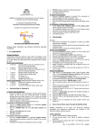
VDS
AIV
User Manual
Cat. No. V01-02-1113
NASBA for the detection of avian influenza virus H1-H16 subtypes
in veterinary and environmental samples
Preparation of Amplification Solution
Add 80 µl reagent sphere diluent to a lyophilised reagent
sphere and immediately vortex well.
Add 14 µl NASBA water and 16 µl KCl stock solution and
vortex.
Add 10 µl AIV primer mix and vortex.
DO NOT centrifuge.
Use within 30 min.
MICROPLATE DETECTION METHOD
Includes main components for 50 reactions
Rev. 0
July 2008
HAI KANG LIFE CORPORATION LIMITED
Preparation of the Enzyme Solution
Add 55 µl enzyme diluent to a lyophilised enzyme sphere.
Leave this enzyme solution to stand for at least 20 min at room
temperature.
Mix gently by flicking the microfuge tube with your finger.
DO NOT vortex any solution containing enzymes.
Centrifuge briefly before use.
Use within one hour.
B. Nucleic Acid Detection
Re-use of detection reagents is possible if the remainder of
opened reagents has been stored at 2 - 8oC.
Bring all reagents to room temperature before use.
Spin down all microfuge tubes to bring liquid to the bottom of the
tube.
PLEASE READ THROUGH THE ENTIRE PROTOCOL BEFORE
STARTING.
1.
1.
2.
KIT COMPONENTS
Storage Conditions
Components should be stored at -20oC with the exception of the
small inner box, which should be stored at 4oC. Expiry dates shown
on the box indicate the date beyond which reagents should no longer
be used.
3.
4.
5.
Nucleic Acid Amplification
5 x 6.5 mg
Enzyme spheres (red cap; contained in a foil
pack with silica gel desiccant)
1 x 0.5 ml
Enzyme sphere diluent (red cap)
5 x 10 mg
Reagent spheres (blue cap; contained in a foil
pack with silica gel desiccant)
1 x 0.6 ml
Reagent sphere diluent (blue cap)
1 x 0.5 ml
KCl stock solution (yellow cap)
1 x 60 µl
AIV primer mix
1 x 25 µl
AIV positive control
2 x 1.5 ml
NASBA water (white cap)
Nucleic Acid Detection
2 x 55 µl
AIV Probe solution
5 x 1.1 ml
Substrate solution (protect from light)
1 x 10 ml
10x TBS Washing buffer, pH 7.4 (store at room
temperature)
12 x 8 well
Microtitre plate strips and support frame (store at
room temperature in the dark)
Small Inner Box Contents (store at 4oC):
3 x 800 µl
Hybridisation buffer
5 x 1.1 ml
Detection solution
1 x 11 µl
Detection concentrate
5 x 1.1 ml
Stop solution
2.
PREPARATION OF REAGENTS
A. Nucleic Acid Amplification
Re-use of reagents: The reconstituted reagent sphere/enzyme
sphere can be re-used once within two weeks of opening
provided they have been stored at -70oC. Re-use of all other
amplification reagents is possible if the remainder of the opened
reagents has been stored at -20oC.
Bring all reagents to room temperature before use.
6.
7.
8.
9.
3.
AIV Probe solution
Thaw to room temperature and mix thoroughly before use.
Hybridisation buffer
Mix thoroughly before use.
Detection solution
Mix thoroughly before use.
Detection concentrate
Dilute 1:500 in Detection solution and mix thoroughly before
use.
Substrate solution
Thaw to room temperature in the dark and mix thoroughly before
use.
Stop solution
May be used without further treatment.
10x TBS washing buffer
Dilute 1:10 with DEPC-treated water and mix thoroughly before
use.
Note: Frequent thawing and freezing of the frozen reagents may
reduce the sensitivity of the kit. Components frozen without
being thawed are stable for 12 months.
Microtitre plate strips
Remove sufficient microtitre strips as are required for the
number of samples to be tested.
Microtitre plate support frame
May be used without further treatment.
PROCEDURES
Remarks:
Wear gloves throughout the procedure to protect your RNA
samples from nucleases.
All procedures should be carried out at room temperature unless
otherwise stated.
DO NOT vortex any solutions containing enzymes.
Nucleic Acid Amplification
1. For each amplification reaction, pipette 5 µl of the nucleic acid
extract into a fresh microfuge tube.
2. Add 10 µl of amplification solution (see section 2).
3. Incubate microfuge tubes for 5 min at 65oC in a heat block.
4. Cool microfuge tubes for 5 min at 41oC in a heat block.
5. Add 5 µl of enzyme solution (see section 2) and mix well by
flicking the microfuge tube with your finger.
6. Immediately return microfuge tubes to 41oC for 5 min.
7. Briefly centrifuge microfuge tubes and incubate for 90 min at
41oC in a water bath.
Rev. 0 (July 2008) | V01-02-1113 | English
8.
Perform the detection or store the RNA amplicons at -20oC for
up to 1 month.
Nucleic Acid Detection
1. Set-up sufficient microtitre strips on the support frame as are
required for the assay.
2. Add 2 µl probe solution, 5 µl NASBA product and 43 µl
hybridisation buffer to a 0.5 ml or 1.5 ml microfuge tube and mix
thoroughly.
3. Repeat for each NASBA assay product.
4. Add hybridisation mixture to separate wells of the microtitre strip.
Avoid introducing bubble into the wells. Cover firmly with plastic
wrap to prevent evaporation.
5. Incubate at 41oC for 1 hr.
6. Wash the wells three times with 250 µl 1x TBS, pH 7.4. Flick dry
the plate each time after discarding solution.
7. Add 100 µl detection solution to each well. Cover firmly with
plastic wrap to prevent evaporation.
8. Incubate at room temperature for 30 min.
9. Wash the wells three times with 250 µl 1x TBS, pH 7.4. Flick dry
the plate each time after discarding solution.
10. Add 100 µl substrate solution to each well (take care to not
introduce bubbles into the wells).
11. Incubate at room temperature for 5 min in the dark.
12. Add 100 µl stop solution and gently shake to stop the colour
development.
13. Load the microtitre frame with microtitre strips into a standard
96-well microtitre plate spectrophotometer and read the
absorbance at 405 nm. Use 100 µl substrate solution with 100 µl
stop solution (without hybridisation and detection reagents) as
background control.
4.
DATA COLLECTION
See your spectrophotometer instruction manual for detailed
guidelines on sample loading and analysis.
Table 1. Typical absorbance readings
Sample
5.
Qualitative result
Signal
(Abs405 + S.D)
Assay negative (all reagents/water instead of RNA)
Negative
0.080 + 0.016
Negative control (non-AIV RNA)
Negative
0.119 + 0.047
Positive control (AIV RNA)
Positive
0.959 + 0.053
DETERMINING THE CUT-OFF LIMIT
The calculation of the most appropriate cut-off limit to adequately
discriminate between genuine positive and negative samples is a
relatively straightforward procedure. However, care must be taken to
calculate the cut-off limit for each set of experiments that are
conducted.
1.
2.
3.
4.
Our laboratory usually obtains a S.D. in the range of 0.15 ± 0.3 for our
negative control samples. The safe-harbour for our cut-off value is,
therefore, approximately 0.45.
Note: If you obtain a cut-off value outside this range we suggest that
you re-evaluate your microplate system or contact Hai Kang Life
Corporation Limited for technical advice.
6.
TROUBLESHOOTING GUIDE
Symptoms
Possible Causes
Low
RNase contamination
Problem solving
The reagents provided are all RNase-free. The other
absorbance
solutions and apparatus used (pipettes, tips and microfuge
signal
tubes, etc) should be treated to ensure they are also
RNase-free.
Poor amplification
Add enzyme solution to the microfuge tubes by taking the
efficiency
microfuge tubes out of the heating block one at a time and
returning them immediately after addition of enzyme.
Maintain the amplification temperature at 41oC ±1oC
Enzymes denatured
DO NOT vortex the enzyme solution. Enzymes are quite
sensitive to physical influences and lose their activity when
vortex mixed.
DO NOT incubate the enzymes above 42oC. It will also
denature enzyme activity.
o
Poor hybridisation
Make sure the incubation temperature (45 C) is correct.
between target RNA and
Mix the probe solution, NASBA product and hybridisation
probe
buffer well before adding the mixture into the well. Avoid
making bubbles when adding the mixture into the well.
7.
TECHNICAL ASSISTANCE
Our technical staff will provide technical assistance you may need in
using this kit. Simply call +(852) 2111 2123 during office hours:
Monday – Friday: 9:00am to 5:30pm
Saturday:
9:00am to 1:00pm
A recorded message (in English, Cantonese or Putonghua) may be
left outside office hours. Alternatively, you may contact our technical
staff by fax or email.
Fax:
Email:
8.
+(852) 2111 9762
[email protected]
WARRANTIES AND LIABILITIES
Hai Kang Life Corporation Limited warrants the products manufactured by it are free of defects in materials and
workmanship when used in accordance with the applicable instructions for a period equal or shorter of one year
from the date of shipment of the product(s) or the expiration date marked on the product packaging under the
storage conditions recommended in the instructions and/or on the package. Application protocols published by
Hai Kang Life Corporation Limited are intended to be only guidelines for the buyers of the products. Buyers are
expected to validate the kit to their individual application. Hai Kang Life Corporation Limited makes no other
warranty, expressed or implied. There is no warranty of merchantability or fitness for a particular purpose.
The sole obligation of Hai Kang Life Corporation Limited with regard to the foregoing warranties shall be, at its
option, to either replace or refund the purchase price of the product(s) or part thereof that proves defective in
materials or workmanship within the warranty period, provided the customer notifies Hai Kang Life Corporation
Limited promptly of any such defect. Hai Kang Life Corporation Limited shall not be liable for any defect,
indirect or consequential damages resulting from economic loss or property damages sustained by the buyer or
any customer from the user of the product(s).
Test 10 known negative samples and 10 known “weak positive”
samples (i.e. dilutions of known positives). Define a cut-off limit
that can correctly differentiate the negative and weak positive
samples tested above.
The cut-off limit will be greater than the highest Abs405 signal
obtained for a known negative sample and lower than the lowest
Abs405 signal obtained for a known weak positive.
Further known negative and weak positive samples can then be
tested to further qualify the appropriateness of the selected
cut-off value.
The chosen cut-off value will be valid for the current set of
experiments only and must be defined again when the assay is
run in the future or if other parameters are altered (e.g. buffer
composition, primer sequence, length of incubation, etc).
A suggested cut-off limit is the mean negative control value plus 10
S.D (i.e. 0.12 + (10 x 0.012) = 0.24).
Rev. 0 (July 2008) | V01-02-1113 | English