1
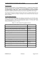
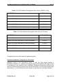
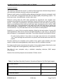
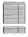
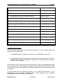
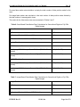
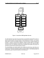
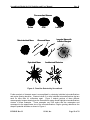
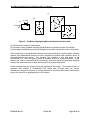
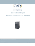
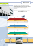
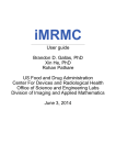
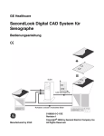
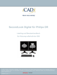
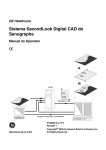
SecondLook Digital for Fuji Labeling and User Manual Rev. B SecondLook Digital for Fuji Labeling and User Manual © 2011, iCAD, Inc. All Rights Reserved. iCAD, the iCAD logo, Never Stop Looking and SecondLook are registered trademarks of iCAD, Inc. Other company, product, and service names may be trademarks or service marks of others. DTM105 Rev. B DTM105, Rev. B iCAD, Inc. Page 1 of 29 SecondLook Digital for Fuji Labeling and User Manual Rev. B This page intentionally left blank. DTM105 Rev. B iCAD, Inc. Page 2 of 29 SecondLook Digital for Fuji Labeling and User Manual Rev. B 98 Spit Brook Rd, Suite 100 Nashua, NH 03062, USA 603 882 5200 The European Representative for iCAD, Inc. is: MDSS GmbH Schiffgraben 41 30175 Hannover, Germany DTM105 Rev. B iCAD, Inc. Page 3 of 29 SecondLook Digital for Fuji Labeling and User Manual Rev. B This page intentionally left blank. DTM105 Rev. B iCAD, Inc. Page 4 of 29 SecondLook Digital for Fuji Labeling and User Manual Rev. B TABLE OF CONTENTS 1 OVERVIEW OF MANUAL ................................................................................................................... 7 2 SECONDLOOK DIGITAL DEVICE LABELING .................................................................................. 7 3 4 2.1 INDICATIONS FOR USE .................................................................................................................... 7 2.2 BRIEF DEVICE DESCRIPTION ........................................................................................................... 7 2.3 WARNINGS .................................................................................................................................... 8 2.4 PRECAUTIONS .............................................................................................................................. 10 2.5 ADVERSE EFFECTS ...................................................................................................................... 10 2.6 CLINICAL STUDIES ........................................................................................................................ 10 2.7 DETAILED DEVICE DESCRIPTION ................................................................................................... 17 2.8 CONFORMANCE TO STANDARDS.................................................................................................... 21 2.9 HOW SUPPLIED ............................................................................................................................ 21 RADIOLOGIST USE OF SECONDLOOK DIGITAL.......................................................................... 22 3.1 RADIOLOGIST REVIEW PRIOR TO VIEWING CAD MARKS................................................................. 22 3.2 RADIOLOGIST REVIEW WITH CAD MARKS...................................................................................... 22 RADIOLOGIST TRAINING WITH SAMPLE CASES ........................................................................ 23 4.1 TRAINING INSTRUCTIONS .............................................................................................................. 23 4.2 SAMPLE CASE.............................................................................................................................. 24 Case History and Mammograms ........................................................................................................ 24 5 SUMMARY OF RADIOLOGIST USE OF SECONDLOOK DIGITAL ................................................ 27 6 REFERENCES ................................................................................................................................... 28 DTM105 Rev. B iCAD, Inc. Page 5 of 29 SecondLook Digital for Fuji Labeling and User Manual Rev. B This page intentionally left blank. DTM105 Rev. B iCAD, Inc. Page 6 of 29 SecondLook Digital for Fuji Labeling and User Manual Rev. B 1 Overview of Manual This manual describes the SecondLook Digital Computer-Aided Detection (CAD) system and provides training to radiologists using the SecondLook Digital system for breast cancer detection. Section 2 provides SecondLook device labeling. Section 3 describes how a radiologist should use SecondLook Digital. Section 4 provides a sample case to familiarize the radiologist with SecondLook Digital. Section 5 provides a summary of the radiologist use of SecondLook Digital. Section 6 provides a list of clinical references. 2 SecondLook Digital Device Labeling 2.1 Indications for Use The SecondLook Computer-Aided Detection (CAD) system for mammography is intended to identify and mark regions of interest on screening and diagnostic mammograms from Fujifilm Medical Systems Computed Radiography system (Fuji CRm) to bring them to the attention of the radiologist after an initial reading has been completed. Thus the system prompts the radiologist to areas on Fuji CRm mammograms for second review only. 2.2 Brief Device Description SecondLook is a mammographic CAD system that prompts radiologists to areas on Fuji CRm mammogram for a second review only. The CAD algorithm version 7.2 includes image processing feature computations, and pattern recognition technology to detect regions of interest. The algorithm was trained on digitized film-screen mammograms and intended to more specifically identify potential breast lesions appearing as clusters of microcalcifications and/or masses. The CAD system was adapted to run on Fuji images, but the CAD algorithm design remained unchanged, and was not otherwise retrained on the Fuji CRm mammograms. DTM105 Rev. B iCAD, Inc. Page 7 of 29 SecondLook Digital for Fuji Labeling and User Manual Rev. B For hardcopy reading, the SecondLook output can be presented on a paper printout showing the CAD marks within the mammogram. How to Use the CAD: SecondLook with the Fuji CRm is intended to be used by a radiologist as follows: The radiologist must always first perform a full conventional read of the mammogram, and only after completing the conventional read, the radiologist may choose to display the CAD marks which may prompt to areas that were or were not examined during the first read. It is crucial to understand that 99.6% of all CAD marks will be placed over areas that are normal breast tissue or benign findings. Be aware that the SecondLook is not a diagnostic device, as the CAD marks are intended to be used to assist only in detection and not to assist with interpretation. 2.3 Warnings Warnings: Radiological Interpretation The radiologist must always first perform a full conventional read of the mammogram, and only after completing the conventional read, the radiologist may choose to display the CAD marks which may prompt to areas that were or were not examined during the first read. The presence or absence of a CAD mark should not in any manner influence your diagnostic decision as to the nature of a mammographic finding, i.e. normal vs. benign vs. malignant, or the clinical action to be taken (e.g. additional imaging or biopsy). Do not rely on the size (or shape) of the CAD mark as it may not be representative of the actual extent (or shape) of the breast lesion. Upon re-evaluation for the original mammogram at the locations indicated by SecondLook, the radiologist must use their interpretative skills to determine if the area should be worked-up based on its mammographic appearance. SecondLook is neither designed nor intended to prompt to: o interval change(s) between mammographic exams o asymmetry between the left and right breast o tubular density/solitary dilated duct o skin thickening, or o nipple retraction. DTM105 Rev. B iCAD, Inc. Page 8 of 29 SecondLook Digital for Fuji Labeling and User Manual Rev. B Warnings: System Operation Do not use the SecondLook system if you suspect any electrical component is defective or inoperable Do not place liquids on or near SecondLook. If a liquid is accidentally spilled on electrical components, immediately turn off the computer, which will automatically shut down the system to prevent any potential electrical shock. Contact your authorized SecondLook service provider for further instructions. Ensure that the system is connected to a properly wired and grounded power receptacle. Ensure that the voltage and current requirements are within system specifications to avoid bodily injury from electrical shock or fire hazard. Warnings: Installation and Maintenance EMC Warning – This SecondLook system has been tested and found to comply with IEC 60950-1, EN 55022 and EN 55024. This system generates, uses and can radiate radio frequency energy and, if not installed and used in accordance with our installation instructions, may cause or be subject to harmful interference with other devices in the vicinity. If the SecondLook system appears to cause or be subject to harmful interference, try the following steps to correct the problem: o o o Reorient or relocate the SecondLook system or the interface device. Increase the separation between the SecondLook system and the interfering device. Plug the SecondLook system into an outlet on a different circuit from the interfering device. Temperature and Humidity Warning – SecondLook system operations must be performed within the following temperature and humidity ranges. o o DTM105 Rev. B Temperature: 50-95 Fahrenheit (10-35 Celsius) Humidity: 20-80% iCAD, Inc. Page 9 of 29 SecondLook Digital for Fuji Labeling and User Manual 2.4 Rev. B Precautions Precautions: System Operation To prevent damage to the system, maintain equipment in a well-ventilated, airconditioned environment. Effectiveness and safety in patients with breast implants has not been established for views that include the implant. When non displaced implant views are analyzed by the system, any resulting CAD marks should not be used by the radiologist in evaluating the patient. Effectiveness and safety have not been established for non-standard mammographic views (e.g., magnification/compression views). When these views are analyzed by the system, any resulting CAD marks should not be used by the radiologist in evaluating the patient. The performance of the SecondLook V7.2 device has not been established for XCC views, ML views and breast implants, and may differ from those derived using conventional views of the breast (i.e. CC and MLO views). Precautions: Installation and Maintenance 2.5 This product contains no independently user serviceable parts. To prevent damage to the system, do not attempt to install or repair the SecondLook system. Only trained personnel are qualified to install or repair the system. For service training, contact iCAD Inc. at 1-866-280-2239. Disconnect power cord before moving or servicing. Adverse Effects SecondLook may increase your false-positive rates for both screening and diagnostic mammography. Increased false-positives may lead to unnecessary additional imaging radiation exposure, biopsy, patient anxiety, etc. 2.6 Clinical Studies Refer to the SecondLook Analog for further details regarding the testing studies used to support the safety and effectiveness of the original approval of the SecondLook analog device for use with digitized film-screen mammograms. DTM105 Rev. B iCAD, Inc. Page 10 of 29 SecondLook Digital for Fuji Labeling and User Manual Rev. B Benchmark testing Benchmark testing consisted of a limited standalone analysis (i.e., analysis of the device without radiologist interaction) on a small sample of Fuji CRm mammograms (53 cancers: 34 masses, 12 microcalcification clusters, and 7 mixed mass/microcalcification). Note that standalone performance testing of SecondLook version 7.2 on Fuji CRm images cannot be directly compared to standalone performance testing of SecondLook on digitized film screen images. The benchmark testing did not measure the effect of the device on radiologist performance and cannot measure or predict any change in radiologist’s cancer detection rates when using the device as intended. Fuji CRm Database Description: Tables 1-3 provide the mammographic and pathologic characteristics on the Fuji CRm cases. This database included 53 cases with cancer and 155 free of cancer. Of the cancer cases, there were 34 mass cases (15 >= 2.0 cm), 12 microcalcification cases (3 >= 2.0 cm), and 7 mixed mass/microcalcification cases (5 >= 2.0cm). Table 1: Fuji CRm Database Demographics (Cancers) TOTAL NUMBER OF CANCER CASES 53 Number of Primarily Mass Cases (Percentage) 34 (64%) Number of Primarily Microcalcification Cases (Percentage) 12 (23%) Number of Mixed Mass/Microcalcification Cases (Percentage) 7 (13%) Average Pathology Mass Lesion size (cm) 2.0 Standard Deviation 1.1 Median 1.8 Range: Minimum – Maximum 0.4 – 5.0 Number of BI-RADS breast density 1 cases (percentage) 9 (17%) Number of BI-RADS breast density 2 cases (percentage) 24 (45%) Number of BI-RADS breast density 3 cases (percentage) 17 (32%) Number of BI-RADS breast density 4 cases (percentage) 3 (6%) DTM105 Rev. B iCAD, Inc. Page 11 of 29 SecondLook Digital for Fuji Labeling and User Manual Rev. B Table 2: Fuji CRm Database Demographics (Non-Cancers, BIRADS 1 Only) DESCRIPTION BI-RADS 1 ONLY Number of Cases 36 Number of BI-RADS breast density 1 cases (percentage) 9 (25%) Number of BI-RADS breast density 2 cases (percentage) 14 (39%) Number of BI-RADS breast density 3 cases (percentage) 7 (19%) Number of BI-RADS breast density 4 cases (percentage) 6 (17%) Table 3: Fuji CRm Database Demographics (Non-Cancers, All Cases) DESCRIPTION ALL CASES Number of Cases 155 Number of BI-RADS breast density 1 cases (percentage) 36 (23%) Number of BI-RADS breast density 2 cases (percentage) 65 (42%) Number of BI-RADS breast density 3 cases (percentage) 44 (28%) Number of BI-RADS breast density 4 cases (percentage) 10 (6%) This dataset does not include cases with global asymmetry. SecondLook Standalone Testing with Fuji CRm Images Standalone testing of the SecondLook version 7.2 with Fuji CRm images provides a performance measure (i.e., sensitivity and average number of false positives per image or case) in the absence of any interaction with a radiologist. Standalone performance measures how often the CAD device places prompts over regions that contain or do not contain known breast abnormalities (i.e., microcalcifications and/or masses) in the absence of radiologist interaction. DTM105 Rev. B iCAD, Inc. Page 12 of 29 SecondLook Digital for Fuji Labeling and User Manual Rev. B Sensitivity Analysis: The sensitivity of SecondLook version 7.2 with Fuji CRm images was estimated using electronic truth and scoring that was manually confirmed by a radiologist. Electronic truth consists of an MQSA certified radiologist drawing an electronic rectangular “truth box” on each lesion, using pertinent data from the mammography and pathology reports. The lesion type (mass, microcalcification, mixed) is also noted. Electronic scoring takes the CAD marks generated by SecondLook and compares their locations to the electronic truth boxes to determine if each CAD mark is a cancer hit or a false positive. A CAD mark is assessed as a true positive detection if the CAD mark of a given type (mass, microcalcification) hits a truth box of the same type; mixed lesions can be hit by a CAD mark of either type. A microcalcification CAD mark is a hit if the CAD mark has any overlap with a microcalcification or mixed truth box. A mass CAD mark is a hit if its center falls within a mass or mixed truth box. Any CAD mark that is not a hit is counted as a false positive. Sensitivity is a count of the true positive detection divided by the total number of cases with mammographically visible cancer, and was calculated separately on a per-case (i.e. per-patient) and per-images (i.e. mammographic view) basis. True positive detection was defined as follows: For each scoring method, if any lesion on an image was scored as a hit (answered “Yes”), then the image was scored as a true positive, and the case was scored as a true positive. Per-case sensitivity was computed as the number of true positive cases divided by the total number of cases with mammographically visible cancer with 95% confidence intervals. Per-image sensitivity was computed as the number of true positive images divided by the total number of images with mammographically visible cancer with 95% CI. The 95% CI were computed using a statistical resampling technique (5000 sample bootstrapping). Results of the sensitivity analysis are summarized in Tables 4 and 5. Table 4: Case-Based Sensitivity Results for SecondLook Digital on Fuji CRm Digital Images Total Cases Enrolled 53 Cases with Clearly Identifiable Lesion(s) 53 Predominant Type of Most Suspicious Lesion: Cases with Masses DTM105 Rev. B 34 iCAD, Inc. Page 13 of 29 SecondLook Digital for Fuji Labeling and User Manual Rev. B Cases with Microcalcifications 12 Cases with Mixed Masses/Microcalcifications 7 Scoring Results (True Positive Cases) With Masses: Percentage Detected – Medium (95% Confidence Interval) 85% (73%, 98%) Percentage Detected – High (95% Confidence Interval) 85% (73%, 98%) With Microcalcifications Percentage Detected – Medium (95% Confidence Interval) 92% (76%, 100%) Percentage Detected – High (95% Confidence Interval) 92% (76%, 100%) With Mixed Masses/Microcalcifications Percentage Detected – Medium (95% Confidence Interval) 100% (100%, 100%) Percentage Detected – High (95% Confidence Interval) 100% (100%, 100%) Overall Percentage Detected – Medium (95% Confidence Interval) 89% (80%, 97%) Percentage Detected – High (95% Confidence Interval) 89% (80%, 97%) Table 5: Image-Based Sensitivity Results for SecondLook Digital on Fuji CRm Digital Images Total Images Enrolled 102 Images with Clearly Identifiable Lesion(s) 102 Predominant Type of Most Suspicious Lesion: Images with Masses 64 Images with Microcalcifications 24 DTM105 Rev. B iCAD, Inc. Page 14 of 29 SecondLook Digital for Fuji Labeling and User Manual Images with Mixed Masses/Microcalcifications Rev. B 14 Scoring Results (True Positive Images) With Masses: Percentage Detected (95% Confidence Interval) 69% (57%, 80%) Percentage Detected (95% Confidence Interval) 70% (58%, 82%) With Microcalcifications Percentage Detected (95% Confidence Interval) 67% (48%, 86%) Percentage Detected (95% Confidence Interval) 71% (52%, 89%) With Mixed Masses/Microcalcifications Percentage Detected (95% Confidence Interval) 79% (57%, 100%) Percentage Detected (95% Confidence Interval) 79% (56%, 100%) Overall Percentage Detected (95% Confidence Interval) 70% (61%, 79%) Percentage Detected (95% Confidence Interval) 72% (62%, 81%) False Marker Rate Analysis: The false marker rate analysis of the SecondLook version 7.2 with Fuji CRm images was measured in two separate ways: 1. Averaging the number of marks in all 4-views of a set of BI-RADS 1 cases: and 2. By averaging the number of marks in all 4-views of cases without proven cancers (all BIRADS). This set includes the BI-RADS 1 cases of set 1, as well as a number of cases of BI-RADS 2-5 with findings. For this analysis, all Fuji CRm images were assumed not to contain a mammographically visible cancer, and therefore all CAD marks were considered false-positive marks. This includes marks on benign or otherwise mammographically interesting regions that are ultimately determined to not be cancer. The false marker rate was calculated per-case (i.e., per patient) and per-image (i.e., per mammographic view), separately. Associated 95% CI were estimated using a statistical resampling technique (5000 sample bootstrapping). DTM105 Rev. B iCAD, Inc. Page 15 of 29 SecondLook Digital for Fuji Labeling and User Manual Rev. B Per-case false marker rate calculation is simply the total number of false positive marks for the case. Per-image false marker rate calculation is the total number of false-positive marks divided by the total number of mammographic views. The results for the false marker rate are summarized in Tables 6 and 7 Table 6: Case-Based False Marker Rate Calculations for SecondLook Digital on Fuji CRm Digital Images Cases Enrolled: Total 155 Contained Standard 4 Views -- All Non-cancers 119 Contained Standard 4 Views -- BIRADS 1 Only 36 Scoring Results: All Non-cancer Cases: Average CAD Marks per Case – Medium (95% Confidence 2.48 (2.15, 2.77) Interval) Average CAD Marks per Case – High (95% Confidence 3.01 (2.66, 3.34) Interval) BIRADS 1 Only Cases: Average CAD Marks per Case – Medium (95% Confidence 1.78 (1.34, 2.22) Interval) Average CAD Marks per Case – High (95% Confidence 2.19 (1.69, 2.68) Interval) Table 7: Image-Based False Marker Rate Calculations for SecondLook Digital on Fuji CRm Digital Images Images Enrolled: Total 620 Contained Standard 4 Views -- All Non-cancers 476 Contained Standard 4 Views -- BIRADS 1 Only 144 DTM105 Rev. B iCAD, Inc. Page 16 of 29 SecondLook Digital for Fuji Labeling and User Manual Rev. B Scoring Results: All Non-cancer Cases: Average CAD Marks Confidence Interval) per Image – Medium (95% 0.62 (0.55, 0.68) Average CAD Marks per Image – High (95% Confidence 0.75 (0.68, 0.82) Interval) BIRADS 1 Only Cases: Average CAD Marks Confidence Interval) per Image – Medium (95% 0.44 (0.34, 0.55) Average CAD Marks per image – High (95% Confidence 0.55 (0.43, 0.67) Interval) 2.7 Detailed Device Description SecondLook uses computer-aided detection (CAD) algorithms to identify regions of interest on mammograms that may contain suspicious finding. The CAD algorithms use advanced image processing, feature computations, and pattern recognition technology to analyze the images for potential areas of concern. These potential areas of concern are displayed for the radiologist by overlaying CAD marks at the appropriate locations of the mammography images within the softcopy review workstation or on a paper printout. The CAD marks are used by the radiologist as an additional tool in breast cancer detection. An overview of the SecondLook CAD algorithms is shown in Figure 1. DTM105 Rev. B iCAD, Inc. Page 17 of 29 SecondLook Digital for Fuji Labeling and User Manual Rev. B Standard Mammography Images MicroCalc Algorithm Density Algorithm Calc Image Enhancement Density Image Enhancement MicroCalc Detector Density Detector Clustering Region Growing MicroCalc Classifier Density Classifier Context Based Patient Evaluation Areas of Concern Highlighted by CAD Marks Figure 1: SecondLook CAD Algorithms Overview The CAD algorithms begin with image enhancement of the digitized mammographic images to accentuate all areas that could be individual microcalcifications and densities. In the case of directly acquired images, the digital images are first transformed into images that resemble digitized film in order to accommodate variations in inter-pixel spacing, gray-level mapping and bit depth. It should be noted that the Modulation Transfer Function (MTF) for Fuji images deviates from the MTF specified for SecondLook in the high frequency range. While the MTF is not directly used in the calculations performed by SecondLook, this deviation may impact the calculation of subtle features along the margins of lesions. The microcalcification and density detectors then identify the areas that are most likely to be individual microcalcifications and densities, based on an initial analysis of morphological and intensity measurements. The types of densities detected are depicted in Figure 2 and include spiculated and non-spiculated masses, architectural distortions, and focal densities. DTM105 Rev. B iCAD, Inc. Page 18 of 29 SecondLook Digital for Fuji Labeling and User Manual Rev. B Circumscribed Masses Round Microlobulated Mass Oval Obscured Mass Spiculated Mass Lobular Irregular Mass with Indistinct Margins Architectural Distortion Figure 2: Densities Detected by SecondLook Further analysis of detected areas is accomplished by clustering individual microcalcifications and region growing densities. Clusters include 3 or more individual microcalcifications that are each no more than 4.1 millimeters apart. Figure 3 depicts portions of three different mammography images showing how the SecondLook system would highlight microcalcifications clusters in these examples. These examples use CAD marks that are rectangular and correspond to the approximate size of the microcalcifications. Region growing determines the shape of potential densities as shown in Figure 4. DTM105 Rev. B iCAD, Inc. Page 19 of 29 SecondLook Digital for Fuji Labeling and User Manual Rev. B c) b) a) 4.1mm 4.1mm Figure 3: CalcMarks Highlighting Microcalcifications Clusters with: (a) The minimum number of calcifications, (b) The extent of the CalcMark enclosing all calcifications considered as part of the cluster, (c) Overlapping CalcMarks are distinctly highlighted even when clusters are close to each other. After clustering for microcalcifications analysis and region growing for density analysis, clinically relevant and mathematical features are then computed to describe each detected cluster of microcalcifications and density. For example, the variability in size and shape of the calcifications in a cluster are good features to describe clusters of microcalcifications. These features are used by microcalcifications and density classifiers, which are specifically designed to select the areas most likely to have features that may be seen with cancer. Further analysis uses the context of all areas selected for the patient. For example, there is a maximum total number of SecondLook CAD marks each 4-image case can include. Simultaneous analysis of all areas of concern detected in the patient allows the locations most likely to be cancer to be highlighted by the CAD marks. DTM105 Rev. B iCAD, Inc. Page 20 of 29 SecondLook Digital for Fuji Labeling and User Manual Rev. B Figure 4: Region Growing to Determine Shape of Density 2.8 Conformance to Standards Refer to the SecondLook Digital Service Manual for the CE Declaration of Conformity (DTB060). 2.9 How Supplied The SecondLook system includes the following components: Computer DTM105 Rev. B iCAD, Inc. Page 21 of 29 SecondLook Digital for Fuji Labeling and User Manual Rev. B 3 Radiologist Use of SecondLook Digital 3.1 Radiologist Review Prior to Viewing CAD Marks The radiologist first reviews the Fuji CR mammograms without viewing the SecondLook Digital CAD marks, following her or his existing procedures of clinical practice. The radiologist will make an initial determination if a work-up is indicated for the patient prior to turning on and viewing the CAD marks with the softcopy review workstation. 3.2 Radiologist Review with CAD Marks The radiologist turns on and views the SecondLook Digital CAD marks with the softcopy review workstation after determining whether or not a work-up is indicated from her or his initial review of the patient mammograms. The radiologist will take a “SecondLook” at the mammograms corresponding to any CAD marks. From this re-evaluation of the mammograms, the radiologist determines if any additional work-up is required. If there are no CAD marks, no re-evaluation of the mammograms is necessary. Work-up decisions are not based solely upon the CAD marks. All work-up decisions are based upon review of the mammograms, supporting clinical information, and CAD marks by the radiologist. Areas of concern marked by SecondLook Digital include suspicious clusters of microcalcifications, spiculated and non-spiculated masses, architectural distortions, and focal asymmetric densities. Below is the recommended case review process with SecondLook Digital: 1. Review patient history and evaluate Fuji CR mammograms prior to turning on and viewing CAD marks with softcopy review workstation 2. Make initial interpretation 3. Turn on and view CAD marks with softcopy review workstation and identify potential areas of concern 4. Review mammograms, re-evaluating areas of concern highlighted by CAD marks with softcopy review workstation 5. Render decision It is very important to remember that it is the radiologist who makes the final decision about a case. When a radiologist decides to work-up a case, the CAD marks must not change the decision; however, the CAD marks can identify locations for further work-up that were initially undetected by the radiologist. DTM105 Rev. B iCAD, Inc. Page 22 of 29 SecondLook Digital for Fuji Labeling and User Manual Rev. B 4 Radiologist Training with Sample Cases 4.1 Training Instructions One sample case demonstrates the use of SecondLook Digital for the radiologist prior to clinical use. This case is intended to familiarize the radiologist with the procedures for using the SecondLook Digital CAD marks. The case review procedures are emphasized. Therefore, the training is accomplished by following the case presentation in Section 4.2 of this manual, without requiring use of the softcopy review station. For the example case in the manual, the procedures for using SecondLook Digital CAD marks are demonstrated to the radiologist with the following steps: 1. The first page will provide the case history and printed versions of the Fuji CR mammograms without CAD marks. During clinical use, the radiologist would first review the mammograms without viewing the CAD marks, following her or his existing procedures of clinical practice. The radiologist would make an initial determination if a work-up were indicated for the patient prior to turning on and viewing the CAD marks with the softcopy review workstation. 2. The second page contains printed versions of the mammograms with CAD marks turned on. During clinical use, the radiologist would “take a SecondLook” at the mammograms corresponding to any CAD marks. From this re-evaluation of the mammograms, the radiologist would determine if any additional work-up was required. If there were no CAD marks, no re-evaluation of the mammograms would be necessary. Work-up decisions are not based solely upon the CAD marks. All work-up decisions are based upon review of the mammograms, supporting clinical information, and CAD marks by the radiologist. 3. The third page then presents a summary of the case, which includes the case history, the mammographic findings, and the resulting pathology. An arrow points to the location of the tumor in printed versions of the mammograms. DTM105 Rev. B iCAD, Inc. Page 23 of 29 SecondLook Digital for Fuji Labeling and User Manual 4.2 Rev. B Sample Case Case History and Mammograms History: 62 yo female with palpable mass in upper outer quadrant of right breast. No family history of breast cancer. **** **** DURING CLINICAL USE, THE INITIAL MAMMOGRAPHY REVIEW AND INITIAL WORK-UP DECISION WOULD BE ACCOMPLISHED DTM105 Rev. B iCAD, Inc. **** **** Page 24 of 29 SecondLook Digital for Fuji Labeling and User Manual Rev. B Mammograms with CAD Marks Note: The softcopy review workstation may use symbols other than rectangles (calcifications) and ellipses (masses) for the CAD marks. **** **** **** **** **** DURING CLINICAL USE, THE AREAS OF CONCERN HIGHLIGHTED BY THE CAD MARKS WOULD BE RE-EVALUATED USING THE SOFTCOPY REVIEW WORKSTATION. FROM THIS RE-EVALUATION OF THE MAMMOGRAMS, THE RADIOLOGIST MAKES THE FINAL WORK-UP DECISION. DTM105 Rev. B iCAD, Inc. **** **** **** **** **** Page 25 of 29 SecondLook Digital for Fuji Labeling and User Manual Rev. B Case Summary History: 62 yo female with palpable mass in upper outer quadrant of right breast. No family history of breast cancer. Mammographic findings: 3 cm circumscribed mass with partially obscured borders in the right breast at 10 o’clock (shown to be a cyst on ultrasound). Linear distribution of pleomorphic calcifications in the right breast at 2 o’clock posteriorly. Pathology: Ductal carcinoma in-situ (arrows show location). DTM105 Rev. B iCAD, Inc. Page 26 of 29 SecondLook Digital for Fuji Labeling and User Manual Rev. B 5 Summary of Radiologist use of SecondLook Digital The radiologist uses the SecondLook Digital CAD marks with mammography according to the following steps: 1) The radiologist first reviews the Fuji CR mammograms without viewing the CAD marks, following her or his existing procedures of clinical practice. The radiologist will make an initial determination if a work-up is indicated for the patient prior to turning on and viewing the CAD marks with the softcopy review workstation. 2) The radiologist turns on and views the CAD marks with the softcopy review workstation after determining whether or not a work-up is indicated from her or his initial review of the patient mammograms. 3) The radiologist will “take a SecondLook” at the mammograms corresponding to any CAD marks. From this re-evaluation of the mammograms, the radiologist determines if any additional work-up is required. If there are no CAD marks, no re-evaluation of the mammograms is necessary. Work-up decisions are not based solely upon the CAD marks. All work-up decisions are based upon review of the mammograms, supporting clinical information, and CAD marks by the radiologist. DTM105 Rev. B iCAD, Inc. Page 27 of 29 SecondLook Digital for Fuji Labeling and User Manual Rev. B 6 References 1 Bird RE, Wallace TW, Yankaskas BC. Analysis of Cancers Missed at Screening Mammography. Radiology, 1992; 184: 613-617. 2 Sickles EA. Auditing Your Practice. RSNA Categorical Course in Breast Imaging 1995, pp. 81-91. 3 Harvey JA, Fajardo LL, Innis CA. Previous Mammograms in Patients with Impalpable Breast Carcinoma: Retrospective vs. Blinded Interpretation. AJR, 1993; 161: 1167-1172. 4 Martin JE, Moskowitz M, Milbrath JR. Breast Cancer Missed by Mammography. AJR, 1979; 132: 737-739. 5 Schmidt RA, Nishikawa RM. Digital Screening Mammography. PPO Updates, 8:7, pp. 1-16, 1994. 6 Thurfjell EL, Lernevall KA, Taube AAS. Benefit of Independent Double Reading in a Population-based Mammography Screening Program. Radiology, 1994; 191: 241-244. 7 Economic Impact Analysis of Regulations Under the Mammography Quality Standards Act of 1992, U.S. Food and Drug Administration and Eastern Research Group, Inc., Task Order No.1, Contract No. 223-94-8031, October 7, 1997. 8 Quality Determinants of Mammography, Clinical Practice Guideline Number 13, Agency for Health Care Policy and Research Publication No. 95-0632: October, 1994. 9 Warren Burhenne LJ, Wood SA, D'Orsi CJ, et al. Potential Contribution of Computer-Aided Detection to the Sensitivity of Screening Mammography. Radiology, 2000; 215:554 – 562. 10 Freer TW, Ulissey MJ. Screening Mammography with Computer-Aided Detection: Prospective Study of 12,860 Patients in a Community Breast Center. Radiology, 2001; 220: 781-786. 11 Gur D, Sumkin JH, Rockette HE, et al. Changes in Breast Cancer Detection and Mammography Recall Rates After the Introduction of a Computer-Aided Detection System. JNCI, 2004; 96(3):185-190. 12 Birdwell RL, Bandodkar P, Ikeda DM. Computer-Aided Detection With Screening Mammography in a University Hospital Setting. Radiology, 2005; 236: 451-457. 13 Cupples TE, Cunningham JE, Reynolds JC. Impact of Computer-Aided Detection in a Regional Screening Mammography Program. AJR, 2005; 185: 944-950. 14 Khoo LAL, Taylor P, Given-Wilson RM. Computer-Aided Detection in the United Kingdom National Breast Screening Programme: Prospective Study. Radiology, 2005; 237: 444-449. 15 Morton MJ, Whaley DH, Brandt KR, et al. Screening Mammograms: Iinterpretation With Computer-Aided Detection – Prospective Evaluation. Radiology, 2006; 239: 375-383. DTM105 Rev. B iCAD, Inc. Page 28 of 29 SecondLook Digital for Fuji Labeling and User Manual Rev. B 16 Dean JC, Ilvento CC. Improved Cancer Detection Using Computer-Aided Detection With Diagnostic and Screening Mammography: Prospective Study of 104 Cancers. AJR, 2006; 187: 20-28. 17 Ko JM, Nicholas MJ, Mendel JB, Slanetz PJ. Prospective Assessment of Computer-Aided Detection in Interpretation of Screening Mammography. AJR, 2006; 187:1483-1491. 18 Fenton JJ, Taplin SH, Carney PA, et al. Influence of Computer-Aided Detection on Performance of Screening Mammography. NEJM, 2007; 356: 1399-1409. 19 Georgian-Smith D, Moore RH, Halpern E, et al. Blinded Comparison of Computer-Aided Detection With Human Second Reading in Screening Mammography. AJR, 2007; 189:11351141. 20 Gromet M. Comparison of Computer-Aided Detection to Double Reading of Screening Mammograms: Review of 231,221 Mammograms. AJR, 2008; 190: 854-859. 21 Brem RF, Baum J, Lechner M, et al. Improvement in Sensitivity of Screening Mammography With Computer-Aided Detection: A Multiinstitutional Trial. AJR, 2003; 181: 687-693. 22 Destounis SV, DiNitto P, Logan-Young W, et al. Can Computer-Aided Detection With Double Reading of Screening Mammograms Help Decrease the False-Negative Rate? Initial Experience. Radiology, 2004; 232: 578-584. 23 Gilbert FJ, Astley SM, McGee MA, et al. “Single Reading with Computer-Aided Detection and Double Reading of Screening Mammograms in the United Kingdom National Breast Screening Program.” Radiology, 2006; 241: 47-53. 24 Balleyguier C, Kinkel K, Fermanian J, et al. Computer-Aided Detection (CAD) in Mammography: Does it Help the Junior or the Senior Radiologist? European Journal of Radiology, 2005; 54:90-96. 25 Marx C, Malich A, Facius M, et al. Are Unnecessary Follow-up Procedures Induced by Computer-Aided Diagnosis (CAD) in Mammography? Comparison of Mammographic Diagnosis With and Without Use of CAD. European Journal of Radiology, 2004; 51:66-72. 26 Hukkinen K, Vehmas T, Pamilo M, Kivisaari L. Effect of Computer-Aided Detection on Mammographic Performance: Experimental Study onReaders With Different Levels of Experience. Acta Radiologica, 2006; 47:257-263. 27 Taplin SH, Rutter CM, Lehman CD. Testing the Effect of Computer-Assisted Detection on Interpretive Performance in Screening Mammography. AJR, 2006; 187:1475-1482. 28 Brem RF, Baum J, Kaplan S. Improvement in Sensitivity of Screening Mammography with Computer-Aided Detection: A Multiinstitutional Trial. AJR, 2003; 181: 687-693. DTM105 Rev. B iCAD, Inc. Page 29 of 29