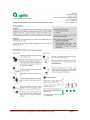
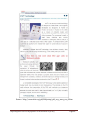
1
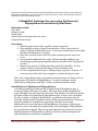
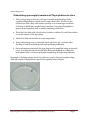
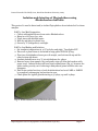
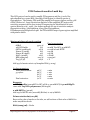
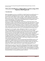
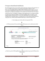
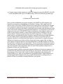
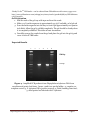
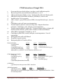
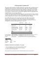
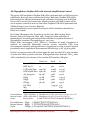
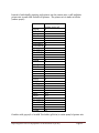
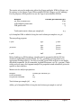
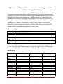
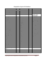
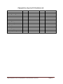
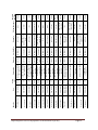
Rapid Diagnostic Tools for Phytophthora on Horticultural Crops Authors Jean Beagle Ristaino, Ph. D. North Carolina State University, Raleigh, North Carolina [email protected] Kelly Ivors, PhD Cal Poly San Luis Obisbo Peter Bonants, PhD Plant Research International, Wageningen, NL [email protected] Monica Blanco, PhD Unversidad de Costa Rica, San Jose Costa Rica [email protected] David Cooke, Ph. D. The James Hutton Institute, Invergowrie, UK [email protected] Pallem Chowdappa ICAR, Banaglore India, cover illustration Rapid diagnostic tools for Phytophthora on horticultural crops 2015 Page 2 Rapid Diagnostic Tools for Phytophthora on Horticultural Crops Table of Contents Isolation, growth, and morphology of Phytophthora p. 4― p. 15 PCR protocols used in Lucid key p. 16 ― p. 20 Molecular identification of Phytophthora p. 21 ― p. 24 Quick NaOH extraction from dried leaf samples p. 25 PCR with Ready-To-Go™ PCR beads p. 26 ― p. 27 CTAB extraction of DNA p. 28 – p. 29 DNEasy plant mini kit extraction p. 30 Phytophthora spp. identification using PCR-RFLP technique p. 31 – p. 32 All Phytophthora Taqman PCR p. 36 – p. 38 12-plex microsatellite genotyping of P. infestans p. 39 – p. 41 Detection of P. ramorum using LAMP p. 42– p. 43 Rapid assay product information p. 44 – p. 52 Phytophthora species ID worksheet p. 53 – p. 54 Phytophthora species characters p. 55 Rapid diagnostic tools for Phytophthora on horticultural crops 2015 Page 3 Protocol derived from this reference: National Plant Diagnostic Network (NPDN) News 4: Vol. 2 Issue 3. Gail Ruhl, Plant and Pest Diagnostic Laboratory, Purdue University. Float incubation technique for Phytophthora and Pythium diagnostics This float incubation technique is an excellent way to induce the production of sporangia as well as mycelial growth from herbaceous tissue for diagnostic purposes. A 1% unsterilized soil extract solution works well for stimulating sporangia production from Phytophthora- infected tissue. This float technique may also be used to stimulate sporangia production from mycelium growing on agar plugs. 1. Prepare a 1% unsterilized soil extract by swirling 10 g soil and 1 L distilled water together in a 2 L flask. 2. Slowly pour extract solution through filter paper lined funnel into media storage bottle and store in refrigerator until needed. Wash funnel and replace filter paper for each bottle. 3. Pour enough refrigerated soil extract solution into Petri dish containing herbaceous roots, stems, and/or leaves to just cover the plant material. 4. Cover Petri dish and incubate on benchtop for 24 hrs. 5. Examine herbaceous material with a compound microscope while it is still floating in the soil extract solution. Look for sexual (oogonia with antheridia or double walled oospores) or asexual (sporangia; Pythium globose and Phytophthora lemon shaped) reproductive structures. Note: Do not confuse protozoans which thrive on herbaceous material floating in unsterilized soil extract solution with sporangia or zoospores. Rapid diagnostic tools for Phytophthora on horticultural crops 2015 Page 4 Protocol from the lab of Jason H. Brock and Glenn H Beard, University of Georgia, 2002. Reference: Miscellaneous Publication 104, The Cooperative Extension Service, University of Georgia College of Agricultural and Environmental Science. A Simplified Technique for recovering Pythium and Phytophthora from infected plant tissue Materials needed: Petri Dish Scalpel or knife Sterile water Acid Fuschin stain (preferred over water) Compound microscope Procedure: 1. Wash the plant tissue under a gentile stream of tap water. 2. Slice multiple sections of tissue from the plant. Select tissue from the border of diseased and healthy areas. To prevent contamination, use a blade that has been flamed over a burner. 3. Place the sectioned tissue in the Petri dish containing sterile water and cover. 4. Leave the dish undisturbed for 24 hr. Pythium and Phytophthora spp. should begin producing sporangia between 24 and 48 hr after being placed in water. 5. Remove one section, including mycelial growth, from the dish. Use the scalpel or blade to macerate the tissue in the acid fuschin stain. 6. Use the compound microscope to find sporangia, oogonia or oospores. If none are preset, allow the tissue samples to continue floating in water. Note: Lack of mycelial growth or reproductive structures does not always rule out the presence of Pythium and Phytophthora. Growth of pathogens can be influenced by temperature and light. Further evaluation may be needed. Identification of Pythium and Phytophthora: A shared morphological characteristic of Pythium and Phytophthora spp. is coenocytic hyphae (lacking cross walls). Most true fungi produce hyphae with septa, although some true fungi also lack septa. The presence of coenocytic hyphae provides evidence for Pythium and Phytophthora but, in itself, is not conclusive. In order to make a conclusive identification, reproductive structures must be observed. Pythium and Phytophthora have similar sexual reproductive structures. Asexual reproductive structures are sporangia, and allow for an easier differentiation between Pythium and Phytophthora. Sporangia of Pythium are globose to oval or may have an irregular shape, while sporangia of Phytophthora are typically lemon-shaped. These are general characteristics that can be used to identify Pythium and Phytophthora; however, diversity within each genus can make identification much more complicated. Rapid diagnostic tools for Phytophthora on horticultural crops 2015 Page 5 General lab protocol Stimulating sporangia formation of Phytophthora in vitro 1. Using a #6 or large cork borer (or larger), transfer multiple plugs of the suspected Phytophthora isolate into an empty Petri dish. It is best to use cultures less than 7 days old; isolates growing on corn meal agar work best. (Cultures on PARP have worked okay sometimes, but many Phytophthora species do not sporulate well on media containing antibiotics). 2. Flood the Petri dish with 2% soil extract solution or dilute V8, until the solution covers the surface of the agar plugs. 3. Label Petri dish and incubate at room temperature. 4. Using a dissecting scope, continually check agar discs 48 – 96 hours after flooding to look for mycelial growth and sporangia production. 5. Once sporangia are detected, the agar plugs can be magnified using an inverted scope, or removed from the flooded Petri dish and smashed on a slide with lacto-phenol blue to observe sporangial morphology at higher magnification. Sporangia of Pythium species tend to be globose to oval or have an irregular shape, while sporangia of Phytophthora species are typically lemon-shaped. Rapid diagnostic tools for Phytophthora on horticultural crops 2015 Page 6 Original protocol published in Inoculum, 56 (6), 2005 by Susan Kaminskyj. Purification of Phytophthora cultures contaminated with bacteria This protocol can be used to purify Phytophthora cultures that are contaminated with bacteria. 1. Choose a plate of media containing antibiotics that was poured thinly (< 1 cm thick). PARP(H) is a good choice, but anything with antibiotics should work. 2. Cut the plate into quarters with a sterile scalpel and set aside. 3. Using a sterile scalpel, remove a thin layer of media from the growing hyphal edge of the contaminated culture. The circumference of the sliver should be about that of a pencil eraser. 4. Place this sliver in the center of an empty, sterile Petri dish. 5. Using a sterile spatula, place a quarter-piece of the antibiotic media on top of the sliver (Fig. 3). 6. Allow 2-4 days for the Phytophthora mycelia to grow up through the media. The bacteria should not be able to move vertically through the media. 7. Using a sterile scalpel, remove a thin layer of media containing uncontaminated hyphae, being careful not to penetrate too deeply through the media. 8. Transfer this wedge onto nutrient agar to make sure that the decontamination was successful. Fig. 1. A quarter-piece of PARP is placed on top of a piece of contaminated Phytophthora culture. Rapid diagnostic tools for Phytophthora on horticultural crops 2015 Page 7 Protocol from the lab of Kelly Ivors, North Carolina State University, 2004. Isolation and detection of Phytophthora using Rhododendron leaf baits This protocol is used to detect and/or isolate Phytophthora from infested soil or tissue samples. PART 1: Leaf Bait Preparation: 1. Collect unblemished leaves from native Rhododendron. 2. Rinse in 10% Clorox for 1 min. 3. Triple rinse with distilled water. 4. Blot dry and place in plastic bag. 5. Store at 4˚C (refrigerator) overnight. PART 2: Leaf Baiting and Isolation: 1. Set incubator temperature at 12˚C both day and night. Turn lights OFF. 2. Place soil or plant tissue to be baited in large gallon ZIPLOC ® bag. 3. Float two rhododendron leaves per soil sample, one bottom side up and the other bottom side down. 4. Incubate baited leaves at 12˚C in total darkness for 4 days. 5. Remove leaves from sample, blot and gently wipe soil from leaf surfaces with paper towel. Infected leaves will have water soaked tissue (Fig. 4), however the water soaking can dry out if culturing is delayed and prime isolation sites can disappear. 6. Plate water-soaked sections of each rhododendron leaf onto PARP or PARPH by completely submerging leaf sections in agar. 7. Check plates for hyphal growth starting at 3-4 days, up until 14 days. Fig. 2. Water-soaked veins of Rhododendron leaf. Rapid diagnostic tools for Phytophthora on horticultural crops 2015 Page 8 Methods of isolation of Phytophthora species A. Introduction to methods of culture. Some species of Phytophthora such as P. capsici are readily cultured via simple surface disinfestation and plating on a selective media. Other species, such as P. infestans from tomato, are more recalcitrant and require passage through a living clean tomato leaf before isolation of sporangia onto antibiotic amended agar. All species can be simply stored on lima bean or corn meal agar disks in sterile water with sterile hemp seed for long term storage. More elaborate and expensive cryostorage in liquid nitrogen can also be done (Tooley, 1988). The following methods and media are provided to assist in the isolation of a Phytophthora species in culture and are not meant to be an inclusive list. Further methods can be found in Erwin and Ribeiro (1996), Gallegly and Hong (2008) and Ribeiro (1978). Contamination by bacteria is common in cultures and should be eliminated before production of asexual and sexual structures for identification. Plating isolates on antibiotic amended media is useful and then transfer to nonamended V-8 or lima bean agar should be done to confirm that bacteria have been eliminated from cultures. Once a clean isolate is obtained, production of asexual sporangia, chlamydospores and sexual structures are needed in order to proceed with morphological identification. See methodologies recommended in Erwin and Ribeiro (1996), Gallegly and Hong (2008) and described briefly below for production of structures for morphological identification. B. Isolation. Most Phytophthora species can be isolated from small pieces of infected plant tissue after surface disinfestation in 0.05% sodium hypochlorite for 3-5 minutes, followed by rinsing in sterile distilled water and blotting on sterile paper towels. Phytophthora species can also be baited from soil or water with rhododendron leaf disks (Erwin and Ribiero, 1996; Larkin et al, 1995). Small pieces of surface disinfested plant tissue are plated on antibiotic amended media such as TPT, PAR(PH) or P10VP media. Phytophthora species are Oomycetes and have coenocytic hyphae. First, mycelium growing from tissue pieces should be observed for coenocytic hyphae. Then proceed to observe morphological characters of asexual and asexual structures. 1. Isolation Media. a. Triple P media (TPT) Add amendments to one liter of corn meal agar (17g/L) after autoclaved and cool. Penicillin G - sodium salt* Polymyxin B Sulfate* Pimaricin (50ai)*(store in dark) PPM 50 mg/L 50 mg/L 30 mg/L Stock 50 mg/ml 50 mg/ml 20 mg/ml Rapid diagnostic tools for Phytophthora on horticultural crops 2015 Use 1.0 ml 1.0 ml 3.0 ml Page 9 *Add amendments separately in a laminar flow hood while stirring to media after sterile and cooled to 50 C. Fungicide and antibiotic stock solutions should be made in sterile bottles with sterile distilled water and stored at 5 C or frozen in sterile plastic vials in aliquots for 1 liter. Pimaricin is sold under the trade name Delvocid (50ai) from Nelson-Jameson, Inc, Marshfield, WI. b. PAR(PH) Media for Phytophthora Add to one liter of corn meal agar (17g/L) or V-8 Agar (see below). Pimaricin (50ai)* Ampicillin* Rifampicin* PPM 10 mg/L 250 mg/L 10 mg/L Stock 20 mg/ml 25 mg/ml 10 mg/ml Use 1.0 ml 10.0 ml 1.0 ml Optional PCNB (96ai)* Hymexazol (99.5ai)** 100 mg/L 50 mg/L 5.2 mg/ml 25 mg/ml 20 ml 2.0 ml *Add amendments separately in a laminar flow hood while stirring to media after sterile and cooled to 50 C. Dissolve ampicillin and rifampicin in 70% ethanol and PCNB in 95% ethanol. All fungicides and antibiotic stock solutions should be prepared aseptically and stored at 5 C or frozen in sterile plastic vials in aliquots for 1 liter. **Hymexazol is added to suppress Pythium species. PCNB is used to suppress soil fungi and is useful for soil dilution plating. Can use Terraclor (PCNB 75%WP) as an alternative to the active ingredient. c. Modified P10VP Corn Meal Agar Add to one liter of corn meal agar (17g/L) Pimaricin* Vancomycin* PCNB(96ai)* PPM 10 mg/L 200 mg/L 100 mg/L Stock 20 mg/ml 50 mg/ml 5.2 mg/ml Use 1.0 ml 4.0 ml 20 ml Optional Hymexazol (99.5ai)** 25 mg/L 25.1 mg/ml 1.0 ml *Add amendments separately in a laminar flow hood while stirring to media after sterile and cooled to 50 C. Dissolve PCNB in 95% ethanol. All fungicides and antibiotics should be prepared aseptically and stored at 5 C or frozen in sterile plastic vials in aliquots for 1 liter. **Hymexazol is added to suppress Pythium species. d. Goodwins Media (for isolation of Phytophthora infestans from plant tissue) Rapid diagnostic tools for Phytophthora on horticultural crops 2015 Page 10 After preparing 10% V-8 Juice Agar add Antibiotics: Rifamycin Polymixin B Sulphate Ampicillin* PPM 20 mg/L 50 mg/L 200 mg/L Stock 10 mg/ml 50 mg/ml 25 mg/ml Use 2.0 ml 1.0 ml 8.0 ml Fungicides: PCNB 75 WP Benomyl 50 WP 50 mg/L 100 mg/L 67 mg/ml 10 mg/ml 1.0 ml 2.0 ml *Dissolve ampicillin in 70% ethanol. Add amendments separately in a laminar flow hood while stirring to media after sterile and cooled to 50 C. All fungicides and antibiotics should be prepared aseptically and stored at 5 C or frozen in sterile plastic vials in aliquots for 1 liter. Surface sterilize tissue in 70% Ethanol (15s) then 10% Clorox (2-5 min). Rinse in distilled water. Plate sporangia from lesions on media and incubate plates at 18 C in the light. 2. Soil Dilution Plating – for isolation of soilborne species Soil dilution plating can be performed to isolate some species with a soilborne phase (ie, P. capsici, P. nicotianae, P. cinnamomi, P. ramorum). Forty grams of soil is added to 160 ml 0.25% sterile water agar, stirred for 5 min, and 1 ml aliquots are plated onto each of 5 plates of Masago’s selective medium. At higher inoculum levels, additional 1:5 serial dilutions are needed. Plates are incubated in the dark at 24 C for 72 hr, rinsed under running water to remove soil residue, and colonies are counted. Gravimetric soil water content (g water /g dry soil) of the soil samples is determined at the time of soil dilution and used to calculate inoculum density per g dry soil. Masago's (Phytophthora isolation from soil) (Masago et al, 1977). Potato Dextrose Agar 39g/L PCNB (96ai)* Benomyl (50 wp) Ampicillin (100ai)* Rifampicin (100ai)* Nystatin (100ai) Hymexazol (99.5ai) PPM 25 mg/L 10 mg/L 500 mg/L 10 mg/L 25 mg/L 25 mg/L Stock Use 5.2 mg/ml 5.0 ml 10 mg/ml 2.0 ml 25 mg/ml 20.0 ml 10 mg/ml 1.0 ml 25 mg/ml 1.0 ml 25.1 mg/ml 1.0 ml *Dissolve ampicillin and rifampicin in 70% ethanol and PCNB in 95% ethanol. Add separately to media after sterile and cooled to 50 C in a laminar flow hood while stirring. All fungicides and antibiotics should be prepared aseptically and Rapid diagnostic tools for Phytophthora on horticultural crops 2015 Page 11 stored at 5 C or frozen in sterile plastic vials in aliquots for 1 liter. Hymexazol will inhibit most Pythium species. 3. Growth media. Most species of Phytophthora will grow on lima bean agar or V8 juice agar. a. Fresh Lima Bean Agar. A good general media for most species of Phytophthora. Boil 200g lima beans for 30 minutes in 1 liter of water. Strain through cheesecloth and bring up to 1 liter. Add 1g glucose and agar (17g/L). Autoclave. Optional: 2 ppm beta- sitosterol (0.8ml of .25% ETOH) for oospore production. Add before autoclaving. b. V-8 CaCO3 Agar. A good growth medium for most species and useful for sporangia formation but difficult to see through media for observation of sexual oospores. V-8 juice (200ml), 800ml distilled water, CaCO3 (2g), and agar (17g/L). All ingredients are mixed and autoclaved for 30 minutes. c. V-8 Broth for Vermiculite Culture. Used to prepare inoculum for soil infestation. Prepare V-8 juice broth; V-8 juice (200ml), distilled water (800ml), CaCO3 (2g). Add 250ml V-8 broth to 500cm3 of vermiculite in 1 qt mason jars and autoclave for 1 hour with a vented lid (plugged with foam stopper) in water-filled pan on two successive days. Alternatively standard laboratory Erlenmeyer flasks can also be used. Cool and cover lid with a plastic bag. Seed vermiculite with inoculum plug of Phytophthora sp. Shake after 2 or 3 weeks. This vermiculite media is useful for inoculation of soil with soilborne species to conduct Koch’s postulates. d. Clarified V-8 Juice Agar. A good medium for observing oospores. Clarify V-8 juice by centrifuging at 4340 g for 10 minutes. Mix 200 ml V8 supernatant, CaCO3 (2g), distilled water (800 ml), and filter through Whatman #1 filter paper. Then add 17g agar and autoclave. Optional: 2 ppm beta- sitosterol (0.8ml of .25% ETOH) for oospore production. Add before autoclaving. e. V-8 Rye Agar. A medium for growth of P. infestans. Rapid diagnostic tools for Phytophthora on horticultural crops 2015 Page 12 Soak 50g rye seed (nonfungicide treated) in 1100ml distilled water at 24 C for 24 36 hours followed by autoclaving for 30 minutes. Filter supernatant through 4 layers of cheesecloth, adjust final volume to 1000 ml with distilled water. Add: 5% V-8, 0.02% CaCO3, 2% Bacto agar. Autoclave. f. Pea Broth. Autoclave 120 g of frozen peas in 1 L of water for five minutes. Filter the supernatant through cheesecloth, pour into bottles and autoclave. This is a general broth for growing mycelia cultures of Phytophthora species. C. Production of structures for morphological identification 1. Growth. Isolates can be tested for growth on lima bean agar at a range of temperatures of 20, 25, 30 and 35 C. 2. Sporangia – Sporangia are the cells or vessels in which zoospores are formed. Agar disks containing mycelium from cultures removed from either lima bean or V-8 juice agar are placed in sterile petri dishes and covered with a thin layer of sterile distilled water or sterile or non-sterile soil extract. Non-sterile soil extract is prepared by adding 1000 ml distilled water to 15 g air-dried field soil. Soil is stirred vigorously for at least 4 hr and allowed to settle overnight. The supernatant is filtered through two layers of cheesecloth, centrifuged at 1935 g for 15 min, filtered through coarse filter paper and can be either autoclaved or used non-sterile. Non-sterile soil extract is more effective then sterile soil extract for sporangia production. Store in the refrigerator at 4 C. (Jeffers and Aldwinkle, 1987). A thin layer of sterile distilled water or soil extract is added to petri plates containing disks removed from cultures. Do not submerge the disks. Plates are incubated under cool white fluorescent light for 1-3 days and observed daily under the dissecting scope for sporangia. Slides can be made and sporangia type (papillate, semipapillate, or nonpapillate), number of apices, shape and size can be observed and measured. Length and breadth (width) of 10 sporangia are measured with an ocular micrometer and length /breath ratio’s are calculated. 3. Sporangiophore. Sporangiosphores are the hyphal strands on which sporangia are borne. Morphology of sporangia can be observed using a binocular microscope from the agar disks described above (2). Sporangiophores can be branched or unbranched to form compound or simple sympodia. The sporangiophore emerges from the base of previous sporangium in either a lax or close manner in a simple sympodium. Sporangia can form in umbels (an inverted umbrella-like cluster of sporangia) on the sporangiophore, or very long irregular branches. Sporangia may proliferate internally through previously formed sporangia on the sporangiophore. Sporangia can be borne in tight or botryose clumps on the sporangiophore. Rapid diagnostic tools for Phytophthora on horticultural crops 2015 Page 13 4. Cauducity. Sporangial disks produced as described above (2) are placed on a microscope slide and agitated to dislodge sporangia in water. Cauducous sporangia will break or fall away from the sporangiophore readily when agitated and can be observed. Pedicel (sporangial stalk) length is measured. The pedicel is the hyphal strand left attached to the sporangium in cauducous species. Pedicel lengths can be grouped into small (< 5 um), medium (5-10 um) and long (> 10 um) categories. 5. Oospores - Oospores of heterothallic isolates are produced by placing an agar disk containing mycelium of an unknown isolate 2-3 cm apart from a tester isolate on clarified V8 (CV8) agar or lima bean agar. Tester isolates of known opposite mating type (A1 or A2) are needed for pairing with each unknown isolate and serve as controls. Homothallic isolates do not require pairing and should produce oospores in single culture. Cultures are incubated in the dark at 20-22 C for approximately 1 mo. Oogonia and antheridia should form within 7 days but oospore formation may take longer. Oospores formed in a distinct band between opposite mating types will confirm heterothallic species. 6. Oogonia and oospore diameter can be measured. Oospore diameter is measured using the outer wall of the oospore. Oogonial diameter is measured using the outer oogonial wall contained within the oospore. Measurements in two directions are usually done and at least 10 oospores or oogonia should be measured. 7. Antheridial characters should be observed. The antheridium is the male gametangium, and is a multinucleate, swollen hyphal tip that can be affixed to the basal side of the oogonium (paragynous) or the oogonial stalk can grow through the antheridium so that the antheridium surrounds the oogonial stalk (amphigynous). Antheridia may be 1 or 2 celled. 8. Hyphal characteristics including hypha swellings and presence of chlamydospores can be observed in thin plates of lima bean agar. E. Stock cultures. Stock cultures of most species can be maintained on cornmeal agar or lima bean agar slants covered with sterilized mineral oil at 20C. Agar disks of most species can also be stored in sterilized water containing autoclaved hemp seed in 1 ml vials at room temperature. Rapid diagnostic tools for Phytophthora on horticultural crops 2015 Page 14 References Erwin, D. C. and Ribeiro, O. K. 1996. Phytophthora diseases worldwide. Amer. Phytopathol. Soc. Press, St. Paul, MN. 562 pp. Gallegy, M. and Hong, C. 2008. Phytophthora: Identifying species by morphology and DNA fingerprints. American Phytopathological Society Press, St. Paul, Mn.158pp. Jeffers, S.N., and Aldwinkle, H. S. 1987. Enhancing detection of Phytophthora cactorum in naturally infested soil. Phytopathology 77:1475-1482. Larkin, R.P., Ristaino, Jean B., and Campbell, C. L. 1995. Detection and quantification of Phytophthora capsici in soil. Phytopathology 85:1057-1063. Masago, H., Yoshikawa, M., Fukada, M., and Nakanishi, N. 1977. Selective inhibition of Pythium spp. on a medium for direct isolation of Phytophthora spp. from soils and plants. Phytopathology 67: 425-428. Ribeiro, O. K. 1978. A source book of the genus Phytophthora. , J. Cramer, Vaduz Liechtenstein. 417 pp. Tooley, P. W. 1988. Use of uncontrolled freezing for liquid nitrogen storage of Phytophthora species. Plant Dis. 72:680-682. Rapid diagnostic tools for Phytophthora on horticultural crops 2015 Page 15 PCR Protocols used in Lucid Key This PCR protocol can be used to amplify ITS sequences and the 5’ end of the mitochondrial cox 1 gene (BOL, Barcode of Life Region) to identify species of Phytophthora. The primers ITS6 and ITS4 amplify both spacer regions and the 5.8S rDNA (White et al., 1990). Restriction digestion of the amplified ITS region with restriction enzymes can be done instead of sequencing for identification of some species (Ristaino et al., 1998). See further methods for use of restriction analysis fingerprints at http://phytophthora-id.org/files/PhytophthoraID%20sequencing%20protocols.pdf. See ITS and BOL maps of gene regions amplified with primers below. Master mix for each 50µl reaction ddH2O 10X PCR buffer dNTPs (2mM each) Primer-F (10µM) Primer-R (10µM) MgCl2 (50mM) BSA (20mg/ml) Taq (5U/µl) 35.25 µl 5 µl 2.5 µl 2 µl 2 µl 1.8 µl 0.25 µl 0.2 µl Final concentration 20 mM Tris-HCl; 50 mM KCl 0.1 mM of each DNTP 0.4 µM 0.4 µM 1.8 mM 0.1mg/ml 1U Add 49 µl of master mix to 1µl template DNA (5-10 ng) Cycling parameters: Initial denaturation: 35 cycles: Final extension: 96 ºC 96 ºC 56 ºC 72 ºC 72 ºC 2 min 1 min 1 min 2 min 10 min Reagents: 10X PCR buffer (200 mM Tris-HCl, pH 8.4; 500 mM KCl) & 50 mM MgCl2 come with Taq DNA polymerase (Invitrogen) 2 mM dNTPs: (300 ml) Add 24 µl of dNTPs mix (100 mM) (Bioline) to 276 µl ddH2O Primer stock soln (100 µM) Base on the value of nmole on the tube, we add 10 times of that value of ddH2O to make 100µM stock soln. BSA (20mg/ml) – Roche Rapid diagnostic tools for Phytophthora on horticultural crops 2015 Page 16 Primers: ITS region – ITS6 – GAAGGTGAAGTCGTAACAAGG ITS4 – TCCTCCGCTTATTGATATGC ITS6 18S rDNA ITS-1 5.5S rDNA ITS-2 28 S rDNA ITS4 BOL region – FM80RC – TTTCAACAAATCATAAAGATATT FM85 – AACTTGACTAATAATACCAAA Cox 1 Cox 1 BOL FM80RC FM85 VI. REFERENCES Blair, J.E., Coffey, M.D., Park, S-Y., Geiser, D.M., and Kang, S. 2008. A multi-locus phylogeny for Phytophthora utilizing markers derived from complete genome sequences. Fungal Genetics and Biology 45: 266–277. Bonants, P.J.M., Hagenaar-de Weerdt, M., van Gent-Pelzer, M., Lacourt, I., Cooke, D.E., and Duncan, J.M. 1997. Detection and identification of Phytophthora fragariae Hickman by the polymerase chain reaction. Eur. J. Plant Pathol. 103: 345-355. Bonants, P.J.M., van Gent-Pelzer, M.P.E., Hooftman, R., Cooke, D.E.L., Guy, D.C., and Duncan, J.M. 2004. A combination of baiting and different PCR formats, including Rapid diagnostic tools for Phytophthora on horticultural crops 2015 Page 17 measurement of real time quantitative fluorescence, for the detection of Phytophthora fragariae in strawberry plants. European Journal of Plant Pathology 110: 689-702. Cooke, D. E. L., Drenth, A., Duncan, J. M., Wagels, G. and Brasier, C. M. 2000. A molecular phylogeny of Phytophthora and related Oomycetes. Fungal Gen. Biol. 30:17-32. Forster, H., M. P. Cummings, and Coffey, M. D. 2000. Phylogenetic relationships of Phytophthora species based on ribosomal ITS I DNA sequence analysis with emphasis on Waterhouse groups V and VI. Mycol. Res. 104:1055-1061. Gallegy, M. and Hong, C. 2008. Phytophthora: Identifying species by morphology and DNA fingerprints. American Phytopathological Society Press, St. Paul, Mn.158pp. Kang, S., Blair, J.E., Geiser, D.M., Khang, C., Park, S., Gahegan, M., O Donnell, K., Luster, D.G., Kim, S.H., Ivors, K.L., Lee, Y., Lee, Y., Grunwald, N.J., Martin, F.N., Coffey, M.D., Veeraraghavan, N., Makalowska, I. 2006. Plant pathogen culture collections: It takes a village to preserve these resources vital to the advancement of agricultural security and plant pathology. Phytopathology. 96:920-925. Kong, P., Hong, C., Richardson, P.A. and Gallegly M. E. 2003. Single-strandconformation polymorphism of the ribosomal DNA for rapid species differentiation in the genus Phytophthora. Fung Gen Biol 39:238-249. Kroon, L.P.N.M., Bakker, F.T., van den Bosch, G.B.M., Bonants, P.J.M., and Flier, W.G. 2004. Phylogenetic analysis of Phytophthora species based on mitochondrial and nuclear DNA sequences. Fungal Genet. Biol. 41: 766-82. Martin, F. N. and. Tooley, P. W. 2003. Phylogenetic relationships among Phytophthora species inferred from sequence analysis of mitochondrially encoded cytochrome oxidase I and II genes. Mycologia 95:269-284. Mills, S.D., Forster, H. and Coffey, MD. 1991. Taxonomic structure of Phytophthora cryptogea and P. dreschsleri based on isozyme and mitochondrial DNA analysis. Mycol Res 95:31-48. Park, J., Park, B., Veeraraghavan, N., Jung, K., Lee, Y., Blair, J., Geiser, D., Isard, S., Mansfield, M., Nikolaeva, E., Park, S., Russo, J., Kim, S., Greene, M., Ivors, K., Balci, Y., Peiman, M., Erwin, D. C., Coffey, M. B., Rossman, A., Farr, D., Cline, E., Grünwald, N. J., Luster, D. G., Schrandt, J., Martin, F., Ribeiro, O., Makalowska, I., and Kang, S. 2008. Phytophthora Database: A Forensic Database Supporting the Identification and Monitoring of Phytophthora. Plant Dis. 92: 966-972. Ristaino, J. B., Madritch, M., Trout, C. L. and Parra, G. 1998. PCR amplification of ribosomal DNA for species identification in the plant pathogen genus Phytophthora. Appl. Environ. Microbiol. 68:948-954. Rapid diagnostic tools for Phytophthora on horticultural crops 2015 Page 18 Ristaino, J. B. 2012. A Lucid Key to the common species of Phytophthora. Plant Disease 96:897-903. Schena, L., Duncan, J.M., and Cooke, D.E.L. 2008. Development and application of a PCR-based ‘molecular tool box’ for the identification of Phytophthora species damaging forests and natural ecosystems. Plant Pathology 57: 64–75. Tooley, P.W., Bunyard, B.A., Carras, M.M., and Hatziloukas, E. 1997. Development of PCR primers from Internal Transcribed Spacer region 2 for detection of Phytophthora species infecting potatoes. Appl. Environ. Microbiol. 63: 1467-1475. Trout, C.L, Ristaino, J.B., Madritch, M, and Wangsomboondee, T. 1997. Rapid Detection of Phytophthora infestans in late blight infected tissue of potato and tomato using PCR. Plant Disease 81: 1042-1048. Wang, H., Qi, M., and Cutler, A. J. 1993. A simple method of preparing plant samples for PCR. Nuc. Acids. Res. 21:4153-4154. White, T.J., Burns, T., Lee, S., and Taylor, J. 1990. Amplification and direct sequencing of fungal ribosomal RNA genes for phylogenetics. Pages 315-322 in: Innis, M.A., Gelfand, D.H., Sninsky, J.J., and White T.J. (eds). PCR Protocols: A guide to Methods and Applications. Academic Press, San Diego, CA. Other useful detection method papers Choi, Y-J., Beakes, G., Glockling, S., Kruse, J., Nam, B., Nigrelli, L., Ploch, S., Shin, HD., Shivas, R.G., Telle, S., Voglmayr, H., Thines, M. 2015. Towards a universal barcode of oomycetes – a comparison of the cox1 and cox2 loci. Mol. Ecol. Res. DOI: 10.1111/1755-0998.12398. Martin, F.N., Abad, Z. G., Balci, Y., Ivors, K. 2012. Identification and detection of Phytophthora: Reviewing our progress, identifying our needs. Plant Disease 96: 1080―1103. Miles, T. D., Martin, F. N., Coffey, M. D. 2015. Development of rapid isothermal amplification assays for detection of Phytophthora spp. in plant tissue. Phytopathology 105: 265-278. Robideau, G. P., de Cockc, A. W. A. M., Coffey, M. D., Voglmayr , H., Bonants , P. J. M., Ristaino, J. B., Chitty, D., Rintoul, T., Désaulniers, N, Eggertson, Q., Bala, K., Gachon , C. M. M., Smith, M. L., Lévesque. A. 2011 DNA barcoding of oomycetes with cytochrome c oxidase subunit I (COI). Mol. Ecol. Res. 11: 1002-1011. Sikora, K., Verstappen, E. Mendes, O., Schoen, C., Ristaino, J. and Bonants, P. 2012. A Universal Micro-array Detection Method for identification of Multiple Phytophthora Species using Padlock Probes. Phytopathology 102:635-645. Rapid diagnostic tools for Phytophthora on horticultural crops 2015 Page 19 Van Doorn, R., Sławiak, M., Szemes, M., Dullemans, A.M., Bonants, P., Kowalchuk, G.A., Schoen, C.D. 2009. Robust detection and identification of multiple oomycetes and fungi in environmental samples by using a novel cleavable padlock probe-based ligation detection assay. Applied and Environmental Microbiology, 75 (12) pp. 41854193. Rapid diagnostic tools for Phytophthora on horticultural crops 2015 Page 20 Protocol from the lab of Seogchan Kang (Michele Mansfield and Seogchan Kang), Penn State University, 2008. Molecular identification of Phytophthora isolates using a DNA Sequence Based Approach I. Introduction Many Phytophthora species can be difficult to identify based on morphology due to a lack of physically distinguishing characteristics and variability of morphological characteristics under different culture conditions. Additionally, it may take several days or weeks for an isolate to mature enough to develop identifying characteristics. Because time is often of the essence in identifying and assessing the potential risk of a newly isolated pathogen, DNA sequence-based identification is frequently used to augment and complement morphological data. To serve as a baseline for identification, classification, and risk assessment of new Phytophthora isolates, PD cataloged genotypic and phenotypic information on isolates of previously described species in a web-accessible and searchable format. To support the identification of new Phytophthora isolates via comparison of their sequences at one or more loci with the corresponding sequences derived from the isolates archived in PD, sequence data from up to nine loci have been generated from more than 2,000 isolates from known and novel species (94 in total) and deposited the data in PD so that these loci can be used for species identification (Blair et al., 2007; Park et al., 2008). The characterized loci include the following: (i) two loci in the nuclear ribosomal RNA (rRNA) encoding genes: the internal transcribed spacer (ITS) regions and the 5’ portion of the large subunit rRNA gene, (ii) nuclear genes encoding 60S ribosomal protein L10, beta-tubulin, enolase, heat shock protein 90, TigA fusion protein, and translation elongation factor 1 alpha, and (iii) a mitochondrially-encoded coxII gene and spacer region between coxI and coxII. PCR reaction conditions for amplifying these marker loci, including the sequences and positions of primers used, can be found in the Genetic Markers section of PD and are hyperlinked from marker names throughout the PD user interface. A comprehensive phylogenetic analysis was performed (using Pythium vexans as an outgroup) to establish evolutionary relationships among the characterized species (Blair et al., 2007), in which sequences of seven loci (all the markers described above except the ITS and cox regions) derived from 228 isolates from 83 species were utilized. The result is shown in the form of a genus-wide phylogenetic tree via individual species pages in PD and will be updated periodically. Sequences employed in this analysis and sequence alignments are available for downloading. Data search and analysis tools in PD (Fig. 7) include BLAST, Phyloviewer (a program for building phylogenetic trees using sequences of selected isolates), and Virtual Gel (a program for generating expected restriction patterns for given sequences). The PD also provides a customized means of storing and sharing data via the web. Rapid diagnostic tools for Phytophthora on horticultural crops 2015 Page 21 II. Sequence-Based Strain Identification The following flowchart protocol will provide a descriptive and pictorial explanation of how to use DNA sequence data along with the Phytophthora Database (PD; http://www.phytophthoradb.org) and GenBank search functions in order to identify new isolates. Detailed descriptions of how to use search functions in PD and of how data can be moved from one analysis tool to another can be found in the user manual at the PD web site. The BLAST tool allows for the identification of an unknown isolate by querying the sequence database in PD (and GenBank) using one or more of the marker sequences described above. Given the comprehensive set of ITS sequences available for the archived isolates, we suggest users to begin the identification process using this locus, which should establish its identity at or near the species level. 1. Grow single-spore isolate and extract genomic DNA 2. PCR using Internal Transcribed Spacer (ITS) primers and conditions described below Fig. 7. 3. Check for positive PCR amplification on an electrophoresis gel, purify PCR products and sequence Rapid diagnostic tools for Phytophthora on horticultural crops 2015 Page 22 4. Manually edit sequence data using appropriate program 5. Compare target isolate sequence to other ITS sequences using the BLAST tool in PD (www.phytophthoradb.org) or GenBank (http://blast.ncbi.nlm.nih.gov/Blast.cgi) 6. Evaluate the search results Once you have submitted your target sequence, the BLAST tool will compare your sequence to sequences contained in your database of choice. The output you will receive will be a list of the CLOSEST but not necessarily EXACT matches (for example if your sequence represents a new species you may not have an exact match). In PD, you will also be provided with links to individual species names and descriptions in the order of the closest matches to your submitted sequence. It is important to look at the output you receive as a COLLECTIVE body of information that suggests what your isolate is most closely related to, rather than a single answer consisting of the top closest match. There are several reasons why this is important: (i) the top closest match may be a misidentified isolate, therefore, using this match as a single identifier for your isolate may be perpetuating a mistake; (ii) even if your isolate has 99% sequence identity to an already described species, several small or singular sequence differences between your isolate and the closest match may suggest that you have a novel species. Given the intensive curation performed on the data archived in PD, the first problem is unlikely (but not impossible). The second situation illustrates why it is often necessary to take the search process further by generating an alignment with the top closest matches and performing a base-by-base visual sequence comparison to determine if there are true differences. If the closest match exhibits substantial sequence differences from your sequence, it is possible that the unknown isolate may belong to a new species. In the latter case, one may sequence all or some of the seven loci used for the genus-wide phylogenetic analysis (Blair et al., 2007) to investigate this possibility further. Characterization of morphological and biological traits (e.g., growth characteristics, pathogenicity on plants) will also be needed to formally describe a new species. Rapid diagnostic tools for Phytophthora on horticultural crops 2015 Page 23 (A) BLAST Search (B) Sequence Alignment (C) Phylogenetic Tree Marker Sequences Database Data Submission (D) Virtual Gel (E) Data Download & Storage Cart & Folder Fig. 3. Overview of the functionality and data flow in PD. This diagram illustrates (A) BLAST, (B) Clustal W: a tool that will align and illustrate a base-by-base comparison between your isolate and the closest matches, (C) Phyloviewer: a tool to visualize the evolutionary relationship between your isolate and related described species, and (D) the Virtual Gel which displays predicted RFLP patterns from selected sequences (your own and the closest sequence matches). Another helpful feature of PD is that your sequence information, along with the closest matches, alignments, phylogenetic trees, and virtual gels, can be stored in a personal folder (E). You can also restrict or share this data with personalized options. III. Literatures Cited Blair JE, Coffey MD, Park S-Y, Geiser DM, Kang S (2007) A multi-locus phylogeny for Phytophthora utilizing markers derived from complete pathogen genomes. Fungal Genet. Biol. 45:266-277. Park J, Park B, Veeraraghavan N, Blair JE, Geiser DM, Isard S, Mansfield MA, Nikolaeva E, Park S-Y, Russo J, Kim SH, Greene M, Ivors KL, Balci Y, Peiman M, Erwin DC, Coffey MD, Jung K, Lee Y-H, Rossman A, Farr D, Cline E, Grünwald NJ, Luster DG, Schrandt J, Martin F, Ribeiro OK, Makalowska I, Kang S (2008) Phytophthora Database: A cyberinfrastructure supporting the identification and monitoring of Phytophthora. Plant Dis. 92: 966-972. Rapid diagnostic tools for Phytophthora on horticultural crops 2015 Page 24 Quick NaOH DNA Extraction from Dried Leaf Samples 1. Using forceps or your fingers, remove a piece of leaf tissue from the healthy leaf that is approximately 2 mm in diameter and place it in a clean 1.5 ml tube. 2. Add 90 µl of 0.5 N NaOH. 3. Grind tissue using a clean konte pestle until the sample is liquified (approximately 1-2 minutes). You will still see fine particles of leaf in the liquid. 4. Immediately transfer 3 µl of the solution with the ground leaf tissue to a new tube containing 300 µl, 100 mM Tris buffer, pH 8.0 (use 1:10 dilution of 1M Tris-HCl (pH 8.0) stock from above). 5. Vortex briefly to mix or shake vigorously until tube contents are well mixed. 6. Place tube on ice. 7. Repeat steps 1-6 for infected leaf tissue. Sample tissue from the visible lesion on the leaf. Make sure you use a clean pellet pestle to grind the sample and also a clean pipette tip each time you work with a new sample. 0.5 N NaOH : 0.5 N NaOH Add to 100 ml H20. 100 ml 2g per L 20g Formula FW NaOH (40) Reference: Wang, H., Qi, M., and Cutler, A. J. 1993. A simple method of preparing plant samples for PCR. Nuc. Acids. Res. 21:4153-4154. Rapid diagnostic tools for Phytophthora on horticultural crops 2015 Page 25 PCR with Ready-To-GoTM PCR beads 1. Obtain 4 tubes containing a Ready-To-GoTM PCR bead. The bead contains: a. Taq polymerase -- the enzyme that catalyzes the reaction b. nucleotides -- the building blocks of DNA c. MgCl2 -- it brings more ions to the reaction solution d. buffer -- it buffers the reaction 2. Add 13 µl sterile water and 2 µl of each primers to each tube. 3. Label 4 tubes and add reagents as follows: a. Add 8 µl of sterile water to tube 1 b. Add 8 µl of diluted DNA from the healthy potato leaf to tube 2 c. Add 8 µl of diluted DNA from the infected leaf tissue to tube 3 d. Add 8 µl of the positive control DNA from the positive control to tube 4 4. Gently mix the reagents in each tube by pipeting the mixture up and down several times. 5. Place on ice. 6. When everyone’s reactions are set up and the machine is programmed and ready to go, place the tubes in the thermal cycler and run the following PCR program: Cycling Parameters: Inititial denaturation 35 Cycles Final extension Hold 94C 94C 55C 72C 72C 4C 2 min. 15 sec 15 sec 15 sec 5 min. Primers: (Primers can be ordered from Invitrogen at http://www.invitrogen.com/) Forward primer -- ITS5: GGAAGTAAAAGTCGTAACAAGG Reverse primer -- PINF2: CTCGCTACAATAGCAGCGTC Preparing Primer stock soln (100 µM): Base on the value of nmole on the tube, we add 10 times of that value of ddH2O to make 100µM stock soln. Preparing 5 µM Primer soln from primer stock soln: Add 10 µl of 100 µM primer stock soln into 190 µl of sterilized distilled water. Rapid diagnostic tools for Phytophthora on horticultural crops 2015 Page 26 TM *Ready-To-Go PCR beads -- can be ordered from GE healthcare with cat# 27-9559-01 at http://www.gelifesciences.com/webapp/wcs/stores/servlet/productById/en/GELifeScience s-us/27955901 Gel Preparation 1. Seal the ends of the gel tray with tape and inset the comb. 2. Make a 1.5% molten agarose at approximately 55-65C and add 5 ul of gel red 3. Pour the molten agarose into the tray to form a gel approximately one quarter inch thick. Allow the gel to solidify completely. The gel should be cloudy when it is completely solidified. This takes at least 20 minutes. 4. Carefully remove the combs from the gel and place the gel into the gel rig and cover it with 1X TBE buffer. Expected Results 1 2 3 4 5 6 1500 bp 600 bp Figure 4. Amplified PCR products from Phytophthora infestans DNA from mycelium and potato leaf tissue. Lanes: 1 and 6 are 100 bp ladder ; 2, negative no template control; 3, P. infestans DNA (positive control); 4, dried, healthy potato leaf; 5, dried potato leaf infected with P. infestans. Rapid diagnostic tools for Phytophthora on horticultural crops 2015 Page 27 CTAB Extraction of Fungal DNA 1. 2. 3. 4. 5. 6. 7. 8. 9. 10. 11. 12. 13. 14. Grow mycelia in pea broth culture 7-10 days or until sufficient mycelia. Harvest mycelia by vacuum filtration and freeze at –20 C. Add 150 µl Extraction Buffer, vortex. Grind mycelia with sterile Konte pestle. Add 150 µl Nuclei Lysis Buffer and 60 µl 5% Sarkosyl, vortex to mix. Incubate at 65 C for 15-30 min. Add 1 volume (~300 µl) Chloroform (CHCl3): Isoamyl Alcohol (24:1), invert to mix. Centrifuge 15 min, 12K rpm, room temperature. Transfer aqueous phase to a new microfuge tube. Repeat chloroform extraction. Centrifuge 15 min, 12K rpm, room temperature. Transfer aqueous phase to a new tube. To aqueous phase add 0.1 volumes 3M Sodium Acetate (NaOAc), pH 8.0 and 2 volumes of cold 100% Ethanol. Allow DNA to precipitate overnight at –20 C. Centrifuge to pellet DNA, 10 min, 12K rpm, room temperature. Pour off supernatant. Wash pellet twice with 70% Ethanol. Dry pellet in speed vacuum. Resuspend pellet in Te buffer, pH 8.0. Extraction Buffer: 250 ml per L Formula (FW) 0.35 M Sorbitol 15.94 g 63.77 g C6H14O6 (182.2) 0.1 M Tris 3.03 g 12.11 g C4H11NO3 (121.1) 0.005 M EDTA(pH 7.5) 0.47 g 1.86 g C10H14N2O8Na2·2H2O (372) 0.02 M Sodium Bisulfite 0.95 g 3.8 g Adjust pH to 7.5 with HCl. Do not autoclave and store at 4 C. CTAB (Nuclei Lysis Buffer): 250 ml 0.2 M Tris 6.05 g 0.05 M EDTA pH 7.5 4.65 g 2.0 M NaCl 29.2 g 2% CTAB 5g (cetyltrimethylammonium bromide) per L 24.2 g 18.6 g 116.88 g 20 g Formula (FW) C4H11NO3 (121.1) C10H14N2O8Na2·2H2O (372.2) NaCl (58.44) C19H42NBr (364.5) 5% Sarkosyl: 5 g N-lauryl sarcosine per 100 ml H2O. Autoclave. 3M Sodium Acetate: 250 ml per L Formula (FW) 3M Sodium Acetate 61.52 g 246.09 g C2H3O2Na (82.03) Adjust pH to 8.0 with HCl and adjust volume to 1 liter. Dispense and autoclave. Store at room temperature. Rapid diagnostic tools for Phytophthora on horticultural crops 2015 Page 28 Buffers 1M Tris HCl (pH 8.0): 100 ml per L Formula (FW) 1M Tris HCl 15.76 g 157.6 g C4H11NO3·HCl (157.6) Add Tris into 80 ml H2O. Adjust pH with HCl. Bring to final 100ml volume. Sterilize by autoclaving 0.5 M EDTA (pH 8.0): 100 ml per L Formula (FW) 0.5 M EDTA 18.6 g 186g C10H14N2O8Na2·2H2O (372) Add EDTA to 70 ml water and stir vigorously. Adjust pH to 8.0 with NaOH. Adjust to 100 ml volume and autoclave. Te Buffer (pH 8.0): 10 mM Tris-HCl 0.1 mM EDTA H2O 50ml 0.5 ml of 1 M Tris-HCl (pH 8.0) 0.01 ml of 0.5M EDTA 49.49 ml TE Buffer (pH 8.0): 10 mM Tris-HCl 0.1 mM EDTA H2O 50ml 0.5 ml of 1 M Tris-HCl (pH 8.0) 0.1 ml of 0.5M EDTA 49.4 ml 70% Ethanol: 73 ml 95% ETOH + 27 ml H20. Rapid diagnostic tools for Phytophthora on horticultural crops 2015 Page 29 DNeasy Plant Mini Kit Extraction Before starting, preheat a water bath or heating block to 65˚C. Make sure 96 100% ethanol has been added to the AW1 and AW2 buffers. Buffer AP1 and AW1 (pre-ethanol) may form precipitates during storage, which can be rectified by warming to 65˚C. 1. 2. 3. 4. 5. 6. 7. 8. 9. 10. 11. 12. 13. 14. 15. 16. Grow mycelia in pea broth culture 7-10 days or until sufficient mycelia. Place approximately 100mg wet weight mycelia into a microcentrifuge tube. Add 400µL Buffer AP1 and 4µL RNAse A solution. Grind mycelia with sterile Konte pestle. Vortex vigorously. Incubate at 65˚C for 10 minutes. Invert the tube two or three times during incubation to mix. Add 130µL Buffer P3, mix, and place on ice for 5 minutes. Centrifuge the lysate for five minutes at 14,000 rpm. Pipet the supernatant into a purple QIAshredder mini spin column (in a 2mL collection tube). Centrifuge for 2 minutes at 14,000 rpm. Transfer the flow through into a new tube without disturbing the cell debris pellet. Add 1.5 volumes of Buffer AW1 to flow through and mix by pipetting (e.g. if there is 450µL of flow through, add 675µL Buffer AW1). Pipet 650µL of the mixture to a clear DNeasy mini spin column (in a 2mL collection tube). Centrifuge for 1 minute at 8000 rpm or greater. Discard flow through and reuse tube and column in next step. Repeat step 10 with the remainder of the mixture. Discard the flow through and collection tube (save the column!). Place the column in a clean 2mL collection tube and add 500µL Buffer AW2. Centrifuge for 1 minute at 8000 rpm or greater. Discard flow through and reuse tube. Add 500µL Buffer AW2 and centrifuge for 2 minutes at 14,000 rpm. This will dry the membrane. Transfer the column to a clean 1.5-2mL microcentrifuge tube. Pipet 100µL Buffer AE directly onto the DNeasy membrane. Incubate for five minutes at room temperature. Centrifuge for 1 minute at 8000 rpm or greater to elute. Repeat step 15 once. Rapid diagnostic tools for Phytophthora on horticultural crops 2015 Page 30 Phytophthora spp identification using PCR-RFLP technique PCR allowed the amplification of DNA fragments. DNA fragments are used to identify species using restriction enzymes. Restriction enzymes cut specific sites inside the amplificated fragments and generate a band pattern that allowed the identification of specific organisms according to the band length. On this laboratory we will identify Phytophthora spp. using the PCR-RFLP technique, using restriction enzymes. For the PCR we will use two specific primers A2 forward and I2 reverse from Drenth et al. (2006) (table 1). Drenth, A., Wagels, G., Smith, B., Sendall, B., O'Dwyer, C., Irvine, G., Irwin, J.A.G. 2006. Development of a DNA-based method for detection and identification of Phytophthora species. Australasian Plant Pathology 35: 147 ― 159. Table 1. Specific primers used for the genera Phytophthora identification. Primer Sequence (5´- 3´) A2 forward ACTTTCCACGTGAACCGTTTCAA I2 reverse GATATCAGGTCCAATTGAGATGC Later we will use the PCR product to make a digestion using the Msp I enzyme. PCR 1) Prepare 2 DNA samples and a negative control (water), each reaction will have a final volume of 25 µl. Prepare the master mix according to the next table (make calculations for 3 reactions: dSH2O Buffer (10X) dNTPs (2 mM each) primer A2 (10 µM) primer I2 (10 µM) MgCl2 (50 mM) Taq polymerase (5 U/µl) Subtotal DNA (add at the end) Total reaction 1x = 25µl 12.6 µl 2.5 µl 2 µl 2 µl 2 µl 1.7 µl 0.4 µl 23.2 µl 2 µl 25 µl 3X x Add to each tube x 2) Close the tubes and put them in the thermocycler using the following program: Initial denaturation 94°C for 5 min Rapid diagnostic tools for Phytophthora on horticultural crops 2015 Page 31 35 Cycles: Denaturation 94°C for 30 s Annealing 65°C for 45 s Extension 72°C for 2 min Final Extension 72°C for 10 min 3) Once the amplification is over, prepare a 1.6% agarose gel (make your own calculations for the agarose to prepare 50 ml TBE 0.5X gel). Dissolve the agarose in the gel and heat in the microwave (be careful it is not boiling and getting out from the tube). Add 5 µl of Gel Red and mix. When is getting warmer (close to 60 ºC), put inside the chamber and put the comb in. 4) Add buffer TBE 0.5X inside the electrophoresis chamber. 5) Use 8 µl from each sample and add 2 µl of loading dye (blue) inside each well. At the end add 6 µl of ladder (molecular marker of 100 bp) on the first one and annotate the order of your samples on the next table: Well 1 2 3 4 5 6 7 8 9 10 11 12 Line top top top top top top top top top top top top Sample Well 1 2 3 4 5 6 7 8 9 10 11 12 Line bottom bottom bottom bottom bottom bottom bottom bottom bottom bottom bottom bottom Sample 6) Connect the cables correctly. Turn on the power and program the electric field to 90V. 7) Once the blue bands get close to the inferior border (close to 1cm) stop the power and look at the gel under UV Light. Take a picture. Rapid diagnostic tools for Phytophthora on horticultural crops 2015 Page 32 RFLP 1) Prepare the enzymatic mixture as follows: dSH2O Tango Buffer Msp I Total For 15 reactions 18 µl 6 µl 6 µl 30 µl 2) Take 2 µl from the enzymatic mixture and add 8 µl from the PCR product. 3) Incubate at 37 ºC for 1 h 4) Repeat steps 3 – 7 from last section. Rapid diagnostic tools for Phytophthora on horticultural crops 2015 Page 34 RFLP Band Patterns for 16 Species of Phytophthora Note: Species 2 - 15 were run using a 100bp ladder; Species 16 - 17 were run using a 50bp ladder Lad 2 2 3 4 5 6 7 8 9 10 11 12 13 14 15 16 17 3 4 5 6 7 8 9 10 11 12 13 14 15 Lad Lad 16 17 Lad P. cactorum P. capsici P. cinnamomi P. citricola P. citrophthora P. drechsleri P. erythrosepitica P. fragariae P infestans P infestans P. nicotianae P. palmivora P. syringae P. tropicalis P. meadii P. boehmeriae Rapid diagnostic tools for Phytophthora on horticultural crops 2015 Page 35 All Phytophthora TaqMan PCR The generic All Phytophthora TaqMan PCR will be performed with 2 µl DNA extract as published by Kox et al (2007) and described below. Real-time (TaqMan) PCR will be performed on the ABI7500 instrument in 96-well plates. In all cases 2 µl of the DNAextract will be used in 30 µl master mix. Negative controls with Milli-Q water will be run as negative controls in each run. Real-time (TaqMan) PCR will be performed with TaKaRa Premix, Lonza (Verviers, Belgium). Positive or negative results will be based upon the cycle threshold (Ct) value (number of cycles after which a positive real-time PCR-signal has been obtained). The CT value will be calculated by the software of the real-time PCR machine (AB7500). Kox, Linda; Heurneman, Ilse; Vossenberg van den, Bart; Beld van den, Ineke; Bonants, Peter and Gruyter de, Hans (2007). Diagnostic values and utility of immunological, morphological and molecular methods for in planta detection of Phytophthora ramorum. Phytopathology 97: 1119-1129. Final conc. Stock x1 Premix TaKaRa ROX Dye II F ITS-15Ph-Phos RITS-279Ph-Phos All_phy probe MQ 1x 1x 250 nM 250 nM 83 nM - 2x 50x 10 µM 10 µM 5 µM - 15 µl 0.6 µl 0.75 µl 0.75 µl 0.5 µl X µl gDNA 100 ng – 1pg 2µl Forward Primer: F ITS-15Ph-Phos 5’- Phosphate-TGC GGA AAG GAT CAT TAC CAC ACC Reverse Primer: RITS-279Ph-Phos 5’- Phosphate-GCGAGCCTAGACATCCACTG Probe: All-phy probe 5’-FAM-TTGCTATCTAGTTAAAAGCA-MGBNFQ-3’ PCR program : 2 min 50°C 10 min 95°C 40 cycles: 95°C 60 sec 60°C 15 sec Samples to run in the 96 well plates - do 3 reps 1. Use a no-template control - buffer and no pathogen DNA 2. Unknown Phytophthora DNA from each of 12 groups 3. Positive control P. infestans DNA One group will set up a dilution series of P infestans DNA at known concentrations Rapid diagnostic tools for Phytophthora on horticultural crops 2015 Page 36 All Phytophthora TaqMan PCR with Internal Amplification Control The generic All Phytophthora TaqMan PCR will be performed with 2 µl DNA extract as published by Kox et al (2007) and described below. Real-time (TaqMan) PCR will be performed on the ABI7500 instrument in 96-well-plates. In all cases 2 µl of the DNAextract will be used in 30 µl master mix. Negative controls with Milli-Q water will be run as negative controls in each run. Real-time (TaqMan) PCR will be performed with TaKaRa Premix, Lonza (Verviers, Belgium). Internal amplification control primers and probe will be included as described by Klerks et al. (2006). Kox, Linda; Heurneman, Ilse; Vossenberg van den, Bart; Beld van den, Ineke; Bonants, Peter and Gruyter de, Hans. 2007. Diagnostic values and utility of immunological, morphological and molecular methods for in planta detection of Phytophthora ramorum. Phytopathology 97: 1119-1129. Klerks, M.M., van Bruggen, A.H.C., Zijlstra, C., Donnikov, M. 2006. Comparison of methods of extracting Salmonella enterica serovar enteritidis DNA from environmental substrates and quantification of organisms by using a general internal procedural control. Applied and Environmental Microbiology 72 (6), pp 3879-3886. Positive or negative results will be based upon the cycle threshold (Ct) value (number of cycles after which a positive real-time PCR-signal has been obtained). The CT value will be calculated by the software of the real-time PCR machine (AB7500). Final conc. Stock Premix TaKaRa ROX Dye II F ITS-15Ph-Phos RITS-279Ph-Phos All_phy probe FPgfp RPgfp PYYgfp IAC DNA MQ 1x 1x 250 nM 250 nM 83 nM 75 nM 75 nM 50 nM - 2x 50x 10 µM 10 µM 5 µM 5 µM 5 µM 5 µM gDNA 100 ng – 1pg x1 15 µl 0.6 µl 0.75 µl 0.75 µl 0.5 µl 0.45 μl 0.45 μl 0.3 μl 1.7 pg/μl 1 μl X µl 2µl Forward Primer: F ITS-15Ph-Phos 5’- Phosphate-TGC GGA AAG GAT CAT TAC CAC ACC Reverse Primer: RITS-279Ph-Phos 5’- Phosphate-GCGAGCCTAGACATCCACTG Probe: All-phy probe 5’-FAM-TTGCTATCTAGTTAAAAGCA-MGBNFQ-3’ Forward Primer: FPgfp 5’-TGGCCCTGTCCTTTTACCAG-3’ Reverse Primer: RPgfp 5’-TTTTCGTTGGGATCTTTCGAA-3’ Probe: PYYgfp 5’-YY-AACCATTACCTGTCCACACAATCTGCCC-3’ Rapid diagnostic tools for Phytophthora on horticultural crops 2015 Page 37 IAC DNA is a mix of genomic DNA from the E.coli host and the gfp containing plasmid DNA PCR program : 2 min 50°C 10 min 95°C 40 cycles: 95˚C 60 sec 60˚C 15 sec Rapid diagnostic tools for Phytophthora on horticultural crops 2015 Page 38 12-plex Microsatellite (SSR) Genotyping of Phytophthora infestans Microsatellites can be used for genotyping lineages of P. infestans. Li and Cooke (2013) have developed a protocol that multiplexes 12 diagnostic SSR primer sets in a single tube for more rapid analysis and genotyping. The protocol uses fluorescently labeled primers, which can then be read by a capillary analyzer for analysis. This protocol is optimized for use with an ABI 3730xl DNA analyzer with a 5 dye set (6FAM, VIC, NED, PET, and LIZ size standard). The following protocol is from Li and Cooke, with modifications implemented by the lab of Bill Fry at Cornell University. Li, Y.; Cooke, D.E.L.; Jacobsen, E.; van der Lee, T. 2013. Efficient multiplex simple sequence repeat genotyping of the oomycete plant pathogen Phytophthora infestans. Journal of Microbiological Methods 92: 316-322. Primers (5’ – 3’) Locus Dye PiG11 Product size range (bp) NED 130-206 Pi02 NED 255-275 PinfSSR11 NED 325-360 D13 FAM 100-210 PinfSSR8 FAM 250-275 PinfSSR4 FAM 280-305 Pi04 VIC 160-175 Pi70 VIC 185-205 PinfSSR6 VIC 230-250 Pi63 VIC 265-280 PinfSSR2 PET 165-180 Pi4B PET 200-295 Primer sequence FwdNED-TGCTATTTATCAAGCGTGGG Rev-GTTTCAATCTGCAGCCGTAAGA FwdNED-ACTTGCAGAACTACCGCCC Rev-GTTTGACCACTTTCCTCGGTTC FwdNED-TTAAGCCACGACATGAGCTG Rev-GTTTAGACAATTGTTTTGTGGTCGC FwdFAM-TGCCCCCTGCTCACTC Rev-GCTCGAATTCATTTTACAGACTTG FwdFAM-AATCTGATCGCAACTGAGGG Rev-GTTTACAAGATACACACGTCGCTCC FwdFAM-TCTTGTTCGAGTATGCGACG Rev-GTTTCACTTCGGGAGAAAGGCTTC FwdVIC –AGCGGCTTACCGATGG Rev-GTTTCAGCGGCTGTTTCGAC FwdVIC – ATGAAAATACGTCAATGCTCG Rev-CGTTGGATATTTCTATTTCTTCG FwdVIC-GTTTTGGTGGGGCTGAAGTTTT Rev - TCGCCACAAGATTTATTCCG FwdVIC – ATGACGAAGATGAAAGTGAGG Rev-CGTATTTTCCTGTTTATCTAACACC FwdPET-CGACTTCTACATCAACCGGC Rev-GTTTGCTTGGACTGCGTCTTTAGC FwdPET – AAAATAAAGCCTTTGGTTCA Rev-GCAAGCGAGGTTTGTAGATT Rapid diagnostic tools for Phytophthora on horticultural crops 2015 Page 39 Instead of individually pipetting each primer into the master mix, a 10X multiplex primer mix is made that includes all primers. The primer mix is made as follows (makes 400µl): Primer PiG11F PiG11R Volume of 100µM primer stock (µl) 2 2 Pi02F Pi02R 2 2 PinfSSR11F 2 PinfSSR11R 2 PinfSSR4F PinfSSR4R 2 2 Pi04F Pi04R 2 2 Pi70F Pi70R 2 2 PinfSSR6F PinfSSR6R 2 2 Pi63F Pi63R 2 2 PinfSSR2F PinfSSR2R 2 2 D13F D13R 6.4 6.4 PinfSSR8F PinfSSR8R 12 12 Pi4BF Pi4BR 12 12 Combine with 303.2µL of 10 mM Tris buffer (pH=8.0) to make 400µl of primer mix. Rapid diagnostic tools for Phytophthora on horticultural crops 2015 Page 40 The master mix can be made using either the Qiagen multiplex PCR kit (Qiagen, cat. No 206145) or the Qiagen Type-it Microsatellite PCR kit (Qiagen, cat. No. 206243). For the purposes of this protocol we use the Type-it microsatellite PCR kit. Reagent 2X Type-it master mix 10X multiplex primer mix PCR grade water Volume per reaction (µl) 6.25 1.25 4 Total reaction mix volume per sample (µl) 11.5 1µl of template DNA is added to bring the total volume per sample to 12.5 µl. Thermocycling program: 1 cycle 95C 5 min. 30 cycles 95C 58C 72C 30 sec. 90 sec. 20 sec. 1 cycle 60C 30 min. Before loading on a DNA analyzer, samples must be prepared with the LIZ size standard (Applied Biosystems LIZ500, cat. No. 4322682) and suspended in an appropriate loading solution. For use on an ABI 3730xl DNA analyzer we use highly deionized formamide (hi di formamide, Applied Biosystems, cat. No. 4311320). Check with your local source for fragment analysis for preparation and submission protocols specific to their facilities. Master mix for analysis preparation: Reagent Hi-di formamide LIZ500 size standard Volume per reaction (µl) 10 0.3 Total volume per sample (µl) 10.3 Add 0.5 µl of template DNA to bring the total volume per sample to 10.8 µl. Rapid diagnostic tools for Phytophthora on horticultural crops 2015 Page 41 Detection of Phytophthora ramorum using loop-mediated isothermal amplification Loop-mediated isothermal amplification (LAMP) is a form of DNA amplification that can be run at a single temperature, instead of requiring a cycling heating and cooling program. As a result, it can be run in either a programmable thermocycler or a water bath held at the appropriate temperature. LAMP protocols are being used for the rapid diagnosis of diseases, including Phytophthora. The following is a protocol for detecting P. ramorum based off the methods of Tomlinson et al (2007). Tomlinson, J.A.; Barker, I.; Boonham, N. 2007. Faster, simpler, more-specific methods for improved molecular detection of Phytophthora ramorum in the field. Applied and Environmental Microbiology 73: 4040-4047. Primers (5’ – 3’) Pram F3 Pram B3 Pram FIP Pram BIP Pram FLoop Pram BLoop CTAAAAACTTTCCACGTGAAC CTTCATCGATGTGCGAGC TCAAGCGCTCGCCATGATAGAGTCAAAACCCTTAGTTGGGGGCT ACTTTTTAAACCCATTCCTAAATACTGAACATCCACTGCTGAAAGTTGC CGAAGCCAGCCGAACAGA GTGGGGACGAAAGTCTCTG LAMP uses a different polymerase known as Bst polymerase (New England Biolabs, cat. No. M0275S). Unlike Taq polymerase, it does not require cycles of heating and cooling to amplify DNA, and is suitable for isothermal PCR techniques. Master Mix Reagent Thermopol buffer MgSO4 dNTP mix Pram F3 Pram B3 Pram FIP Pram BIP Pram F-loop Pram B-loop Betaine Bst polymerase ddH2O Initial Concentration 10x Final Concentration 1x Volume per Reaction (µl) 2.5 50 mM 10 mM each 10 µM 10 µM 100 µM 100 µM 100 µM 100 µM 5M 8 U/µl - 6 mM 1.4 mM 200 nM 200 nM 2 µM 2 µM 1 µM 1 µM 1.2 M 0.32 U/µl - 3 3.5 0.5 0.5 0.5 0.5 0.25 0.25 6 1 5.5 Rapid diagnostic tools for Phytophthora on horticultural crops 2015 Page 42 Add 1 µl of DNA to 24 µl of master mix per reaction. To run the reaction, incubate at 65˚C for an hour in either a thermocycler or a water bath, and then increase to 80˚C for 5 minutes to inactivate the Bst polymerase. Samples can be visualized on a gel or through the use of a visual dye such as SYBR green, which turns from orange to green in the presence of dsDNA. Add 1µl of 1000X SYBR green directly to the product for visualization. Rapid diagnostic tools for Phytophthora on horticultural crops 2015 Page 43 Rapid diagnostic tools for Phytophthora on horticultural crops 2015 Page 44 Rapid diagnostic tools for Phytophthora on horticultural crops 2015 Page 45 Rapid diagnostic tools for Phytophthora on horticultural crops 2015 Page 46 Rapid diagnostic tools for Phytophthora on horticultural crops 2015 Page 47 Source: http://www.nfstc.org/pdi/Subject03/pdi_s03_m04_02_d.htm Rapid diagnostic tools for Phytophthora on horticultural crops 2015 Page 48 Source: https://orders.agdia.com/InventoryD.asp?loc=IN&collection=ISK%2092601&attribut e_Size=25 Source: http://plant.neogeneurope.com/product.asp?strParents=&CAT_ID=161&P_ID=614& numCurrencyID=2 Rapid diagnostic tools for Phytophthora on horticultural crops 2015 Page 49 Source: https://www.gelifesciences.com/gehcls_images/GELS/Related%20Content/Files/131 4750913712/litdoc11002607AB_20110831023628.pdf Rapid diagnostic tools for Phytophthora on horticultural crops 2015 Page 50 Source: http://www.biotium.com/product/product_info/flyer/GelRed%20&%20GelGreen%2 0Flyer.pdf Rapid diagnostic tools for Phytophthora on horticultural crops 2015 Page 51 Source: http://www.qiagen.com/Products/Catalog/Sample-Technologies/DNASample-Technologies/Genomic-DNA/DNeasy-Plant-Mini-Kit Source: http://www.qiagen.com/Products/Catalog/Sample-Technologies/DNASample-Technologies/Genomic-DNA/Gentra-Puregene-YeastBact-Kit Rapid diagnostic tools for Phytophthora on horticultural crops 2015 Page 52 Phytophthora Species ID Worksheet Name: Morphological Identification Asexual Structures Sexual Structures Sporangia Papillate Semipapillate Nonpapillate Reproductive behavior Homothallic Heterothallic Chlamydospores, hyphae, culture characteristics Chlamydospores Present Absent Antheridia Amphigynous Paragynous Hyphal Swellings Present Absent Oogonia Size <30 um >30-40 um >40-50 um >50 Culture growth habit rosette not rosette Number apices one more than one Caducous yes no Pedicel Length short - up to 5 um medium - up to 10 um long - up to Sporangium shape spherical ellipsoid ovoid obpyriform obturbinate obovoid distorted Oogonium features ornamented tapered base Oospore size <20 um 20-30 uim >30 um >40 um Culture growth rate slow not slow Temperature Optimum low < 22 C moderate 22-28 C high > 28 C Oospores plerotic aplerotic Sporangium base tapered not tapered Sporangium length/breadth ratio <1.6 >1.6 Rapid diagnostic tools for Phytophthora on horticultural crops 2015 Page 53 Phytophthora Species ID Worksheet ctd. Asexual Structures Ctd. Sporangium length less than 45 um 45-75 um >75 um Sporangiophore features sympodia compound sympodia simple long irregular umbellate internal proliferation basal swelling intercallary swelling clumps Rapid diagnostic tools for Phytophthora on horticultural crops 2015 Page 54 Rapid diagnostic tools for Phytophthora on horticultural crops 2015 Page 55 24-28 28-31 27.5 18-22 27-32 heterothallic heterothallic homothallic homothallic heterothallic homothallic/ heterothallic P. citrophthora P. drechsleri P. erythroseptica P. fragariae P. nicotianae P. meadii P. infestans P. tropicalis 24-28 20 heterothallic heterothallic 15-20 plerotic aplerotic amphigynous aplerotic noncaducous, nonpapillate noncaducous, nonpapillate noncaducous, nonpapillate caducous, semipapillate caducous, papillate tapered bases spherical slightly tapered N/A spherical; funnelshaped base amphigynous paragynous aplerotic amphigynous plerotic plerotic aplerotic amphigynous globose smooth-walled; spherical; brown pigment round spherical spherical to pyriform aplerotic amphigynous round and smooth paragynous amphigynous amphigynous paragynous plerotic noncaducous, semipapillate spherical rare present some Chlamydospore absent present absent absent absent absent terminal and intercalary absent absent present small, in clumps absent absent absent absent coralloid hyphae rare absent absent Hyphal Swelling rare terminal and intercalary absent absent some some rare amphigynous funnel-shaped base grape-like clusters amphigynous noncaducous, papillate noncaducous, papillate spherical Oogonia spherical or paragynous tapering toward the base mostly aplerotic plerotic plerotic plerotic amphigynous Antheridia noncaducous, semipapillate heterothallic 27.5-30 caducous, papillate homothallic Oospores 25-30 cauducous, papillate, aplerotic amphigynous medium pedicels 25-28 28 homothallic P. syringae caducous, papillate, short pedicels caducous, papillate, very long pedicels, variable in shape noncaducous, non24-28 papillate 25 P. citricola P. palmivora Sporangia 25-32 cauducous,, papillate, plerotic short pedicels Temp (˚C) heterothallic heterothallic homothallic homothallic Sex P. cinnamomi P. capsici P. cactorum P. boehmeriae Species


![[Phytophthora infestans (Mont) de Bary] EN CHAPINGO, MÉXICO](http://vs1.manualzilla.com/store/data/006207786_1-5a8924e6b4674eefa2af9dcbcb4dc0ac-150x150.png)