1
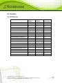
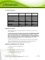
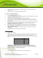
EPIGENTEK Complete Solutions for Epigenetics EpiNext™ ChIP-Seq High-Sensitivity Kit Base Catalog # P-2030 PLEASE READ THIS ENTIRE USER GUIDE BEFORE USE Uses: The EpiNext™ ChIP-Seq High Sensitivity Kit is designed to selectively enrich a chromatin fraction containing specific DNA sequences from various species, particularly mammals, and to prepare a ChIP-Seq library for next generation sequencing using Illumina platforms such as Illumina Genome Analyzer II, HiSeq and MiSeq systems. The optimized protocol and components of the kit allow capture of low abundance protein/DNA complexes with minimized non-specific background levels and the ability to construct both non-barcoded (singleplexed) and barcoded (multiplexed) ChIPSeq libraries quickly with reduced bias. Input Amount of Tissue/Cells: In general, the amount of cells and tissues for each reaction can be 1 x 10⁵ to 1 x 10⁶ and 5 mg to 50 mg, respectively. For optimal preparation, the input amount should be 4 to 5 x 10⁵ cells or 20 to 30 mg tissues so that the amount of DNA enriched from the ChIP reaction can range from at least 1 ng to 100 ng. Starting Materials: Starting materials can include various tissue or cell samples such as culture cells from a flask or plate, fresh and frozen tissues, etc. Antibodies: Antibodies should be ChIP-grade in order to recognize fixed and native proteins that are bound to DNA or other proteins. If you are using antibodies which have not been validated for ChIP, then appropriate control antibodies such as RNA Polymerase II (Cat. # A-2032) should be used to demonstrate that the antibody and prepared chromatin are suitable for ChIP. Internal Controls: Both negative and positive ChIP controls are provided in this kit. Precautions: To avoid cross-contamination, carefully pipette the sample or solution into the strip wells. Use aerosol-barrier pipette tips and always change pipette tips between liquid transfers. Wear gloves throughout the entire procedure. In case of contact between gloves and sample, change gloves immediately. 110 Bi County Blvd. Ste. 122, Farmingdale, NY 11735 Tel: 1-877-374-4368 ■ Fax: 1-718-484-3956 ■ E-mail: [email protected] ■ Web: www.epigentek.com © Epigentek Group Inc. All rights reserved. Products are for research use only. Page 1 Printed 2014-07-29 P-2030 EPIGENTEK Complete Solutions for Epigenetics KIT CONTENTS For ChIP Reaction Component 12 reactions P-2030-12 24 reactions P-2030-24 Storage Upon Receipt WB (Wash Buffer ) 12 ml 25 ml 4°C AB (Antibody Buffer) 1 ml 2 ml 4°C LB (Lysis Buffer) 7 ml 14 ml RT CB (ChIP Buffer) 6 ml 12 ml 4°C DRB (DNA Release Buffer) 7 ml 14 ml RT DBS (DNA Binding Solution) 7 ml 14 ml RT BS (Blocker Solution) 1 ml 2 ml 4°C DEB (DNA Elution Buffer) 0.5 ml 1 ml RT Enrichment Enhance* 25 µl 50 µl -20°C Protease Inhibitor Cocktail (PIC)* 15 µl 30 µl 4°C Non-Immune IgG (1 mg/ml)* 5 µl 10 µl 4°C Anti-RNA Polymerase II (1 mg/ml)* 5 µl 10 µl 4°C Proteinase K (10 mg/ml)* 30 µl 60 µl 4°C RNase A (10 mg/ml)* 15 µl 30 µl -20°C GAPDH Primer - Forward (20 µM)* 5 µl 10 µl 4°C GAPDH Primer - Reverse (20 µM)* 5 µl 10 µl 4°C 8-Well Assay Strips (With 1 Frame) 2 4 4°C 8-Well Strip Caps 2 4 RT Adhesive Covering Film Strip 4 8 RT F-Spin Column 15 30 RT F-Collection Tube 15 30 RT 110 Bi County Blvd. Ste. 122, Farmingdale, NY 11735 Tel: 1-877-374-4368 ■ Fax: 1-718-484-3956 ■ E-mail: [email protected] ■ Web: www.epigentek.com © Epigentek Group Inc. All rights reserved. Products are for research use only. Page 2 Printed 2014-07-29 P-2030 EPIGENTEK Complete Solutions for Epigenetics For Library Preparation Component 12 reactions Cat. #P-2030-12 24 reactions Cat. #P-2030-24 Storage Upon Receipt 10X End Polishing Buffer* End Polishing Enzyme Mix* End Polishing Enhancer* 2X Ligation Buffer* T4 DNA Ligase* Adaptors (50 µM)* MQ Binding Beads* 2X HiFi PCR Master Mix* Primer U (10 µM)* Primer I (10 µM)* Elution Buffer* User Guide 30 µl 13 µl 13 µl 250 µl 15 µl 15 µl 1.6 ml 160 µl 15 µl 15 µl 1000 µl 1 60 µl 26 µl 26 µl 500 µl 30 µl 30 µl 3.2 ml 320 µl 30 µl 30 µl 2000 µl 1 -20°C -20°C -20°C -20°C -20°C -20°C 4°C -20°C -20°C -20°C -20°C RT * Spin the solution down to the bottom prior to use. SHIPPING & STORAGE The kit is shipped in two parts: the first part at ambient room temperature and the second part on frozen ice packs at 4°C. Upon receipt: Store the following components at -20°C immediately: Enrichment Enhancer, RNase A, 10X End Polishing Buffer, End Polishing Enzyme Mix, End Polishing Enhancer, 2X Ligation Buffer, T4 DNA Ligase, Adaptors, 2X HiFi PCR Master Mix, Primer U, Primer I, and Elution Buffer. Store the following components at 4°C: WB, AB, CB, BS, Protease Inhibitor Cocktail, NonImmune IgG, Anti-RNA Polymerase II, Proteinase K, GAPDH Primer – Forward, GADPH Primer – Reverse, and 8-Well Assay Strips (With 1 Frame) and MQ Binding Beads. Store all other components at room temperature. All components of the kit are stable for 6 months from the date of shipment, when stored properly. Note: Check if WB and CB contain salt precipitates before use. If so, briefly warm at room temperature or 37°C and shake the buffer until the salts are re-dissolved. MATERIALS REQUIRED BUT NOT SUPPLIED Sonicator or enzymes for DNA fragmentation Vortex mixer Dounce homogenizer with small clearance pestle Variable temperature waterbath or incubator oven Thermocycler with 48 or 96-well block 110 Bi County Blvd. Ste. 122, Farmingdale, NY 11735 Tel: 1-877-374-4368 ■ Fax: 1-718-484-3956 ■ E-mail: [email protected] ■ Web: www.epigentek.com © Epigentek Group Inc. All rights reserved. Products are for research use only. Page 3 Printed 2014-07-29 P-2030 EPIGENTEK Complete Solutions for Epigenetics Centrifuge including desktop centrifuge (up to 14,000 rpm) Agilent® Bioanalyzer® or comparable method to assess the quality of DNA library Orbital shaker Magnetic stand (96-well PCR plate format) Adjustable pipette and pipette tips 0.2 ml or 0.5 ml PCR vials 1.5 ml microcentrifuge tubes 15 ml conical tube Antibodies of interest Cells or tissues 100% ethanol Distilled water Cell culture medium 37% formaldehyde (if cross-linked) 1.25 M glycine solution (if cross-linked) 1X PBS GENERAL PRODUCT INFORMATION Quality Control: Each lot of the EpiNext™ ChIP-Seq High Sensitivity Kit is tested against predetermined specifications to ensure consistent product quality. Epigentek guarantees the performance of all products in the manner described in our product instructions. Product Warranty: If this product does not meet your expectations, simply contact our technical support unit or your regional distributor. We also encourage you to contact us if you have any suggestions about product performance or new applications and techniques. Safety: Suitable lab coat, disposable gloves, and proper eye protection are required when working with this product. Product Updates: Epigentek reserves the right to change or modify any product to enhance its performance and design. The information in this User Guide is subject to change at any time without notice. Thus, only use the User Guide that was supplied with the kit when using that kit. Usage Limitation: The EpiNext™ ChIP-Seq High Sensitivity Kit is for research use only and is not intended for diagnostic or therapeutic application. Intellectual Property: The EpiNext™ ChIP-Seq High Sensitivity Kit and methods of use contain proprietary technologies by Epigentek. 110 Bi County Blvd. Ste. 122, Farmingdale, NY 11735 Tel: 1-877-374-4368 ■ Fax: 1-718-484-3956 ■ E-mail: [email protected] ■ Web: www.epigentek.com © Epigentek Group Inc. All rights reserved. Products are for research use only. Page 4 Printed 2014-07-29 P-2030 EPIGENTEK Complete Solutions for Epigenetics A BRIEF OVERVIEW Protein-DNA interaction plays a critical role for cellular functions such as signal transduction, gene transcription, chromosome segregation, DNA replication and recombination, and epigenetic silencing. Identifying the genetic targets of DNA binding proteins and knowing the mechanisms of protein-DNA interaction on a genome-wide scale is important for understanding cellular processes. Chromatin immunoprecipitation (ChIP) followed by next generation sequencing (ChIP-Seq) offers an advantageous tool for studying genome-wide protein-DNA interactions. It allows for detection that a specific protein binds to specific sequences in living cells. In particular, ChIP antibodies targeted against various transcriptional factors (TF) for genome-wide transcription factor binding site analysis by Chip-Seq is in high demand. Such analysis requires that ChIPed DNA contain minimal background for reliably identifying true TF-enriched regions. Currently used ChIP-Seq methods play an important role in identifying genome-wide protein-DNA interaction. However, these methods still have several drawbacks: 1) large amounts of cell/tissues are needed for obtaining a sufficient yield of library DNA, therefore these methods cannot be used for biological samples such as tumor biopsy and embryonic tissues whose amounts are limited; 2) the background levels of ChIPed DNA are high; and 3) the procedures are time consuming (>3 days) and inconvenient. To address this issue, Epigentek developed the EpiNext™ ChIP-Seq High Sensitivity Kit by combining its microplate-based ultra ChIP and high sensitive DNA library construction technologies. This kit has the following features: Optimized buffers and protocol allow minimal ChIP background by overcoming the weaknesses that cause non-specific enrichment, thereby increasing sensitivity and specificity of the ChIP reaction. Increased antibody selectivity and capture efficiency through the use of unique chimeric proteins containing the maximum number of IgG binding domains coated on the strip-wells. This allows strong binding of any IgG subtype antibodies within a wide pH range regardless of monoclonal or polyclonal form. Highly efficient enrichment of targeted DNA. Enrichment ratio of positive to negative control > 500. High sensitivity and flexibility: Can be used for both non-barcoded (singleplexed) and barcoded (multiplexed) DNA library preparation. The input cell number can be as few as 50,000 cells with a range from 50,000 to 1,000,000 cells. Broad range of cell/tissue samples can be used, including samples with limited amount. Fast and streamlined procedure: The procedure from cell/tissues to library DNA is less than 7 hours. No clean-up is required between each step from ChIPed DNA to size selection, and all reactions take place in the same tube, thereby saving time and preventing handling errors or loss of valuable samples. Gel-free size selection further reduces the preparation time. Highly convenient for use: The kit contains all required components for each step of ChIP-Seq, which are sufficient for both ChIP and ChIPed DNA library preparation, thereby allowing the ChIPSeq to be the most convenient with reliable and consistent results. Minimized bias: Ultra HiFi amplification and optional PCR-free step allow achievement of reproducibly high yields of DNA library with minimal sequence bias and low error rates. PRINCIPLE & PROCEDURE 110 Bi County Blvd. Ste. 122, Farmingdale, NY 11735 Tel: 1-877-374-4368 ■ Fax: 1-718-484-3956 ■ E-mail: [email protected] ■ Web: www.epigentek.com © Epigentek Group Inc. All rights reserved. Products are for research use only. Page 5 Printed 2014-07-29 P-2030 EPIGENTEK Complete Solutions for Epigenetics The EpiNext™ ChIP-Seq High Sensitivity Kit contains all necessary reagents required for carrying out a successful ChIP-Seq starting from mammalian cells or tissues. In the ChIP reaction, chromatin is isolated from cell/tissues and the target protein-DNA complex is immunoprecipitated using the antibody of interest. Immunoprecipitated DNA is then cleaned, released, and eluted. Included in the kit are a positive control antibody (RNA polymerase II), a negative control non-immune IgG, and GAPDH primers, which can be used as a positive control to demonstrate the efficacy of the kit reagents and protocol. RNA polymerase II is considered to be enriched in the GAPDH gene promoter that is expected to be undergoing transcription in most growing mammalian cells and can be immunoprecipitated by RNA polymerase II antibody but not by non-immune IgG. In the library preparation, ChIPed DNA fragments are end repaired and dA tailed (end polishing) simultaneously. Adaptors are then ligated to both ends of the polished DNA fragments for amplification and sequencing. Ligated fragments are size selected and purified using MQ binding beads, which allows quick and precise size selection of DNA. Size-selected DNA fragments are amplified with a high-fidelity PCR mix which ensures maximum yields from minimum amounts of starting material and provides highly accurate amplification of library DNA with low error rates and minimum bias. Chromatin Isolation and Shearing ChIP Reaction Crosslink Reversal and DNA Purification DNA End Polishing Adaptor Ligation Size Selection Amplification NGS (Illumina) Fig 1. Workflow of the EpiNext™ ChIP-Seq High Sensitivity Kit 110 Bi County Blvd. Ste. 122, Farmingdale, NY 11735 Tel: 1-877-374-4368 ■ Fax: 1-718-484-3956 ■ E-mail: [email protected] ■ Web: www.epigentek.com © Epigentek Group Inc. All rights reserved. Products are for research use only. Page 6 Printed 2014-07-29 P-2030 EPIGENTEK Complete Solutions for Epigenetics Fig 2. High sensitive ChIP: The sheared chromatin isolated from different number of MBD-231 cells was used fo ChIP-qPCR analysis of RNA polymerase II enrichment in GAPDH promoters. Fig3. Size distribution of library fragments. Ten nanograms of DNA was ChIPed by RNA polymerase II enrichment and used for DNA library preparation. ASSAY PROTOCOL For the best results, please read the protocol in its entirety prior to starting your experiment. ChIP Reaction 110 Bi County Blvd. Ste. 122, Farmingdale, NY 11735 Tel: 1-877-374-4368 ■ Fax: 1-718-484-3956 ■ E-mail: [email protected] ■ Web: www.epigentek.com © Epigentek Group Inc. All rights reserved. Products are for research use only. Page 7 Printed 2014-07-29 P-2030 EPIGENTEK Complete Solutions for Epigenetics 1. Preparation of Working Buffers and Solutions a. Prepare Working Lysis Buffer by adding 6 µl of Protease Inhibitor Cocktail to every 10 ml of LB (Lysis Buffer) b. Prepare Working CB ChIP buffer by adding 1 µl of Protease Inhibitor Cocktail to every 1 ml of CB (ChIP buffer). 2. Antibody Binding to Strip Wells. a. Predetermine the number of strip wells required for your experiment. Carefully remove unneeded strip wells from the plate frame and place them back in the bag (seal the bag tightly and store at 4°C). b. Setup the antibody binding reactions by adding the reagents to each well according to the following chart: Reagents Sample Positive Control Negative Control AB (Antibody Buffer) 50-80 µl 50-80 µl 50-80 µl Your Antibodies Anti-RNA Polymerase II 0.5-2 µl 0 0 0 0.8 µl 0 Non-Immune IgG 0 0 0.8 µl Note: The final amount of each component should be (a) antibodies of interest: 0.8 µg/well; (b) RNA Polymerase II: 0.8 µg/well; and (c) non-immune IgG: 0.8 µg/well. The amount of the positive control (RNA polymerase II) and negative control (Non-Immune IgG) are sufficient for matched use with samples if two antibodies are used for each sample or one antibody is used for two of the same samples. If using one antibody of interest for each sample with matched use of the positive and negative control, extra RNA polymerase II, non-immune IgG and 8-well strips are required and can be separately obtained from Epigentek. c. Seal the wells with Adhesive Covering Film Strips and incubate the wells at room temperature for 60-90 min on an orbital shaker (100 rpm). Meanwhile, perform the steps from Section 3 “Cell Collection and Cross-Linking” to Section 5 “Chromatin Shearing”. 3. Cell Collection and Cross-Linking 3.1. For Monolayer or Adherent Cells: a. Grow cells (treated or untreated) to 80%-90% confluence on a 6-well plate or 100 mm dish (the number of cultured MDA-231 cancer cells on an 80-90% confluent plate is listed in the table below as a reference), then trypsinize and collect them into a 15 ml conical tube. Count the cells in a hemocytometer. 110 Bi County Blvd. Ste. 122, Farmingdale, NY 11735 Tel: 1-877-374-4368 ■ Fax: 1-718-484-3956 ■ E-mail: [email protected] ■ Web: www.epigentek.com © Epigentek Group Inc. All rights reserved. Products are for research use only. Page 8 Printed 2014-07-29 P-2030 EPIGENTEK Complete Solutions for Epigenetics Container 96-well plate 24-well plate 12-well plate 6-well plate 60 mm dish 100 mm dish 150 mm dish Cell Number (x 105) 0.3-0.6/well 1-3/well 3-6/well 5-10/well 20-30 50-100 150-180 b. Centrifuge the cells at 1000 rpm for 5 min. Discard the supernatant. c. Wash cells with 10 ml of PBS once by centrifugation at 1000 rpm for 5 min. Discard the supernatant. Note: For cells that are not cross-linked, go directly to Step 3.1.i after Step 3.1.c. d. Add 9 ml fresh cell culture medium containing formaldehyde to a final concentration of 1% (i.e., add 270 µl of 37% formaldehyde to 10 ml of cell culture medium) to cells. e. Incubate at room temperature (20-25°C) for 10 min on a rocking platform (50-100 rpm). f. Add 1 ml of 1.25 M glycine for every 9 ml of cross-link solution. g. Mix and centrifuge at 1000 rpm for 5 min. h. Remove medium and wash cells once with 10 ml of ice-cold PBS by centrifuging at 1000 rpm for 5 min. i. Add Working Lysis Buffer to re-suspend the cell pellet (200 µl/1x106 cells) and incubate on ice for 10 min. Note: If the total solution volume is less than 1.5 ml, transfer the solution to a 1.5 ml microtube. j. Vortex vigorously for 10 sec then centrifuge at 3000 rpm for 5 min. Go to Step 4a. 3.2. For Suspension Cells: a. Collect cells (treated or untreated) into a 15 ml conical tube (2 x10⁵ to 5x10⁵ cells are required for each ChIP reaction). Count cells in a hemocytometer. b. Centrifuge the cells at 1000 rpm for 5 min. Discard the supernatant. c. Wash cells with 10 ml of PBS once by centrifugation at 1000 rpm for 5 min. Discard the supernatant. Note: For cells that are not cross-linked, go directly to Step 3.2.i after Step 3.2.c. d. Add 9 ml fresh cell culture medium containing formaldehyde to a final concentration of 1% (i.e., add 270 µl of 37% formaldehyde to 10 ml of cell culture medium) to cells. e. Incubate at room temperature (20-25°C) for 10 min on a rocking platform (50-100 rpm). f. Add 1 ml of 1.25 M glycine for every 9 ml of cross-link solution. g. Mix and centrifuge at 1000 rpm for 5 min. 110 Bi County Blvd. Ste. 122, Farmingdale, NY 11735 Tel: 1-877-374-4368 ■ Fax: 1-718-484-3956 ■ E-mail: [email protected] ■ Web: www.epigentek.com © Epigentek Group Inc. All rights reserved. Products are for research use only. Page 9 Printed 2014-07-29 P-2030 EPIGENTEK Complete Solutions for Epigenetics h. Remove medium and wash cells once with 10 ml of ice-cold PBS by centrifuging at 1000 rpm for 5 min. i. Add Working Lysis Buffer to re-suspend the cell pellet (200 µl/1x106 cells) and incubate on ice for 10 min. j. Vortex vigorously for 10 sec and centrifuge at 3000 rpm for 5 min. Then go to Step 4a. 3.3. For Tissues: a. Put the tissue sample into a 60 or 100 mm plate. Remove unwanted tissue such as fat and necrotic material from the sample. b. Weigh the sample and cut the sample into small pieces (1-2 mm3) with a scalpel or scissors. Note: For tissues that are not cross-linked, go directly to Step 3.3.j after Step 3.3.b. c. Transfer tissue pieces to a 15 ml conical tube. d. Prepare cross-link solution by adding formaldehyde to cell culture medium to a final concentration of 1%. (e.g., add 270 µl of 37% formaldehyde to 10 ml of culture medium). e. Add 1 ml of cross-link solution for every 50 mg tissues. f. Incubate at room temperature for 15-20 min on a rocking platform. g. Add 1 ml of 1.25 M glycine for every 9 ml of cross-link solution. h. Mix and centrifuge at 800 rpm for 5 min. Discard the supernatant. i. Wash cells with 10 ml of ice-cold PBS once by centrifugation at 800 rpm for 5 min. Discard the supernatant. j. Transfer tissue pieces to a Dounce homogenizer. k. Add 0.5 ml Working Lysis Buffer for every 50 mg of tissues. l. Disaggregate tissue pieces by 20-40 strokes. m. Transfer homogenized mixture to a 15 ml conical tube and centrifuge at 3000 rpm for 5 min at 4°C. If total mixture volume is less than 2 ml, transfer mixture to a 2 ml vial and centrifuge at 5000 rpm for 5 min at 4°C. Then go to Step 4a. 4. Cell Lysis and Chromatin Extraction a. Carefully remove supernatant. b. Add CB (ChIP Buffer) to re-suspend the chromatin pellet (100 µl/1x106 cells or 50 mg tissue, 500 µl maximum for each vial). c. Transfer the chromatin lysate to a 1.5 ml vial and incubate on ice for 10 min and vortex occasionally. 110 Bi County Blvd. Ste. 122, Farmingdale, NY 11735 Tel: 1-877-374-4368 ■ Fax: 1-718-484-3956 ■ E-mail: [email protected] ■ Web: www.epigentek.com © Epigentek Group Inc. All rights reserved. Products are for research use only. Page 10 Printed 2014-07-29 P-2030 EPIGENTEK Complete Solutions for Epigenetics 5. Chromatin Shearing a. Resuspend the chromatin lysate by vortexing. b. Shear chromatin with one of the following methods: Waterbath Sonication: Epigentek EpiSonic 1100 (Epigentek Cat No. EQC-1100): Use 50 ul of chromatin lysate per 0.2 ml tube or per PCR plate well. Shear 20 cycles under cooling condition, 30 seconds ON, 30 seconds OFF, each at 170-190 watts. For more detailed information of use, please see the “Chromatin Shearing Protocol” for EpiSonic 1100. If using other waterbath sonicators, please follow the supplier’s instruction. Probe-based Sonication: Use 300 ul of chromatin lysate per 1.5 ml microcentrifuge tube. As an example, sonication can be carried out with a microtip attached to a Branson 450 sonifier, set to 25% power output. Sonicate 3-4 pulses of 10-15 seconds each, followed by 30-40 seconds rest on ice between each pulse. (The conditions of cross-linked DNA shearing can be optimized based on cells and sonicator equipment). Note: When probe-based sonication is carried out, the shearing effect may be reduced if foam is formed in the chromatin sample solution. Under this condition, discontinue sonication and centrifuge the sample at 4°C at 12,000 rpm for 3 min to remove the air bubbles then continue with sonication. The isolated chromatin can also be sheared with various enzyme-based methods. Optimization of the shearing conditions, for example enzyme concentration and incubation time, is needed in order to use enzyme-based methods. c. Centrifuge at 12,000 rpm at 4°C for 10 min after shearing. d. Transfer supernatant to a new vial. The chromatin solution can now be used immediately or stored at –80°C after aliquoting appropriately until further use. Avoid multiple freeze/thaw cycles. Note: The size of sonicated chromatin should be verified before starting immunoprecipitation step. The length of sheared DNA should be between 100-700 bps with a peak size of about 300 bps. The following steps can be carried out to isolate DNA for gel analysis of DNA fragment size: (1) add 25 µl of each chromatin sample to a 0.2 ml PCR tube followed by adding 25 µl of DRB (DNA Release Buffer) and 2 µl of Proteinase K; (2) incubate the sample at 60°C for 30 min followed by incubating at 95°C for 10 min; (3) spin the solution down to the bottom; (4) transfer supernatant to a new 0.2 ml PCR vial. Use 30-40 µl for DNA fragment size analysis along with a DNA marker on a 1-2% agarose gel; and (5) stain with ethidium bromide or other fluorescent dye for DNA and visualize it under ultraviolet light. 6. Preparation of ChIP Reaction a. Peel away the Adhesive Covering Film on the antibody binding wells (from Step 2b) carefully to avoid contamination between each well. b.. Remove the antibody reaction solution and non-immune IgG solution from each well and wash the wells one time with 150 µl of CB (ChIP Buffer). 110 Bi County Blvd. Ste. 122, Farmingdale, NY 11735 Tel: 1-877-374-4368 ■ Fax: 1-718-484-3956 ■ E-mail: [email protected] ■ Web: www.epigentek.com © Epigentek Group Inc. All rights reserved. Products are for research use only. Page 11 Printed 2014-07-29 P-2030 EPIGENTEK Complete Solutions for Epigenetics c. Setup the ChIP reactions by adding the reagents to the wells that are bound with antibodies (sample and positive control wells) or IgG (negative control well) according to the following chart: Reagents Sample Positive Control Negative Control CB (ChIP Buffer) 50-80 µl 50-80 µl 50-80 µl Chromatin 10-40 µl 10-40 µl 10-40 µl Enrichment Enhancer 2 µl 2 µl 2 µl BS (Blocker Solution) 10 µl 10 µl 10 µl Note: The final amount of chromatin should be 2 µg/well (2 x 10⁵ cells may yield 1 µg of chromatin); Sonicated chromatin can be further diluted with CB (ChIP Buffer) to desired concentration. For histone samples containing sufficient chromatin (> 0.5 ug), the Enrichment Enhancer is not required and 5080 µl of CB (ChIP Buffer) can be used. For low abundance targets, 2 µl of Enrichment Enhancer and 88 µl of chromatin can be used without adding CB (ChIP Buffer). Freshly prepared chromatin can be directly used for the reaction. Frozen chromatin samples should be thawed quickly at RT and then placed on ice before use. Store remaining chromatin samples at -20°C or at -80°C if they will be not used within 8 hours. An input DNA control is only used for estimating the enrichment efficiency of ChIP and is generally not necessary since the positive and negative control can be used for estimating the same objective more accurately. If you would like to include the input DNA control, the purified input DNA prepared at Step 5d Note can be used. d. Cap wells with strip cap and incubate at room temperature for 60-90 min on an orbital shaker (100 rpm). For low abundance targets, incubation time should be extended to 2-3 hours or at 4°C overnight. 7. Washing of the Reaction Wells a. Carefully remove the solution using a pipette and discard from each well. b. Wash each well with 200 µl of fresh WB each time for 4 times. Allow 2 minutes on an orbital shaker (100 rpm) for each wash. Pipette wash buffer out from the wells. c. Wash each well with 200 µl of DRB one time by pipetting DRB into the well and then removing it. 8. Reversal of Cross-Links, Release and Purification of DNA a. Prepare RNase A solution by adding 1 µl of RNase A to 400 µl of DRB. b. Add 40 µl of DRB-RNase A to each well, and then cover with a strip cap. c. Incubate the wells at 42°C for 30 min. d. Add 2 µl of Proteinase K to each well and re-cap the wells. e. f. Incubate the wells at 60°C for 30 min. Quickly transfer the DNA solution from each well to 0.2 ml strip PCR tubes. Cap the PCR tubes. 110 Bi County Blvd. Ste. 122, Farmingdale, NY 11735 Tel: 1-877-374-4368 ■ Fax: 1-718-484-3956 ■ E-mail: [email protected] ■ Web: www.epigentek.com © Epigentek Group Inc. All rights reserved. Products are for research use only. Page 12 Printed 2014-07-29 P-2030 EPIGENTEK Complete Solutions for Epigenetics g. Incubate the PCR tubes containing DNA solution at 95°C for 15 min in a thermolcycler. h. Place the PCR tubes at room temperature. If liquid is collected on the inside of the caps, briefly spin the liquid down to the bottom. i. Place a spin column into a 2 ml collection tube. Add 200 µl of DBS (DNA Binding Solution) to the samples and transfer mixed solution to the column. Centrifuge at 12,000 rpm for 30 seconds. j. Add 200 µl of 90% ethanol to the column, centrifuge at 12,000 rpm for 30 seconds. Remove the column from the collection tube and discard the flowthrough. k. Replace column to the collection tube. Add 200 µl of 90% ethanol to the column and centrifuge at 12,000 rpm for 30 seconds. l. Remove the column and discard the flowthrough. Replace column to the collection tube and wash the column again with 200 µl of 90% ethanol at 12,000 rpm for 1 min. m. Place the column in a new 1.5 ml vial. Add 11 µl of DEB (DNA Elution Buffer) directly to the filter in the column and centrifuge at 12,000 rpm for 30 seconds to elute purified DNA. Purified DNA is now ready for ChIPed DNA library preparation after verifying the quality and amount of the ChIPed DNA by qPCR or a suitable fluorescence method. Note: For real time PCR analysis, we recommend the use of 1µl of eluted DNA in a 20 µl PCR reaction. If input DNA will be used, it should be diluted 10 fold before adding to PCR reaction. Control primers (110 bp, for human cells) included in the kit can be used as a positive control. In general, the amplification difference between “normal IgG control” and “positive control” may vary from 3 to 8 cycles, depending on experimental conditions. Optimally, 10 ng of ChIPed DNA are required for ChIP DNA library construction. This amount can be easily generated for high abundance targets from a single ChIP reaction well. However, this may be difficult for low abundance target enrichment. We recommend pooling the DNA solution from several low abundance ChIP reaction wells to gain 10 ng or more of DNA. ChIPed DNA Library Preparation 9. DNA End Polishing a. Prepare end repair reaction in a 0.2 ml PCR tube according to Table 1: Table 1. End Polishing Reaction b. Component ChIPed DNA Volume 10 µl 10X End Polishing Buffer End Polishing Enzyme Mix End Polishing Enhancer Distilled Water Total Volume 1.5 µl 1 µl 1 µl 1.5 µl 15 µl Mix and incubate for 20 min at 25°C and 20 min at 72°C in a thermocycler (without heated lid). 110 Bi County Blvd. Ste. 122, Farmingdale, NY 11735 Tel: 1-877-374-4368 ■ Fax: 1-718-484-3956 ■ E-mail: [email protected] ■ Web: www.epigentek.com © Epigentek Group Inc. All rights reserved. Products are for research use only. Page 13 Printed 2014-07-29 P-2030 EPIGENTEK Complete Solutions for Epigenetics Note: The amount of fragmented DNA can be 0.2-100 ng with an optimal amount of 10-50 ng. 10. Adaptor Ligation a. Prepare a reaction mix for adaptor ligation according to Table 2. Add the following reagents to a 0.2 ml PCR tube containing end repaired/dA tailing (end polished) DNA from Step 9. Table 2. Adaptor Ligation Component End polished DNA (from step 9) Volume 15 µl 2X Ligation Buffer 17 µl 1 µl 1 µl 34 µl T4 DNA Ligase Adaptors Total volume b. Mix and incubate for 15 min at 25°C in a thermocycler (without heated lid). Note: (1) The pre-annealed adapters included in the kit are suitable for both non-barcoded (singleplexed) and barcoded (multiplexed) DNA library preparation and are fully compatible with Illumina platforms, such as MiSeq® or HiSeq™ sequencers. (2) If using adaptors from other suppliers (both single-end and barcode adaptors), make sure they are compatible with Illumina platforms and add the correct amount (final concentration 1.5-2 µM, or according to the supplier’s instruction). 11. Size Selection/Clean-up 11.1. Size Selection of Ligated DNA Note: If the starting DNA amount is less than 50 ng, the size selection is not recommended and alternatively, clean-up of ligated DNA can be performed prior to PCR amplification according to 11.2. protocol. a. b. c. d. e. f. g. h. i. j. Resuspend MQ Binding Beads by vortex. Add 14 µl of resuspended MQ Binding Beads to the tube of ligation reaction. Mix well by pipetting up and down at least 10 times. Incubate for 5 minutes at room temperature. Put the tube on an appropriate magnetic stand until the solution is clear (about 2 minutes). Carefully transfer the supernatant containing DNA to a new tube. (Caution: Do not discard the supernatant.) Discard the beads that contain the unwanted large fragments. Add 10 µl resuspended beads to the supernatant, mix well and incubate for 5 minutes at room temperature. Put the PCR tube on an appropriate magnetic stand until the solution is clear (about 2 minutes). Carefully remove and discard the supernatant. (Caution: Be careful not to disturb or discard the beads that contain DNA). Keep the PCR tube in the magnetic stand and add 200 μl of freshly prepared 90% ethanol to the tube. Incubate at room temperature for 1 min, and then carefully remove and discard the ethanol. Repeat Step g one time, for total of two washes. Open the PCR tube cap and air dry beads for 10 minutes while the tube is on the magnetic stand. Resuspend the beads in 12 µl Elution Buffer, and incubate at room temperature for 2 minutes to release the DNA from the beads. 110 Bi County Blvd. Ste. 122, Farmingdale, NY 11735 Tel: 1-877-374-4368 ■ Fax: 1-718-484-3956 ■ E-mail: [email protected] ■ Web: www.epigentek.com © Epigentek Group Inc. All rights reserved. Products are for research use only. Page 14 Printed 2014-07-29 P-2030 EPIGENTEK Complete Solutions for Epigenetics k. l. Capture the beads by placing the tube in the magnetic stand for 4 minutes or until the solution is completely clear. Transfer 11 µl to a new 0.2 ml PCR tube for PCR amplification. 11.2. Clean-up of Ligated DNA (Optional) a. b. c. d. e. f. g. h. i. j. Resuspend MQ Binding Beads by vortex. Add 34 μl of resuspended beads to the PCR tube of ligation reaction. Mix thoroughly on a vortex mixer or by pipetting up and down at least 10 times. Incubate for 5 minutes at room temperature to allow DNA to bind to beads. Put the PCR tube on an appropriate magnetic stand until the solution is clear (about 2 minutes). Carefully remove and discard the supernatant. (Caution: Be careful not to disturb or discard the beads that contain DNA. Keep the PCR tube in the magnetic stand and add 200 μl of freshly prepared 90% ethanol to the tube. Incubate at room temperature for 1 min, and then carefully remove and discard the ethanol. Repeat Step e two times for total of three washes. Open the PCR tube cap and air dry beads for 10 minutes while the tube is on the magnetic stand. Resuspend the beads in 12 µl Elution Buffer, and incubate at room temperature for 2 minutes to release the DNA from the beads. Capture the beads by placing the tube in the magnetic stand for 4 minutes or until the solution is completely clear. Transfer 11 µl to a new 0.2 ml PCR tube for PCR amplification. 12. Library Amplification a. Prepare the PCR reactions. Thaw all reaction components including master mix, DNA/RNA free water, primer solution and DNA template. Mix well by vortexing briefly. Keep components on ice while in use and return to -20˚C immediately following use. Add components into each PCR tube/well according to the following table: Component Size (:l) Final Concentration Component Size (µl) HiFi Master Mix (2X) 12.5 µl Primer U 1 µl Primer I 1 µl Adaptor Ligated DNA Total Volume 10.5 µl 25 µl Important Note: Use of Primer I included in the kit will generate a singleplexed library. For multiplexed library preparation, replace Primer I with one of the12 different barcodes (indexes) contained in the EpiNextTM NGS Barcode (Index) Set-12 (Cat. No. P-1060). You can also add userdefined barcodes (Illumina compatible) instead of Primer I. b. Program the PCR reactions. Place the reaction plate in the instrument and set the PCR conditions as follow: 110 Bi County Blvd. Ste. 122, Farmingdale, NY 11735 Tel: 1-877-374-4368 ■ Fax: 1-718-484-3956 ■ E-mail: [email protected] ■ Web: www.epigentek.com © Epigentek Group Inc. All rights reserved. Products are for research use only. Page 15 Printed 2014-07-29 P-2030 EPIGENTEK Complete Solutions for Epigenetics Cycle Step Temp Time Cycle Activation 98°C 30 sec 1 Cycling 98°C 55°C 72°C 20 sec 20 sec 20 sec Variable* Final Extension 72°C 2 min 1 * PCR cycles may vary depending on the input DNA amount. In general, use 8 PCR cycles for 100 ng, 10 cycles for 50 ng, 13 cycles for 5 ng, 15 cycles for 1 ng and 18 cycles for 0.2 ng DNA input. Further optimization of PCR cycle number may be required. 13. Clean-Up of Amplified Library DNA a. b. c. d. e. f. g. h. i. j. Resuspend MQ Binding Beads by vortex. Add 25 μl of resuspended beads to the PCR tube of amplification reaction. Mix thoroughly on a vortex mixer or by pipetting up and down at least 10 times. Incubate for 5 minutes at room temperature to allow DNA to bind to beads. Put the PCR tube on an appropriate magnetic stand until the solution is clear (about 2 minutes). Carefully remove and discard the supernatant. (Caution: Be careful not to disturb or discard the beads that contain DNA.) Keep the PCR tube in the magnetic stand and add 200 μl of freshly prepared 80% ethanol to the tube. Incubate at room temperature for 1 min, and then carefully remove and discard the ethanol. Repeat Step e two times for total of three washes. Open the PCR tube cap and air dry beads for 10 minutes while the tube is on the magnetic stand. Resuspend the beads in 22 µl Elution Buffer, and incubate at room temperature for 2 minutes to release the DNA from the beads. Capture the beads by placing the tube in the magnetic stand for 4 minutes or until the solution is completely clear. Transfer 20 µl to a new 0.2 ml PCR tube. Quality of the prepared library can be assessed using an Agilent® Bioanalyzer® or other comparable methods. Library fragments should have the correct size distribution (e.g., 300-400 bps at peak size) without adaptors or adaptor-dimers. To check the size distribution, dilute library 5-fold with water and apply it to an Agilent® high sensitivity chip. If there is presence of <150 bp adaptor dimers or of larger fragments than expected, they should be removed. To remove fragments below 150 bps or above 500 bps, use 0.8X MQ Binding Beads according to sub-steps a through l of Step 11.1 – “Size Selection of Ligated DNA”. Store the prepared library at -20ºC until ready to use for sequencing. TROUBLESHOOTING For ChIP Reaction: Problem Possible Cause Suggestion Little or no PCR products Poor chromatin quality due to insufficient amount of cells, or The optimal amount of chromatin per ChIP reaction should be 2-4 µg (about 110 Bi County Blvd. Ste. 122, Farmingdale, NY 11735 Tel: 1-877-374-4368 ■ Fax: 1-718-484-3956 ■ E-mail: [email protected] ■ Web: www.epigentek.com © Epigentek Group Inc. All rights reserved. Products are for research use only. Page 16 Printed 2014-07-29 P-2030 EPIGENTEK Complete Solutions for Epigenetics generated from both sample and positive control wells 5 insufficient or over cross-linking. 2-4 x 10 cells). Appropriate chromatin cross-linking is also required. Insufficient or over-crosslinking will cause DNA loss or increased background. During the cross-linking step of chromatin preparation, ensure that the cross-linking time is within 1015 min, the final concentration of formaldehyde is 1%, and the quench solution is 0.125 M glycine. Poor enrichment with antibody; some antibodies used in ChIP might not efficiently recognize fixed protein. Increase the antibody amount and use ChIP-grade antibodies validated for use in ChIP. Inappropriate DNA fragmenting condition. If chromatin is from specific cell/tissue types, or is differently fixed, the shearing conditions should be optimized to allow DNA fragment size to be between 100-700 bp. Incorrect temperature and/or insufficient time during DNA release. Ensure the incubation times and temperatures described in the protocol are followed correctly. Improper PCR conditions, including improper PCR programming, PCR reaction solutions, and/or primers. Ensure the PCR is properly programmed. If using a homebrew PCR reaction solution, check if each component is correctly mixed. If using a PCR commercial kit, check if it is suitable for your PCR. Confirm species specificity of primers. Primers should be designed to cover a short sequence region (70-150 bp) for more efficient and precise amplification of the target DNA region (the binding sites of the protein of interest). Improper sample storage. Chromatin sample should be stored at –80°C for no longer than 6 months, preferably less than 3 months. Avoid repeated freeze/thaw cycles. DNA samples should be stored at – 20°C for no longer than 6 months, preferably less than 3 months. No difference in signal intensity between negative and positive control wells Insufficient washing. Check if washing recommendations at each step is performed according to the protocol. If the signal intensity in the negative control is still high, washing stringency can be increased in the following ways: 110 Bi County Blvd. Ste. 122, Farmingdale, NY 11735 Tel: 1-877-374-4368 ■ Fax: 1-718-484-3956 ■ E-mail: [email protected] ■ Web: www.epigentek.com © Epigentek Group Inc. All rights reserved. Products are for research use only. Page 17 Printed 2014-07-29 P-2030 EPIGENTEK Complete Solutions for Epigenetics 1. Increase wash time at each wash step: after adding WB, leave it in the wells for 3-4 min and then remove it. 2. Add an additional one wash with WB, respectively: The provided volume of WB is sufficient for 4 extra washes for each sample. Too many PCR cycles: Plateau phase of amplification caused by excessive number of PCR cycles in endpoint PCR may mask the difference of signal intensity between negative contol and positive control. Decrease the number of PCR cycles (i.e., 32-35 cycles) to keep ampification at the exponential phase. This will reduce high background in endpoint PCR and allow differences in amplification to be seen. Real time PCR is another choice in such cases. Little or no PCR products generated from sample wells only Poor enrichment with antibody: some antibodies used in ChIP might not efficiently recognize fixed protein. Increase the antibody amount and use ChIP-grade antibodies validated for use in ChIP. PCR primers are not optimized. Confirm species specificity of primers. Primers should be designed to cover a short sequence region (70-150 bp) for more efficient and precise amplification of target DNA region (the binding sites of the protein of interest). For ChIPed DNA Library Preparation: Problem Possible Cause Suggestion Low yield of library Insufficient amount of starting DNA. To obtain the best results, the amount of input DNA should be 100-200 ng. For a library directly used for sequencing without amplification, 500 ng or more is needed. Insufficient purity of starting DNA. Ensure that RNA is removed by RNase A treatment before starting library preparation protocol. Improper reaction conditions at each reaction step. Check if the reagents are properly added and incubation temperature and time are correct at each reaction step including DNA End Polishing, 110 Bi County Blvd. Ste. 122, Farmingdale, NY 11735 Tel: 1-877-374-4368 ■ Fax: 1-718-484-3956 ■ E-mail: [email protected] ■ Web: www.epigentek.com © Epigentek Group Inc. All rights reserved. Products are for research use only. Page 18 Printed 2014-07-29 P-2030 EPIGENTEK Complete Solutions for Epigenetics Adaptor Ligation, Size Selection and Amplification. Unexpected peak size of Agilent® Bioanalyzer® trace: presence of <150 bp adaptor dimmers or presence of larger fragments than expected Improper storage of the kit. Ensure that the kit has not exceeded the expiration date. Standard shelf life, when stored properly, is 6 months from date of receipt. Improper ratio of MQ Binding Beads to DNA volume in size selection. Check if the correct volume of MQ Binding Beads is added to DNA solution accordingly. Proper ratios should remove the fragments with unexpected peak sizes. Insufficient ligation. Too much and too little input DNA may cause insufficient ligation, which can shift peak size of the fragment population to be shorter or larger than expected. Make sure that ligation reaction is properly processed with the proper amount of input DNA. Over-amplification of library. PCR artifacts from over-amplification of the library may cause the fragment population to shift higher than expected. Make sure to use proper PCR cycles to avoid this problem. RELATED PRODUCTS Chromatin Preparation P-2001 ChromaFlash™ Chromatin Extraction Kit P-2023 ChromaFlash™ Chromatin Isolation/Shearing Kit DNA Isolation and Cleanup P-1003 FitAmp™ General Tissue Section DNA Isolation Kit P-1004 FitAmp™ Plasma/Serum DNA Isolation Kit P-1006 DNA Concentrator Kit P-1007 FitAmp™ Gel DNA Isolation Kit P-1009 FitAmp™ Paraffin Tissue Section DNA Isolation Kit P-1017 FitAmp™ Urine DNA Isolation Kit P-1018 FitAmp™ Blood and Cultured Cell DNA Extraction Kit Sonication Instruments EQC-1100 EpiSonic™ Multi-Functional Bioprocessor 1100 DNA Enrichment Reaction P-1015 Methylamp™ Methylated DNA Capture Kit P-1038 EpiQuik™ Hydroxymethylated DNA Immunoprecipitation (hMeDIP) Kit P-1052 EpiQuik™ MeDIP Ultra Kit P-2002 EpiQuik™ Chromatin Immunoprecipitation Kit P-2003 EpiQuik™ Tissue Chromatin Immunoprecipitation Tissue Kit P-2014 EpiQuik™ Plant ChIP Kit 110 Bi County Blvd. Ste. 122, Farmingdale, NY 11735 Tel: 1-877-374-4368 ■ Fax: 1-718-484-3956 ■ E-mail: [email protected] ■ Web: www.epigentek.com © Epigentek Group Inc. All rights reserved. Products are for research use only. Page 19 Printed 2014-07-29 P-2030 EPIGENTEK Complete Solutions for Epigenetics P-2025 P-2026 P-2027 ChromaFlash™ One-Step ChIP Kit ChromaFlash™ One-Step Magnetic ChIP kit ChromaFlash™ ChIP Ultra Kit PCR Analysis P-1029 EpiQuik™ Quantitative PCR Kit DNA Library Prep P-1051 EpiNext™ DNA Library Preparation Kit (Illumina) P-1053 EpiNext™ High Sensitive DNA Library Prep Kit (Illumina) NGS Barcode P-1060 EpiNext™ NGS Barcode (Index) Set-12 For ChIP-grade antibodies, search “chip-grade” at www.epigentek.com 110 Bi County Blvd. Ste. 122, Farmingdale, NY 11735 Tel: 1-877-374-4368 ■ Fax: 1-718-484-3956 ■ E-mail: [email protected] ■ Web: www.epigentek.com © Epigentek Group Inc. All rights reserved. Products are for research use only. Page 20 Printed 2014-07-29 P-2030