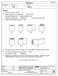
1
Policy and Procedure BACTEC™ 9000MB I. PRINCIPLE Since the mid-1980’s and the spread of the AIDS epidemic, the incidence of mycobacterial infections due to Mycobacterium tuberculosis (MTB) and mycobacteria other than tuberculosis (MOTT), especially Mycobacterium avium complex (MAC), have experienced a resurgence. From 1985 to 1992, the number of reported MTB cases increased 18%. Tuberculosis still kills an estimated 3 million persons a year worldwide, making it the leading infectious disease cause of death. Between 1981 and 1987, AIDS case surveillance indicated that 5.5% of the patients with AIDS had disseminated nontuberculous mycobacterial infections; e.g., MAC. By 1990, the increase in cases of disseminated non-tuberculous mycobacterial infections had resulted in a cumulative incidence of 7.6%. In addition to the resurgence of MTB, multidrug-resistant MTB (MDRTB) has become an increasing concern. Laboratory delays in the growth, identification and reporting of these MDR-TB cases contributed at least in part to the spread of the disease. The Centers for Disease Control and Prevention (CDC) has recommended that every effort be made by laboratories to use the most rapid methods available for diagnostic mycobacterial testing. These recommendations include the use of both a liquid and a solid medium for mycobacterial culture.1 The BACTEC 9000MB System is designed for the rapid detection of mycobacteria in clinical specimens. The system includes liquid culture media (BACTEC MYCO/F-Sputa culture vial, BACTEC MYCO/F LYTIC culture vial), a growth supplement (BACTEC Supplement/F) and an antibiotic supplement (BACTEC™ PANTA™/F). BACTEC Supplement/F contains growth enhancers for mycobacteria and is used to reconstitute BACTEC PANTA/F. BACTEC PANTA/F contains a mixture of antimicrobial agents used to suppress the growth of contaminating or normal flora which may survive the decontamination process. Microorganisms present in these specimens metabolize nutrients and oxygen in the culture media. The culture vials contain a fluorescent sensor that responds to the concentration of oxygen in the culture medium. The sensor is monitored by the instrument for increasing fluorescence, which is proportional to a decreasing dissolved oxygen content. A positive reading indicates the presumptive presence of viable mycobacteria in the vial. II. MATERIAL A. MEDIA 1. BACTEC MYCO/F-Sputa Culture Vials:2 When supplemented with BACTEC Supplement/F and BACTEC PANTA is used for the in vitro culture and recovery of mycobacteria from digested decontaminated clinical specimens and sterile body fluids other than blood. a. Each vial contains the following active ingredients prior to processing: Policy and Procedure page 2 • • • • • 40 mL Processed Water Middlebrook 7H9 Broth Base Casein Hydrolysate Supplement H Glycerol • • • • Ammonium Sulfate Ferric Ammonium Citrate Polysorbate 80 Hemin All BACTEC media are dispensed with added CO2 . b. o o Store at 2 - 25 C, in a dry location out of direct sunlight. DO NOT use after expiration date. 2. BACTEC™ PANTA™/F Antibiotic Supplement:3 a. Each vial contains the following lyophilized drugs: • • • • • b. Polymyxin B Amphotericin Nalidixic Acid Trimethoprim Azlocillin 10,000 units 1,000 g 4,000 g 1,000 g 2,000 g Store unreconstituted at 2 - 8C. Once reconstituted with 10 mL of Supplement/F, use immediately or freeze aliquots at -20C to -70C for up to 6 months. DO NOT thaw and refreeze. Protect from light. Overheating must be avoided. DO NOT use after expiration date. 3. BACTEC SUPPLEMENT/F Media Supplement:4 a. Each vial contains the following ingredients: • • • • b. Processed Water Lactic Acid Polyoxyethylene Stearate Bovine Serum Albumin • • • Dextrose Biotin Catalase Store Supplement/F at 15 - 25C. DO NOT use after expiration date. c. Supplement/F, reconstituting fluid for PANTA/F Antibiotic Supplement, is a required supplement to the MYCO/F-Sputa culture vials for recovery of mycobacteria. Supplement/F must be added to the MYCO/F-Sputa medium prior to inoculation of specimen or organism suspension. Refer to the BACTEC SUPPLEMENT/F Media Supplement package insert. 4. BACTEC™ MYCO/F LYTIC:5 A non-selective culture medium for the qualitative culture and recovery of mycobacteria from blood specimens. a. Each vial contains the following active ingredients prior to processing: Rev A May 1998 Policy and Procedure page 3 • • • • • • • • 40 mL Processed Water Middlebrook 7H9 Broth Base Brain Heart Infusion Casein Hydrolysate Supplement H Glycerol Sodium Polyanetholsulfonate Inositol • • • • • • • Ammonium Sulfate Pyridoxal HCl Ferric Ammonium Citrate Polysorbate 80 Potassium Phosphate Saponin Antifoam The BACTEC medium is dispensed with added CO2 and O2. b. Store at 2° - 25° C in a dry location out of direct sunlight. B. MATERIALS REQUIRED BUT NOT PROVIDED Centrifuge; Biological Safety Cabinet; Autoclave; CO2 incubator at 37°C; Falcon™ BRAND 50 mL centrifuge tubes (Cat. No 2070); 4% sodium hydroxide; 2.9% sodium citrate solution; N-acetyl-L-cysteine powder; sterile phosphate buffer, pH 6.8; or BBL™ MycoPrep™ Specimen Digestion/Decontamination Kit; vortex mixer; sterile transfer pipettes; sterile tuberculin syringe with a permanently attached 25 gauge needle; mycobactericidal agent; 70% isopropyl alcohol; mycobacterial agar or egg-based medium; 7H9 broth, sterile saline; venting unit; Quality Control organisms (Mycobacterium tuberculosis, ATCC™ 27294; Mycobacterium fortuitum, ATCC 6841; Mycobacterium kansasii, ATCC 12478; and Mycobacterium intracellulare, ATCC 13950); microscope and materials for staining slides and subculturing vials. C. INSTRUMENT 1. BACTEC 9000MB (BACTEC Fluorescent Series) When microorganisms are present, nutrients in the BACTEC MYCO/F-Sputa culture vial or BACTEC MYCO/F LYTIC culture vial are metabolized resulting in the depletion of oxygen in the medium. Analysis of the rate of oxygen decrease as measured by increasing fluorescence enables the BACTEC 9000MB to determine if the vial is instrument positive. A positive determination indicates the presumptive presence of viable microorganisms in the vial. Rev A May 1998 Policy and Procedure page 4 2. Computer and Peripherals The system computer stores all the system software, including the application software that controls instrument operations and the user interface, which enables the user to enter patient information, view results, print reports, identify errors, etc. NOTE: The microprocessor associated with each rack is responsible for the actual testing of the vials and positivity analysis. 3. Barcode Scanner The barcode scanner facilitates the performance of routine operation such as vial entry, removal of positives, removal of negatives, and resolving errors. III. SPECIMEN A. TYPE 1. BACTEC MYCO/F-Sputa culture vials with BACTEC PANTA/F, reconstituted with BACTEC Supplement/F are to be used for the culture of digested and decontaminated clinical specimens and sterile body fluids other than blood. 2. BACTEC MYCO/F LYTIC culture vials are to be used for the rapid detection of mycobacteria from non-processed blood specimens. B. PROCESSING 1. Sputum or other respiratory specimens must be digested, decontaminated and concentrated prior to inoculation into MYCO/F- Sputa culture vials. The N-acetyl-L1 cysteine-sodium hydroxide method is recommended. Alternatively, the BBL MycoPrepϑ Kit may be used for processing the specimen. Other decontamination methods have not been tested in conjunction with MYCO/F Sputa medium. After centrifugation (>/= 3000 x g, 15 min.) the sediment should be re-suspended in 1 - 2 mL of sterile phosphate buffer, pH 6.8. 2. Processing of non-respiratory specimens other than blood should be conducted according to the Clinical Microbiology Handbook6, CDC Manual7, or as defined in your laboratory's procedural manual. 3. Blood specimens must be collected using sterile technique. The range of blood volume to be cultured in BACTEC MYCO/F LYTIC medium is 1.0 mL to 5.0 mL. It is recommended that the medium be inoculated with the specimen at bedside. The BACTEC vial should be transported to the laboratory as quickly as possible and placed in the instrument. Rev A May 1998 Policy and Procedure page 5 C. SPECIMEN LABELING 1. Each vial should be labeled with the appropriate patient information: • • • • • Patient’s name Hospital number (Patient ID) Patient’s location (room and bed #) Date and time of collection Site of respiratory specimen 2. Each request slip should also have all the information above. IV. QUALITY CONTROL A. MEDIA Each case of media has a Quality Control certificate indicating the organisms tested and the acceptibility of those tests. It is recommended that each new shipment or lot of BACTEC MYCO/F - Sputa culture medium and/or BACTEC MYCO/F LYTIC culture medium be tested with the ATCC control organisms identified below as positive controls and an uninoculated vial as a negative control. A. BACTEC MYCO/F Sputa culture medium: Organism tuberculosis, Mycobacterium H37Rv, 27294 Mycobacterium fortuitum, ATCC 6841 Mycobacterium kansasii, ATCC 12478 ATCC Range of Time-to-detection (days) 8 to 12 1 to 3 6 to 12 To prepare the positive control vial: 1. Grow the organism in 7H9 broth or on solid media. 2. Prepare a 1.0 McFarland suspension of the organism using sterile saline. NOTE: For suspensions prepared from solid media, add 6-8 sterile glass beads to help break up the clumps of the organism. Vortex 20-30 seconds.1 4 3. Dilute the suspension to 10 CFU/mL using sterile saline. 4. Inoculate 1 mL of this suspension into a MYCO/F-Sputa culture vial which has been supplemented with 2.0 mL of reconstituted BACTEC™ PANTA/F. Inoculum of 1 mL is to be used for Quality Control organisms only. Rev A May 1998 Policy and Procedure page 6 B. BACTEC MYCO/F LYTIC medium: Organism Mycobacterium intracellulare ATCC 13950 Mycobacterium kansasii ATCC 12478 Mycobacterium fortuitum ATCC 6841 Range of Time-to-detection (days) 8 to 16 3 to 13 1 to 3 To prepare the positive control vial: 1. Grow the organism in 7H9 broth or on solid media. 2. Prepare a 1.0 McFarland suspension of the organism using sterile saline. NOTE: For suspensions prepared from solid media, add 6-8 sterile glass beads to help break up the clumps of the organism. Vortex 20-30 seconds.1 3. Dilute the suspension 1:100 using sterile saline. 4. Inoculate 0.1 mL of this suspension into a MYCO/F LYTIC culture vial. The positive and negative control vials should be scanned into the instrument and tested. The positive control vials should be detected as instrument positive within the range given in the above chart. The negative control should remain negative. If these vials do not give the expected results, do not use the media until you have contacted Technical Services, Becton Dickinson Microbiology System at 1-800-638-8663, prompt 2. B. INSTRUMENT The following procedures should be performed at the start of each day’s testing and recorded on the maintenance log. 1. Check temperature readout of each rack and cabinet on the instrument’s o o temperature controller. Verify that each rack is currently at 37 C ± 1.5 C and the o o cabinet temperature is at 34 C ± 1.0 C. Also verify that the calibrated internal o o temperature probe is at 37 C + 1.5 C. If any rack or cabinet is not within temperature range, call Becton Dickinson Microbiology Systems Field Service at 1800-544-7434. 2. Check rack indicator operation by opening the instrument door and using the barcode scanner: a. Scan the selection, ILLUMINATE GREEN RACK INDICATORS. Listen for a beep indicating the scan is successful. The GREEN lamps at each station should illuminate. If any lamp does not, the station should be removed from Rev A May 1998 Policy and Procedure page 7 service. Refer to User’s Manual (section 4.3) for how to remove a station from service. b. Repeat step “a”, but this time scan the selection, ILLUMINATE RED RACK INDICATORS. c. Scan the selection, ILLUMINATE FRONT PANEL INDICATORS. All four of the indicator lamps on the front of the instrument should illuminate together, then one at a time. If any lamp does not, refer to User’s Manual (section 6.7) for instructions on replacing the burned out lamp. d. Scan the selection, AUDIBLE ALARM TEST. The instrument’s audible alarm will sound three times. The pattern “BD” is also displayed in the station LED’s (light emitting diodes) followed by the instrument number. If the alarm does not sound, contact Becton Dickinson Microbiology Systems Field Service at 1-800544-7434. 3. Perform System Backup a. Use a 3½ inch, high density, formatted, and not write-protected, diskette. b. From the main menu, press [F5] Utilities. c. From the Utilities menu, press [F5] Backup. d. Insert the diskette in the disk drive. e. Press [F10] to begin the backup process. f. When the backup is completed, remove the diskette and store it in a safe place. g. Use a different disk every day. (Maintain a set of seven disks with each disk labeled with a different day of the week.) 4. Weekly, check the air filter at the rear of the instrument. Clean and replace the filter monthly. Refer to the User’s Manual (Section 6.6). Rev A May 1998 Policy and Procedure page 8 V. PROCEDURE A. GENERAL SAFETY CONSIDERATIONS Always observe “Universal Safety Precautions” handling and disposing of infectious materials. 9, 10 and institutional guidelines when 1. Biosafety Level 2 practices, containment equipment, and facilities are recommended for preparing acid-fast stains and for culturing clinical specimens. For activities involving the propagation and manipulation of Mycobacterium tuberculosis or Mycobacterium bovis grown in culture, Biosafety Level 3 practice, containment 1, 7 equipment, and facilities are required as recommended by the CDC. 2. Perform all specimen processing and positive vial staining/subculturing in a Biological Safety Cabinet. 3. DO NOT USE vials showing evidence of contamination, damage or incomplete crimping of the cap. 4. To minimize the potential of leakage during inoculation of specimen into culture vials, use a tuberculin syringe with a permanently attached 25-gauge needle. A one-handed technique and suitable vial holder should be used to prevent accidental needle sticks. Needles larger than 20-gauge must not be used. For supplement addition, pressure release or subculturing, use of larger needles may permanently damage the vial septum, and may cause leakage. 5. LEAKING OR BROKEN VIALS: If an inoculated vial leaks or is accidentally broken, use your laboratory’s established safety procedure for dealing with mycobacterial spills. As a minimum, “Universal Precautions” should be employed. An approved form of respiratory protection is recommended. Any vial found to have minimal leakage confined to the septum and cap of the vial may be topically disinfected using a mycobacterial disinfectant, followed by 70% isopropyl alcohol. If this should occur within the instrument, a potential for an aerosol exposure exists. Turn off the instrument and close the doors immediately. Vacate the affected area. Contact your facility’s Safety or Infection Control Officer(s). Determine the necessity of turning off or modifying the settings of the air handling units serving the affected area. Do not return to the area until any potential aerosols have settled or have been removed by appropriate ventilation. Becton Dickinson Microbiology Systems should be notified by calling 1-800-544-7434 in the USA. Guidelines for proper handling of accidental mycobacterial contamination due to breakage of culture tubes or broth suspensions 1, 7 have been issued by the CDC. 6. Perform all specimen processing and positive vial staining/subculturing in a biological safety cabinet and appropriate protective clothing, including gloves and masks, should be worn. Rev A May 1998 Policy and Procedure page 9 7. Properly dispose of all contaminated materials. Place syringes, needles, sharps and other contaminated materials in a puncture proof container. Never attempt to recap a needle. 8. Before discarding, autoclave all inoculated MYCO/F-Sputa and MYCO/F LYTIC culture vials. B. PROCESSING NEW CULTURES 1. INOCULATION OF CULTURE VIALS a. MYCO/F - Sputa (1) Remove the flip-off cap from the BACTEC™ MYCO/F-Sputa culture vial and inspect the vial for cracks, contamination, excessive cloudiness, bulging or indented septum and incomplete crimping of the cap. DO NOT USE if any defect is noted. (2) Label culture vial with specimen identification. (3) Reconstitute the lyophilized BACTEC PANTA/F vial with 10 mL of BACTEC Supplement/F using a syringe with a LUER-LOK™ BRAND tip fitted with a 25 gauge needle. Mix the contents well. Visually inspect the contents to insure dissolution of the BACTEC PANTA/F Antibiotic Supplement. (4) For respiratory and non-sterile specimens: MYCO/F-Sputa medium vials must be supplemented with 2.0 mL of the reconstituted BACTEC PANTA/F solution. Supplemented vials should be inoculated within two hours of the BACTEC PANTA/F solution addition. Before inoculating with BACTEC PANTA/F solution, swab the MYCO/F LYTIC vial septum with 70% isopropyl alcohol. A tuberculin syringe fitted with a permanently attached 25-gauge needle is recommended. A one-handed inoculation technique and a suitable vial holder should be employed to prevent accidental needle stick injury. For tissue or other particulate specimens. it is recommended to use a larger needle (20 - 22 gauge) and a syringe with a LUER-LOK™ brand or similar type tip. For sterile body fluids other than blood (e.g. synovial fluid, peritoneal): BACTEC MYCO/F-Sputa medium vials must be supplemented with 2.0 mL of BACTEC Supplement/F prior to inoculation. Before inoculating with BACTEC Supplement/F, swab the septum with 70% isopropyl alcohol. A tuberculin syringe equipped with a 25-gauge permanently attached needle is recommended. A one-handed inoculation technique and a suitable vial holder should be used to prevent accidental needle stick injury. (5) Aseptically inject 0.5 mL of processed specimen per vial using a tuberculin syringe with a permanently attached 25-gauge needle. The insertion and Rev A May 1998 Policy and Procedure page 10 withdrawal of the needle should be done in a straight-line motion, avoiding any side-to-side motion which could permanently damage the septum. Swab the septum with a mycobactericidal agent, allow to dry and follow with a 70% isopropyl alcohol wipe. (6) At this time, conventional solid media such as Lowenstein-Jensen or 7H10/7H11 should be inoculated. b. MYCO/ F LYTIC (1) Remove the flip-off cap from the vial top. Inspect the vial for cracks, leaks, contamination, excessive cloudiness, and bulging or indented septum. DO NOT USE if any defect is noted. (2) Label the culture vial with specimen identification and mark the medium fill graduation line on the vial label. (3) Before inoculating, swab the septum with alcohol. Aseptically inject with a syringe or draw directly with the aid of the graduation lines on the vial label 1 to 5 mL of specimen per vial (see the section of Limitations of the Procedure). Inoculated vials should be placed into the BACTEC 9000MB instrument as soon as possible for incubation and monitoring. (4) Vials entered into the instrument will be automatically tested for the duration of the testing protocol. 2. ENTERING DATA AND LOADING INSTRUMENT8 a. Entering Data into the Computer: • From the Main menu, press [F3] Culture • Complete the following fields: Ν Ν Ν Ν Ν Ν • Rev A May 1998 Patient’s ID Patient’s Name Accession Number Collection Date Collection Time Hospital Service Advance the cursor to the sequence number field. With the computer’s barcode scanner, scan the vial barcode label. The sequence number, media type, station number, and the status field are then filled in automatically. Policy and Procedure page 11 • Check the protocol field. (Default protocol is 42 days). NOTE: To change the length of the testing protocol, use the [↓] to advance the cursor to the protocol field and enter the desired value (42 to 56 days). • Press [F10] to save the entry. • Enter a different Accession number for each culture. b. Loading the Instrument: • Take the new vials to the instrument and open the instrument doors. • Scan the vial’s barcode label. Listen for the beep indicating a successful scan and look for the station with the illuminated GREEN and RED LEDs. • Insert the vial into that station. • Repeat the above step for each of the new vials. NOTE: • Close the instrument’s doors. NOTE: Rev A May 1998 If you haven’t already entered the patient’s data into the computer, be sure to write down the station number on the patient’s requisition slip to use for logging patient’s information into the computer at a later time. Avoid placing vials into the instrument without scanning the barcode. If vials are not scanned into the instrument, they will become ANONYMOUS VIALS. An anonymous vial must be identified as soon as possible so that the instrument can display the vial’s current status (i.e. ongoing, positive, etc.) Identifying an anonymous vial involves the removal and re-insertion of vials, which can result in an increase of false positive rates. Refer to User’s Manual (Section 4.9) for identifying anonymous vials. Policy and Procedure page 12 2. POSITIVE CULTURES a. The system will indicate the presence of presumptive positive vials in several ways: • An audible alarm sounds (if configured) and the computer’s instrument status display Title and Cabinet windows flash. • The Positive Indicator Lamp on the front of the instrument illuminates (yellow). • On the computer’s instrument status display, the station number and the total positives in the summary window are displayed in FLASHING GREEN, FLASHING RED. b. To Remove the Positive Vials: • Press [F2] to acknowledge the alarm. • Open the instrument doors, and using the instrument barcode scanner scan the menu option REMOVE POSITIVES. Listen for a beep indicating that the item was scanned successfully. • Find the station with the FLASHING GREEN and RED LEDs. Remove the vial and scan its barcode using the instrument barcode scanner. Listen for the beep and the LEDs will extinguish. • Repeat the appropriate steps above for each additional positive. The acknowledged alarm condition is not clear until all the positive vials are removed. NOTE: An AFB stain and a subculture should be performed from each presumptive positive vial. c. To Return ‘Smear Negative’ Positive Vials: Rev A May 1998 • If a presumptive positive vial is determined to be smear negative for either mycobacteria or contaminants, the vial may be re-entered into the instrument. • To return a smear negative "positive" vial to the instrument, open the doors and scan the vial’s barcode using the instrument barcode scanner. • The original station will be indicated by FLASHING GREEN and RED LEDs changing to solid GREEN and RED LEDs. Return the vial to the station. Policy and Procedure page 13 NOTE: The original station location will be ‘held’ for five hours after a positive vial has been removed from the instrument. The data from the returned positive vial is maintained until it is explicitly removed as a positive or a final negative. 3. NEGATIVE CULTURES a. Negative cultures may exist as ongoing negatives (in protocol) and out-ofprotocol negatives. • The system will indicate ongoing negatives by displaying the station number as solid green digits on the computer’s instrument status screen. • Out-of-protocol negatives will be displayed in FLASHING GREEN digits on the computer’s instrument status display. Also the negative count in the summary window of the computer will be FLASHING GREEN. b. To Remove Out-of-Protocol Negatives: • Open the instrument’s doors. • Scan the menu option, REMOVE NEGATIVES using the instrument barcode scanner. Listen for a beep from the scanner indicating the item was scanned successfully. • Find the station(s) with FLASHING GREEN LEDs and remove the vial. • Scan the vial barcode label using the instrument barcode scanner. Listen for the beep indicating a successful scan and the GREEN LEDs will extinguish. • Repeat the above three steps to remove additional negatives. 4. PROCESSING OF POSITIVE CULTURES a. Remove vial from instrument and transport to an area using BSL Level III practices and containment facilities. b. Invert vial to mix contents. c. In a biological safety cabinet, vent the vial to equilibrate vial pressure and atmosphere. d. Remove an aliquot from the vial (approx. 0.1 mL) for stain preparation (AFB and Gram stains). Rev A May 1998 Policy and Procedure page 14 d. Inspect smear and preparations. Report preliminary results only after acid-fast stain evaluation. e. Follow the established procedure(s) for reporting results. Recommended actions may be : • If AFB positive, subculture to solid media and report as: instrument positive, AFB positive, ID pending. Subculture and identify organisms according to your laboratory protocol. • If microorganisms other than acid-fast bacilli are present, report as: instrument positive, AFB Negative, Contaminated. Refer to the User’s Manual, Appendix F for vial decontamination procedure. • If no microorganisms are present on the smears, re-enter the vial into the instrument as an ongoing negative and allow the vial to complete testing protocol. No reportable results. VI. LIMITATIONS Contamination Care must be taken to prevent contamination of the sample during collection, digestion, decontamination, and inoculation into the BACTEC™ MYCO/F Sputa culture vials. Due to the richness of the medium and the nonselective nature of fluorescent detection of oxygen consumption, breakthrough contamination may occur. Digestion and decontamination procedures must be carefully performed to reduce breakthrough contamination by nonmycobacterial organisms. A contaminated vial will give a positive instrument reading, but may not indicate a relevant clinical result. Such a determination must be made by the user, based on such factors as acid-fast smear result, type of organism recovered, occurrence of the same organism in multiple cultures, patient history, etc. (Refer to the BACTEC 9000MB User’s Manual, MA-0092, for vial re-decontamination procedure). For respiratory cultures, decontamination with N-acetyl-L-cysteine/sodium hydroxide method is recommended. Other decontamination methods have not been tested. Care must be taken to prevent contamination of the blood sample during collection and inoculation into the BACTEC MYCO/F LYTIC vial. A contaminated vial will give a positive instrument reading, but will not indicate a relevant clinical result. Such a determination must be made by the user, based on such factors as, stain results, type of organism recovered, occurrence of the same organism in multiple cultures, patient history, etc. Specimen Type MYCO/F-Sputa culture vials with BACTEC™ PANTA™/F, reconstituted with BACTEC Supplement/F is to be used for the culture and recovery of mycobacteria. Acceptable Rev A May 1998 Policy and Procedure page 15 specimens to be used are digested decontaminated clinical specimens and sterile body fluids other than blood. The minimum detection level of this test may vary according to the mycobacterial species present. Optimum recovery of isolates from MYCO/F Sputa vials will be achieved by adding 0.5 mL of processed and re-suspended specimen inoculum to each vial. The severity of the digestion and decontamination protocol used to process the specimen can also affect recovery and time-to-detection. Optimum recovery of isolates from MYCO/F LYTIC vials will be achieved by adding 1 to 5 mL of blood to each vial of BACTEC MYCO/F LYTIC culture medium. Use of higher or lower volumes may adversely affect recovery, detection times and/or specificity. False positivity most likely will increase when the blood volume is greater than 5 mL. General Considerations Detection of mycobacterial species in clinical specimens is dependent on the number of organisms present in the specimen, specimen collection methods, patient factors such as presence of symptoms, prior treatment, and the method of processing. Adherence to procedural instructions is critical for optimum recovery of mycobacteria. Contamination with saprophytic mycobacteria in tap water or other laboratory reagents and equipment may cause false positive results. Mycobacteria may vary in acid-fastness depending on strain, age of culture and other variables. All positive vials as indicated by the instrument or appearing turbid at end of testing protocol should be subcultured to both selective and nonselective mycobacterial media. Non-mycobacterial species may overgrow mycobacteria present. These culture vials should be re-decontaminated and re-cultured. (Refer to the BACTEC 9000MB User’s Manual, MA-0092, for vial re-decontamination procedure.) Colonial morphology and pigmentation characteristics can only be determined on solid media. Further identification tests may be performed to determine mycobacterial speciation from a positive MYCO/F-Sputa vial. BACTEC MYCO/F-Sputa vials which appear positive may contain one or more species of mycobacteria and/or other non-mycobacterial species. Identification of mycobacteria present requires subculture to a solid medium and additional procedures to identify organisms present. The consistency of microscopic morphology in BACTEC MYCO/FSputa vials has not been established. o BACTEC MYCO/F-Sputa vials are incubated at 37 C potentially precluding the recovery of mycobacteria requiring other incubation temperatures (e.g., M. marinum, M. ulcerans M. haemophilum). Recovery of such organisms requires additional culture methods. Organisms with special growth requirements (e.g., M. haemophilum) may not be recovered in BACTEC MYCO/F-Sputa when incubated at the appropriate temperature. The following isolated were detected as positive in the BACTEC 9000MB System using BACTEC MYCO/F-Sputa during both internal studies and/or clinical trials: M. abcessus, M. bovis, M. Rev A May 1998 Policy and Procedure page 16 celatum, M. chelonae, M. fortuitum, M. gordonae, M. intracellulare, M. kansasii, M. malmoense, M. simiae, M. szulgai, M. terrae, M. tuberculosis and M. xenopi. The use of BACTEC PANTA/F Antibiotic Supplement, although necessary for all nonsterile specimens, may have inhibitory effects on some mycobacteria. BACTEC MYCO/F LYTIC vials are not selective and will support the growth of other aerobic organisms besides mycobacteria. Positive vials may contain one or more species of mycobacteria and/or other non-mycobacterial species. If present, fast growing organisms may mask the detection of slower growing mycobacteria. Subculture and additional procedures are required. The consistency of microscopic morphology in BACTEC MYCO/F LYTIC has not been established. o BACTEC MYCO/F LYTIC vials are incubated at 37 C potentially precluding the recovery of mycobacteria requiring other incubation temperatures (e.g., M. marinum, M. ulcerans M. haemophilum). Recovery of such organisms requires additional culture methods. The following isolates were detected as positive in the BACTEC 9000MB System using BACTEC MYCO/F LYTIC during internal studies and/or clinical trials: M. abcessus, M. bovis, M. celatum, M. fortuitum, M. gordonae, M. intracellulare, M. kansasii, M. malmoense, M. simiae, , M. terrae, M. tuberculosis. During internal studies, M. xenopi and M. szulgai exhibited unsatisfactory recovery with BACTEC MYCO/F LYTIC culture medium. Blood may contain antimicrobials or other inhibitors which may slow or prevent the growth of microorganisms. Any vial assigned to a new station (i.e., in the event of a bad station) should have an acidfast stain performed immediately and at the end of the testing protocol. Rev A May 1998 Policy and Procedure page 17 VII. REFERENCES 1. Kent, P.T., and G.P. Kubica. et al. Public Health Mycobacteriology; A Guide for the Level III Laboratory, US Department of Health and Human Services, Public Health Service/Centers for Disease Control, Atlanta, GA 30333, 1985. 2. BACTEC MYCO/F-Sputa Culture Vials Insert. August, 1997. Becton Dickinson Microbiology Systems. 3. BACTEC PANTA /F Antibiotic Supplement Insert. August, 1995. Becton Dickinson Microbiology Systems. 4. BACTEC SUPPLEMENT/F Media Supplement Insert. Dickinson Microbiology Systems. 5. BACTEC MYCO/F LYTIC Culture Vials Insert. February, 1998. Becton Dickinson Microbiology Systems. 6. Isenberg, H.D., editor in chief. 1992. Clinical Microbiology Procedures Handbook. American Society for Microbiology, Washington, DC 7. Biosafety in microbiological and Biomed Laboratories. US Department of Health and Human Services, Public Health Service/Centers for Disease Control and Prevention, Atlanta, GA. May 1993. 8. BACTEC 9000MB User’s Manual. Document Number MA-0092. Becton Dickinson Microbiology Systems. August, 1995. Becton 9. Recommendations for preventing transmission of Human Immunodeficiency Virus and Hepatitis B Virus to patients during exposure-prone invasive procedures. MMWR 1991, Vol. 40, No. RR-8. 10. Bloodborne pathogens. Code of Federal Regulations, Title 29, Part 19910.1030, Federal Register 1991, 56:64175 - 64182. BACTEC, VACUTAINER, SAFETYLOK and LUER-LOK are trademarks of Becton Dickinson and Company. ATCC is a registered trademark of the American Type Culture Collection. Rev A May 1998