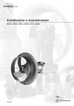
1
University of Nebraska Medical Center
Division of Laboratory Science
Clinical Laboratory Science Program CLS 418/CLS 419
BLOOD & CATHETER TIP CULTURES – GENERAL PROCEDURE
I.
Principle
Microorganisms present in the circulating blood are a threat to every organ in the body. This is
true even if the organisms are present continuously, intermittently, or transiently in the blood.
For this reason, blood cultures are one of the most important specimens submitted to the
Microbiology Laboratory for examination. Blood cultures are important in the diagnosis and
treatment of many conditions including bacteremia, fungemia, sepsis, infections of native and
prosthetic heart valves, superlative thrombophlebitis, and infections of vascular grafts. The
presence of microorganisms in the patient’s blood can be indicative of septicemia, a serious life
threatening disease. Therefore, rapid detection and identification of bloodborne pathogens is
very important, as well as notification of the clinician.
Common pathogenic organisms in blood culture sites include coagulase-negative
Staphylococcus, Staphylococcus aureus, Streptococcus pneumoniae, Alpha hemolytic
Streptococcus viridans group, Enterococcus species, Enterobacteriaceae, Pseudomonas
species, Haemophilus influenzae, Bacteroides fragilis, and Candida albicans. Other bacteria
yeast, molds and mycobacteria have also been associated with blood infections. All organisms
are potential pathogens in an immunosuppressed host. Several sources of bacterial sepsis
include infective endocarditis, infected arteriovenous fistulas, mycotic aneurysms, superlative
thrombophlebitis, infected intravenous catheters, infected indwelling arterial catheters,
genitourinary tract, respiratory tract, abscesses, surgical wounds, and biliary tract. Understanding
the circumstances in which bacteremia and sepsis can occur is helpful in interpreting results of
blood cultures.
From adult patients it is recommended that two sets of blood cultures should be collected per
febrile episode to help distinguish probable pathogens from possible contaminants, some sources
indicate three sets be drawn. A set includes both an aerobic bottle and an anaerobic bottle.
Collection of multiple sets of blood cultures is especially useful when determining the significance
of coagulase-negative staphylococcus, diptheroids (Corynebacterium species), Streptococcus
viridians group, and Bacillus species. These organisms can potentially be skin contaminants
associated with improper collection techniques.
Automated blood cultures machines incubate routine blood cultures for 5 days, this is sufficient
time to recover most bacteria while decreasing the isolation of contaminants. Blood cultures that
are incubated manually should be held a minimum of 7 days with a series of blind subcultures.
Blind subcultures are when you subbed the bottle to appropriate media and perform a gram stain
even though there are no visual signs of growth. When an unusual organism is suspected such
as Mycobacterium special media is used and extended incubation times are utilized.
Catheter tips are collected and cultured most often to diagnosis catheter-related bacteremia. The
most common organisms associated with catheter related sepsis are organisms found on the skin
as part of the normal flora. For this reason, semiquantitative cultures of catheter tips can help to
differentiate significant isolates from contamination organisms. Various indwelling catheter tips
are cultured in an attempt to isolate the possible source of infection in bacteremic patients.
Coagulase negative staphylococcus, in particular Staphylococcus epidermidis, appear to be
uniquely suited for causing catheter-related infections due production of a slime layer that allows
the organism to adhere to Teflon and plastics. Foley catheters are not suitable for culture
because they are usually highly colonized with bacteria from the urethra.
CLS 418/ CLS 419 Procedure – Blood & Catheter Tip Cultures
Student use only
Page 1
II. Specimen Collection, Transport and Handling
A. Indication for drawing blood cultures:
1. Increased temperature
2. Chills
3. Leukocytosis - especially with a left shift
4. Suspected meningitis
5. Suspected pneumonia
6. Fever associated with a heart murmur (suspected endocarditis)
7. Typhoid fever
8. Brucellosis
B. Specimen types and collection
1. Timing of Blood Culture Drawing
a. Generally blood cultures are drawn as a set (1 aerobic, 1 anaerobic) times 2 or 3
(obtaining more than three blood cultures within 24 hours does not result in a
significant increase in positive results), with at least a 45-60 minute interval between
each set drawn.
b. The ideal time to draw a set of blood cultures is one-half hour before an expected
chill or temperature spike as the patient’s blood will contain the highest numbers of
bacteria at this time. Once the temperature spikes the numbers of bacteria present
will rapidly decrease.
c. Ideally blood cultures should be drawn prior to administering antibiotic therapy. If this
is not possible, draw blood in bottles containing resin beads that nonspecifically
absorb antibiotics allowing for better growth of organisms that may be present.
2. Volume of Blood Culture Drawing
a. Volume is system specific.
b. In general, for adults 10-30 ml per draw is recommended.
c. For children, since they have a smaller blood volume, 1-5 ml is recommended.
d. When added to the broth medium, the final dilution of blood should be 1:5, 1:10 or
1:20 to dilute out antibacterial substances.
3. Procedure for Drawing Blood Cultures
a. After locating a vein, cleanse the site with isopropyl alcohol followed by an iodine
solution.
b. Repeat cleansing procedure two more times.
c. Allow the site to dry completely before drawing the specimen.
d. Do not palpate the site further after cleansing unless your finger has been cleaned in
the same manner. It is important not to introduce skin contaminants into a blood
culture as they can make interpretation of results difficult.
e. When sufficient blood has been collected for culture, introduce blood into appropriate
bottles.
f. Mix the bottles well to prevent clotting of the specimen and transport to the lab
immediately.
g. Do not refrigerate after drawing, hold at room temperature while transporting.
h. Considerations must be made for those patients allergic to iodine. In those
cases cleanse the site with isopropyl alcohol only.
4. Catheter tip cultures
a. After the tip is aseptically pulled and cut, it is placed in a sterile container for
transport.
b. Do not add any saline or other transport media to the specimen.
III. Direct Examination
A. Not routinely performed – gram stain not sensitive enough.
CLS 418/ CLS 419 Procedure – Blood & Catheter Tip Cultures
Student use only
Page 2
IV. Culture Setup
A. Manual (Conventional) Blood Culture Systems
1. Blood cultures should be incubated at 35ºC and examined visually for evidence of growth
(hemolysis, turbidity, gas production, colony formation) for a minimum of 7 days.
2. Blind subcultures and microscopic examination of the bottles should be performed at 1224 hours, 48 hours, and some time before the bottles are discarded.
3. Some systems have paddles of agar incorporated in the lid of the blood culture bottles.
Subculturing can be accomplished by tipping the broth over the paddles of agar.
B. Lysis-Centrifugation Blood Culture System (Isolator System)
1. The Isolator is a special tube that contains saponin, a chemical that lyses cells.
2. Approximately 7.5 to 10 ml of blood is placed in the tube.
3. The tube is then centrifuged for 15 minutes at 3000 rpm to concentrate any
microorganisms that may be present.
4. After centrifugation, the sediment is subcultured to appropriate media.
5. Literature indicates this system increases the yield of fungemia, yet may have an
increased contamination rate due to increased (over other systems) processing.
C. Non-radiometric Bactec (i.e., NR660)
1. Metabolizing organisms produce CO2. CO2 absorbs infrared light. At specified intervals,
the gas in each bottle is tested via infrared spectrophotometry.
2. The more infrared light passing through the gas sample from the bottle, the less CO2 in
the sample.
3. Threshold levels are set. The instrument will flag any bottle with a CO2 reading over the
specified threshold.
4. Metabolizing RBCs and WBCs will also give off CO2 (i.e., false positive).
5. Organisms that don’t produce large amounts of CO2 as a metabolic by-product may be
difficult to detect (i.e., false negative).
D. Examples of Continuous Monitoring Blood Culture Systems
1. BacT/Alert
a. Detects CO2 via a CO2 sensitive sensor at the bottom of each bottle (the sensor is
separated from the blood/media mixture via a CO2 permeable membrane).
b. The sensor visibly turns from green to yellow when CO2 is present.
c. A light-sensitive detector in the instrument continually monitors each bottle’s sensor.
2. BACTEC 9440/9120
a. Detects CO2.
b. Similar to the BacT/Alert except fluorescent, rather than spectral, light is used to
detect increased CO2 levels.
c. As CO2 is produced in each bottle, its sensor emits a fluorescent light that passes an
emission filter on the way to a light sensitive diode.
3. Difco ESP (Extra Sensing Power) Blood Culture System
a. Both gas production and consumption are monitored.
b. Changes in the concentration of CO2, H2, N2, and O2 are monitored via detecting any
change in pressure in the headspace of each bottle.
E. Blood Culture Media
1. The media used in most blood culture systems is multi-purpose and nutritionally
enriched.
2. Most commercially available media use the anticoagulant SPS (sodium
polyanetholsulfonate) in concentrations varying from 0.025% to 0.05%.
a. SPS also inactivates neutrophils, certain antibiotics (aminoglycosides & polymyxin),
and inactivates complement.
b. SPS may inhibit growth of Peptostreptococcus anaerobius, Neisseria gonorrhoeae,
Neisseria meningitidis, and Gardnerella vaginalis.
CLS 418/ CLS 419 Procedure – Blood & Catheter Tip Cultures
Student use only
Page 3
3. Synthetic antibiotic-removing resin beads can be added to bind antibiotics. The literature
is inconclusive as to whether or not these beads are effective.
F. Catheter tips are aseptically rolled on the surface of a single blood agar plate that is then
incubated aerobically.
G. Pathogens commonly isolated in blood.
1. Staphylococcus aureus
2. Coagulase negative Staphylococcus species – the most common cause of SBE
associated with prosthetics devices
3. Beta hemolytic strep including Group B Streptococcus - in infants
4. Streptococcus pneumoniae
5. Alpha hemolytic Streptococcus viridans group - the most common cause of SBE
6. Haemophilus influenza
7. Enteric gram-negative rods
8. Pseudomonas species
9. Enterococcus species
10. Yeasts and molds
11. Anaerobes – Bacteroides and Clostridium species
12. Neisseria species
H. Pathogens commonly isolated in catheter tips
1. Staphylococcus epidermidis
2. Staphylococcus aureus
3. Enteric gram-negative rods
4. Pseudomonas species
I.
Common contaminants for both blood and catheter cultures due to improper collection
techniques or culture set up (Normal skin flora)
1. Coagulase negative Staphylococcus species
2. Propionibacterium acnes
3. Alpha hemolytic Streptococcus viridans group
4. Bacillus species
V. Culture Interpretation
A. Reporting Results
1. An initial report is sent out at 24 or 48 hours and the final report is sent out at 5 days for
all no growth specimens that are incubated with a continuous monitoring system. It is
recommended that cultures not monitored continuously be held for 7 days before a final
report is sent out.
2. Cultures requested Brucella species are held for 3 weeks due to the growth rate of these
organisms. Brucella can be isolated in as little as 3 days but due to its slow growth rate it
is recommended that cultures be held for 21 days.
3. Cultures requested for yeast should be held for 7 days; those for fungus are held for 2128 days (recommended).
B. Positive Cultures
1. Gram stain the bottle to determine the morphology of the organisms present.
a. Call the results of the gram stain to the physician so that appropriate therapy can be
initiated if necessary.
2. Subculture to appropriate media based on gram stain results.
a. Always include an anaerobic subculture when indicated.
b. Include selective media when appropriate (i.e. MacConkey for gram negative rods)
c. Include direct Coagulase when appropriate (inoculating 100 ul of well mixed blood)
CLS 418/ CLS 419 Procedure – Blood & Catheter Tip Cultures
Student use only
Page 4
d. Include differential discs on media when appropriate for quick identification of
organisms. (Optochin disk, Bacitracin disk and/or Vancomycin disk)
e. Perform susceptibility on appropriate isolates from the isolation plates the next day.
3. Some facilities set up a sensitivity directly from the blood culture bottle. However, these
results are only preliminary and must be repeated from the isolation plates the next day
for confirmation. They are often inaccurate and do not correlate with repeat results so
many facilities have discontinued there use.
4. Isolation of an organism in multiple sets suggests bacteremia or fungemia rather than
contamination when isolating common skin contaminants.
C
Catheter Tips
1. Quantitate, identify and perform sensitivities on all potential pathogens with > 15 colonies
in pure culture (See Sec. III G).
2. Mixed cultures with > 15 colonies must be evaluated by a case by case basis.
3. Isolation of < 15 colonies is usually associated with skin contamination.
4. Correlation of catheter tips results with blood culture results is helpful in determining the
significance of isolated organisms.
V. References
A. Textbook of Diagnostic Microbiology, Mahon & Manuselis, 2nd edition, Chapter 30, pages
997-1010.
B. Color Atlas and Textbook of Diagnostic Microbiology, Koneman, 5th edition, Chapter 3, pages
153-162.
C. Bailey & Scott’s Diagnostic Microbiology, Forbes, 11th edition, Chapter 55, pages 865-883.
CLS 418/ CLS 419 Procedure – Blood & Catheter Tip Cultures
Student use only
Page 5
Learning About the ESP Culture System II
Learning About the ESP Culture System II
Overview
The Difco ESP Culture System II is a walk-away instrument designed for the culture and rapid
detection of microorganisms in blood and other sterile body fluids. The modular automated
system incubates, monitors and signals when microbial growth occurs in specially formulated
culture media that have been inoculated with patient specimens.
The ESP System is intended for use in a clinical laboratory setting by trained laboratory
personnel.
ESP Technology
Monitoring Microbial Growth
Once an inoculated culture bottle is loaded into the instrument, incubation and monitoring begins
and can continue from 5-60 days, depending on the test type selected. The culture medium in
each bottle contains specific ingredients that promote growth and result in gas consumption
and/or gas production. An instrument sensor connected to each bottle monitors the resulting
changes in headspace gases.
Detection
The detection technology is based on the sensitive measurement of gas production and gas
consumption within the headspace of a sealed culture bottle. During bottle loading, each bottle is
attached to a sensor through a connector. The sensor monitors changes in gas consumption
and/or gas production that are recorded by the instrument at 12 and 24-minute intervals for
aerobic/myco and anaerobic locations, respectively. This information is used to generate a curve
for each bottle. Figure 1-1 illustrates a typical curve showing gas production.
Figure 1-1. Typical curve generated by the ESP System
CLS 418/ CLS 419 Procedure – Blood & Catheter Tip Cultures
Student use only
Page 6
Learning About the ESP Culture System II
Algorithm
An internal algorithm analyzes the changes in gas consumption and/or gas production as data
points are collected from each culture bottle. The algorithm uses the information to determine the
status of each specimen. When a certain set of conditions is met, a bottle is flagged as positive.
Advantages of the Technology
Detection Beyond CO2 Production
The system detects the consumption and /or the production of metabolic gases rather than the
production of a single gas, such as carbon dioxide. Certain microorganisms that produce nitrogen
or hydrogen gas as a major metabolic by-product, rather than carbon dioxide, are reliable
detected.
Fast Detection Time
When both gas consumption and production occur in a growth cycle, gas consumption always
occurs before gas production. Since the ESP System detects more than gas production, earlier
detection can result.
Culturing Mycobacteria on the ESP Culture System II
The unique technology and modular design of the ESP Culture System II combine to provide a
single instrument for both blood culture and mycobacteria testing. Any testing module can be
configured for either blood or mycobacterial testing. This configuration is controlled entirely within
the software. The only requirements are that the module must be non-shaking and the routine
blood culture adapter cups must be replaced by cups specially adapted for the smaller ESP Myco
bottles. Myco modules have a green label on the faceplate the module numbers are followed by
an M instead of an A or N.
ESP Systems capable of both blood and mycobacterial testing are designated “Combi”
instruments. Four types of “Combi” instruments are available and are known as the Myco Combi
I, II, III and IV respectively. The roman numeral refers to the number of myco modules that have
been added to the base ESP System. These “Combi” instruments have 256 blood culture
locations and 16 myco locations for each added myco module. ESP Systems dedicated to myco
testing exclusively are known as the ESP Culture System II Myco 128,k 256, and 384. The
numbers refer to the total testing capacity of each unit. The modules in these systems are all nonshaking. Converting an anaerobic module of an ESP System II Blood Culture instrument to a
myco module means that the opposing aerobic module becomes unpaired. There is an option
available only to the users of such systems that allows them to select these unpaired aerobic
locations during specimen entry.
The detection method for mycobacteria is the same as for routine blood cultures. The ESP
monitors the headspace of the specimen bottle for any changes in pressure that would indicate
gas production or consumption. In the case of mycobacteria, the net effect of their metabolic
activity is gas consumption resulting in a decrease in pressure. This causes a positive signal and
notification of the user.
The ESP Myco bottle is capable of testing blood, body fluids, respiratory, urine, gastric aspirates,
and tissue specimens. The ESP Myco bottle contains small cellulose sponges that increase the
reactive surface area and enhance recovery. Non-sterile specimens require decontamination
prior to inoculation. Mucoid specimens also require digestion and decontamination treatment. A
growth supplement, ESP Myco GS (Middlebrook OADC enrichment), and an antibiotic, ESP
Myco PVNA (Polymyxin B, Vancomycin, Nalidixic acid, and Amphotericin B) are aseptically
added to each specimen bottle just prior to incubation. A connector is attached to each bottle and
the specimens are loaded according to whichever ESP entry mode is preferred.
CLS 418/ CLS 419 Procedure – Blood & Catheter Tip Cultures
Student use only
Page 7
Learning About the ESP Culture System II
Positive specimens signal in the same way as with routine blood culture specimens. The following
is a graph of a microorganism that consumes oxygen. (Figure 1-2).
Figure 1-2. Sample graph showing O2 consumption
The user can remove positive culture material from the ESP Myco bottle and perform
confirmatory testing. Negative bottles not exhibiting signs of turbidity can be discarded according
to routine laboratory procedures.
ESP Modular Design
The ESP system has a modular design. Up to five incubator units in any combination can be
connected to a central computer system for control, monitoring and data management (Figure 13). Each incubator is referred to as a “unit” with a letter designation A, B, C, D, or E.
Figure 1-3. Complete ESP System
CLS 418/ CLS 419 Procedure – Blood & Catheter Tip Cultures
Student use only
Page 8
BACTEC Fluorescent Series User’s Manual
Introduction
Fluorescent Series Overview
The BACTEC fluorescent series instruments, such as the BACTEC 9240 and 9120, are
designed for the rapid detection of bacteria and fungi in clinical cultures of blood. Samples
are drawn from patients and injected directly into BACTEC culture vials.
When microorganisms are present, they metabolize nutrients in the culture medium,
releasing carbon dioxide into the medium. A dye in the sensor reacts with CO2. This
modulates the amount of light that is absorbed by a fluorescent material in the sensor. The
instrument’s photo detectors measure the level of fluorescence, which corresponds to the
amount of CO2 released by organisms. Then the measurement is interpreted by the system
according to preprogrammed positivity parameters. (See Figure 1-1.)
At system startup, the computer performs diagnostics and downloads operating instructions
to the instrument(s). Then the instrument(s) begin continuous automated testing. Light
Emitting Diodes (LEDs) behind the vials illuminate the rack(s), activating the vials’ fluorescent
sensors. After a warm-up period, the instrument’s photo detectors then take the readings. A
test cycle of all racks is completed every ten minutes. Positive cultures are immediately
flagged by an indicator light on the front of the instrument and displayed on the monitor.
When positive vials are identified, the lab technologist pulls them from the instrument for
confirmation of results, and for isolation and identification of the organism.
Figure 1-1. BACTEC Fluorescent Test Technology
1. Organisms metabolic activity
releases CO2...
2. Which reacts with dye in vial
sensor.
3. LED, modulated by dye,
activates fluorescent material in
sensor.
4. Photo detector reads
fluorescence.
5. Raw data from detector is sent
to rack microprocessor....
6. Where positivity analysis is
performed.
7. Positive vial lamp illuminates,
audible alarm sounds, positive
stations are displayed.
1.
Positive
Vial
2.
7.
LED
Photo
detector
4.
3.
Microprocessor
Raw data
5.
CLS 418/ CLS 419 Procedure – Blood & Catheter Tip Cultures
Student use only
Test Results
Positivity Analysis
6.
Page 9
Figure 1-2. BACTEC System Functions
A single 9240 Instrument is capable of monitoring a total of 240 BACTEC culture vials, and a
9120 instrument can monitor a total of 120 vials. A multi-instrument system, with up to five
9240s, can monitor up to 1,200 culture vials. The vials are arranged in racks (six in the 9240,
three in the 9120), each of which holds up to 40 vials. The racks are continuously incubated
at 35 C and are agitated for maximum recovery of organisms. The practical capacity of an
instrument system is from 12 to 120 new culture sets per day with a five-day test protocol.
Major features of the BACTEC fluorescent series instruments include:
• Automated, continuous, unattended testing of cultures through non-invasive
fluorescent technology
• Up to five instruments in a multi-instrument configuration
• Minimum user interaction and handling
• Immediate notification of positives through an instrument indicator lamp, display on
the monitor, and an audible alarm (if configured)
• Independent microprocessors for each of the instrument racks insure that testing can
continue even if one or more racks fail
• Simple user interface, with barcoded commands at the instrument for routine
operations
• Incubation and shaking for all cultures
• Proven BACTEC culture media
CLS 418/ CLS 419 Procedure – Blood & Catheter Tip Cultures
Student use only
Page 10