1
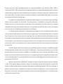
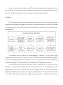
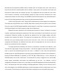
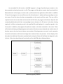
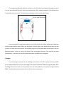
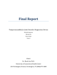
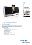
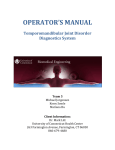
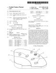
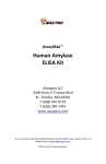
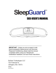
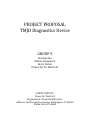
PROJECT PROPOSAL
TMJD Diagnostics Device
GROUP 5
Mariana Hu
Michael Jorgensen
Kerry Semle
Project for Dr. Mark Litt
CLIENT CONTACT:
Name: Dr. Mark Litt
Organization: UConn Health Center,
Address: 263 Farmington Avenue, Farmington, CT 06030
Phone: 860-679-4680
Executive Summary
The purpose of this project is to develop a wearable device to record bruxism (tooth grinding and
clenching) events and joint sounds during sleep through the use of electromyography.
Patients with temporomandibular joint disorder (TMJD) frequently engage in bruxism, which
wears down the fibrocartilage disc in the temporomandibular joint leading to chronic problems. Popping
and clicking sounds are audible and frequently cause significant discomfort. Patients often have a
difficult time in completely opening and closing the jaw, making it difficult to adequately masticate food.
Over time, the condition may worsen and pain may develop in the temporomandibular joint region.
TMJD often goes unnoticed until the fibrocartilage disc is finally worn down to the point where the
symptoms become self-evident.
Current screening methods are ineffective at diagnosing this condition. This project is an
improvement on current screening methods by allowing the patient to wear a comfortable device in
their home environment and acquire data consistent with their natural sleeping habits.
1. Introduction
1.1 Background
Temporomandibular Joint Disorder (TMJD) affects approximately 35 million people in the United
States. It can cause problems with eating, drinking, swallowing, talking, making facial expressions, and
even breathing in people who suffer from it. Bruxism can wear down the fibrocartilage discs in the jaw,
which makes it difficult to open and close the mouth. Bruxism, the act of grinding or clenching of the
teeth, is a common symptom of TMJD that clinicians use to diagnose it.
Currently patients who are suspected to have TMJD or exhibit signs of bruxism through wear on
the teeth are subject to spend a night in a sleep lab, in which they are connected to electrodes while
sleeping. Those electrodes are connected to machines through wires that record and analyze the activity
of the masseter and temporalis. A trip to the sleep lab is inconvenient for many patients, many of whom
also have sleep apnea and typically use a breathing machine.
1.2 Purpose of the Project
The TMJD Diagnostics Device aims to make sleep labs for TMJD diagnosis superfluous. It will
make it possible for the muscular activity associated with bruxing to be recorded in the patient’s home.
This would take the necessity of transporting a breathing machine out of the diagnosis of TMJD for
patients with sleep apnea, and be more cost effective for patients as well as their insurance companies.
1.3 Previous work done by others
1.3.1 Products
A current low-cost bruxism diagnostic device is in the market. The BiteStrip is a single-use
disposable device used by the patients at home. It is a miniature electronic system consisting of: two
pre-gelled skin EMG electrodes and an EMG amplifier, for acquiring nocturnal EMG signals from the
masticatory muscles; a CPU for running real-time software that analyzes EMG strength patterns and for
detecting and counting each bruxing episode; a permanent chemical display for presenting the study
outcome in the morning and a lithium battery. All the elements are integrated onto a small lightweight
plastic film that is attached to the patient’s cheek.
Components of the BiteStrip
Its indications correlate well with comparable indications from formal sleep lab studies. It is available
through health providers worldwide at a very low price to the public as a self-administered or
prescription test. Relevant patents were applied for, and clinical studies are in process in well-known
sleep labs to prove its accuracy.
In using Bitestrip, the patient is required to perform at least two maximal voluntary clenches
during the first 30 minutes by biting on a wooden spatula provided in order to establish the threshold
which is set at 30% of the average EMG amplitude. The BiteStrip counts each EMG peak stronger than
the threshold for a period of up to six hours. The patient then returns the Bitestrip to the clinician for
interpretation of the results.
Despite the advantages offered by Bitestrip, the device presents some limitations. In the first
place, the study requires a minimum of 5 hours. If the Bitestrip is removed earlier than this minimum
period, an error will occur. Moreover, this device does not save the EMG signals, which may be useful
for the clinician in order to assess an accurate diagnosis. Bitestrip only provides the number of bruxing
events. This information will not allow the clinician to distinguish between teeth grinding and clenching,
nor record the intensity of the clenching or grinding or the time when these events occur.
Sakagami, Horii, Ino, Matoba, Kato and Kawanami developed a portable nocturnal bruxism
monitoring and analysis device in 2002 and verified its accuracy and easiness of use. Their bruxism
diagnostic system consisted of two parts: the portable bruxism monitoring and analysis device, and the
software that was installed to a computer.
The monitoring and analysis device (Morita MFG) was equipped with a microcomputer to autoanalyze the data. Due to the small size of this device and its light weight, it could be either placed on the
forehead, or inserted in a pocket of a jacket. The device had two sets of data import connectors for pickup cables, which were hooked to the disposable electrodes (Duotrode®, Myotronics Inc., USA) attached
either to both sides of the cheek over the masseter muscles, or to both sides of the temple over the
temporalis muscles. In order to launch the device, the user would push the start button on the side of
the device. After amplification and processing of the EMG signals, these were compared to a manually
set threshold level. The device provided the total number of bruxing events as well as the lasting time of
each bruxing event. Finally, the recorded data was transmitted into the computer via serial port. The
device was equipped with EEPROM and the data is recorded on the memory to the maximum of 676
data sets in one night.
Although the authors who carried out the study verified the accuracy of the device, and the
subjects of the study declared that it was easy to use, this device does not provide comfort and mobility
to the patients due to the presence of wires. Moreover, serial communication limits the speed of data
acquisition.
Components of the bruxism monitoring and analysis device
developed by Sakagami, Horii, Ino, Matoba, Kato and Kawanami.
Left: The temporalis and masseter muscles. Center: Electrodes attached to the cheek for the data
acquisition from the masseter muscles, and to the chin for earthing. Right: Electrodes attached to the
temple for the data acquisition from the temporalis muscles, and to the forehead for earthing.
1.3.2 Patent search results
The Bitestrip described above has applied for relevant patents and clinical studies are in process
in well-known sleep labs to prove the accuracy of this device.
In addition, after patent search, only one more device was vaguely related to the purpose of our
project. It is a “Measuring device for quantifying the severity of bruxism”, US patent #5,911,576,
comprising of a thin shell formed to the shape and elastically retained to one or more teeth. The shell
consists of many layers of different colors. The outer layer of the shell, when worn out by clenching and
grinding, reveals an inner layer. This way, the regions of wear may be analyzed to determine the extent
of the bruxing activity. However, this device does not provide the EMG signals for accurate diagnosis and
may not be comfortable to use since it may interfere with normal breathing.
2. Project Description
2.1 Objective
The TMD Diagnostics Device utilizes electromyography (EMG) to obtain biosignals from the
muscles involved in mastication, including the left and right temporalis muscles, and the left and right
masseter muscles. These EMG signals will be used to determine if the patient is bruxing or clenching
during sleep, two major contributing factors to temporomandibular joint disorder (TMD). EMG is
currently utilized in TMD sleep studies with significant efficacy in determining bruxing events. However,
sleep studies are expensive and uncomfortable for the patient, resulting in data that is inconsistent with
their sleep habits. This device allows the patient to perform a comfortable EMG study in their home
environment and acquire accurate data.
The signals will be obtained from reusable electrodes without the use of adhesive gel, providing a
superior level of comfort for the patient. The setup process is simplified by using these electrodes,
allowing the user to slip the device on and make minor adjustments to ensure optimal fit, without
individually attaching each adhesive electrode. The electrodes will also not have to be interchanged
after each use, thus simplifying the maintenance of the device.
The device will be comprised of a wearable hat-like apparatus. The reusable electrodes will be
securely placed inside the device over the muscles of mastication. The electrode leads will be connected
to the internal circuitry so that no wires are exposed. The circuit will be enclosed and located on the top
of the device so that the patient does not damage it during sleep. Both the electrodes and the circuit will
be removable so that the patient can wash the exterior case. The device will be comprised of fleece to
increase the comfort for the patient. Velcro® will be utilized to allow for minor adjustments and achieve
an optimal fit.
The EMG signals will be sent wirelessly to a notebook personal computer. A LabVIEW program
will automatically load each time the computer is powered on. The interface will be easy to use and will
provide sufficient help information in addition to a detailed user manual. The user will simply run the
program and initiate the data acquisition process. The program will have the capability to analyze, send,
store and display data. The raw data will be processed into useful diagnostic information that may be
presented to the clinician.
Wireless communication (Bluetooth®) between the device and the notebook personal computer
is part of the optimal design, because it reduces the complexity of the device and also improves the
patient’s comfort while sleeping. The device will contain an additional serial (RS-232) port, providing an
optional connection that is compatible with numerous computer configurations, not just computers with
Bluetooth® connectivity. The selected notebook computer will have both Bluetooth® connectivity and a
serial port to account for this.
In order to test the device, subjects with TMD and control subjects will be employed in clinical
trials. Therefore, an institutional review board (IRB) must be applied for in consideration of clinical trials.
This will be possible with the help of the UConn Health Center and School of Dentistry.
2.2 Methods
Electromyography will be used to obtain physiological data from the muscles of mastication and
therefore proper analog circuit design is necessary for the successful operation of the device. Electrodes
specifically designed to obtain EMG signals will be connected to an enclosed circuit represented by the
flowchart below.
EMG circuit flowchart
The frequency range of interest for electromyography is between 25 Hz and 500 Hz, with
amplitudes from 100 μV to 90 mV. However, the use of surface electrodes (surface electromyography)
can decrease the signal amplitudes to a maximum of 1 mV. Taking these parameters into consideration,
it is possible to design a circuit that conditions and amplifies the biological signals of interest.
An RC lowpass and RC highpass combination to form a bandpass filter is necessary for each
electrode to remove unwanted frequencies that will cause distortion. The cutoff frequency for the
highpass filter was chosen to be 25 Hz, but may be reduced to around 15 Hz to allow more low
frequency EMG signals to pass. Likewise, the lowpass cutoff frequency for the lowpass filter was chosen
to be 500 Hz, but it may be increased to pass higher frequency signals. A one-pole RC filter was chosen
here to reduce the amount of integrated circuits necessary for active filters. Each muscle requires two
electrodes, thus there would need to be at least eight operational amplifiers to provide the filtering
capabilities. Since space is a design constraint, RC filters are sufficient at this point in the circuit, as more
filtering can be done after amplification and in the LabVIEW program as well.
Protection circuitry is an absolutely critical portion of the design of the overall device, since
numerous electrodes will be attached to the patient during sleep with the potential for electrical shock.
Diodes are excellent for simple protection against small voltages. Low leakage diodes will be constructed
in parallel so that if the voltage near the electrode ever reaches 700 mV, the diodes will reverse and
forward biased, allowing the potentially harmful current to run directly to ground. Electromyography
voltages typically do not exceed 200 mV, however it is still important to protect the patient against any
possibility of electrical shock.
An instrumentation amplifier was chosen as a means of amplifying the signal due to several
reasons. The input impedance of a three-stage instrumentation amplifier is very high, which is desirable
that minimizes loading effects. The output impedance is low with respect to the input impedance,
another desirable characteristic. The gain of an instrumentation amplifier can be adjusted simply by
changing one resistor value, allowing for a flexible gain factor that can be adjusted accordingly. The
design also incorporates a high common-mode rejection ratio (CMRR), which is a desirable characteristic
when dealing with biological signals. The AD620 instrumentation amplifier from Analog Devices is
initially selected since it provides a superior performance to the classic three-stage instrumentation
amplifier, has a gain adjustable from 1 to 10,000 by changing one external resistor, exhibits low noise
(280 nV p-p noise from 0.1 Hz to 10 Hz) at a frequency below that of the EMG signals. It is specified on
the manufacturer’s data sheet that it is applicable to ECG and medical instrumentation, and works well
with battery operation and portable equipment, with a maximum supply current of just 1.3 mA. Because
the AD620 has the three-stage instrumentation built into a small package, it saves space on the printed
circuit, which is another desirable parameter for consideration.
An active bandpass filter following the instrumentation amplifier is implemented in the design to
further suppress frequencies outside of the EMG signal range. An op amp design will be used to provide
for a higher order circuit with a high Q-factor. Constructing the filter circuit after the instrumentation
amplifier also allows for the suppression of noise created by the previous electrical components. It is
desirable that the operation amplifier used for filtration does not introduce noise, thus a low-noise op
amp will be selected. A quad amplifier will be utilized to save space on the printed circuit board and
eliminate the need to have four separate op amps. This configuration will also reduce the noise of the
circuit because it eliminates cross-talk between multiple op amps and noise introduced from other
components utilized in the filter design. The LM3900 by National Semiconductor was selected because it
meets all of the design parameters and is a low cost quad operational amplifier.
A 60 Hz notch filter with a twin T design will be implemented to remove power line interference.
This design has a high Q-factor and an infinitely deep notch, ensuring that only 60 Hz noise is filtered.
Since this is part of the EMG signal bandwidth, proper design of a notch filter is necessary to ensure that
the biological signals in the lower frequency range are not suppressed.
Bluetooth has been selected as the communication protocol of choice. It allows for a wireless
interface, significantly reducing the complexity of the device and making it user-friendly for set up and
maintenance. Bluetooth modules can generally be powered from low voltage supplies and work
excellent when powered by battery. The range can be anywhere up to 350 feet or more, which is
desirable since the receiving laptop may be across the user’s room. Since not all computers are
compatible with Bluetooth, an additional serial port will be incorporated into the circuit so that the
device can transfer data to other computers.
The laptop would be provided by the clinician to the patient. It should be cost effective, small
size and have a light weight for easy transportation. It will also be required to have built-in Bluetooth in
order to receive the processed signal from the circuit. In addition, it must meet the requirements
needed to launch a LabVIEW application: operating systems Windows XP, Windows Vista or Windows 7,
Pentium III/Celeron 866 MHz or equivalent, at least 255 MB of RAM memory, a screen resolution of
1024x768 pixels and 1.6 GB of disk space. The laptop will be strictly configured to be user-friendly,
ensure proper functionality and prevent any modifications by the user.
For example, it will be
configured so that when the user turns on the laptop, the LabVIEW application will launch automatically.
Also, it will prevent the user from installing or deleting applications. Ideally, the user will only have to
click a Start button in the LabVIEW application to launch the program and click a Stop button at the end
of the session. This will ensure an easy use of the device and prevent errors of operation by the user.
An executable file will contain a LabVIEW program in charge of performing an analysis on the
data received and saving the results in a file. The program will be able to receive data from the built-in
Bluetooth device of the laptop. In order to do so, a Bluetooth client application will be built in LabVIEW.
To launch the program, the user will have to click a Start button. A window will pop up asking to enter
the name of the file where the data corresponding to that session will be saved. The user will be
required to enter the current date as the name of the file. Next, the program will initiate a loop that will
constantly add the data with their corresponding time as elements of a 2D vector. As the data is being
acquired, it will be continuously saved in the specified file and displayed in a waveform graph in the
front panel. In addition, the program will perform analysis on the data contained in the vector. These
will include calculating and displaying the total number of bruxism events detected, the total duration of
bruxism events, the time interval where most number of bruxing events occur and a unit conversion
from Volts to Newtons and Pascals to display then maximum force and pressure. A clear display of the
data plus the additional information provided by the analysis of data will facilitate the diagnosis by the
clinician. A flow chart describing the functionality of the LabVIEW code is displayed below.
LabVIEW program flowchart
The proposed product will look like a winter hat. It will house nine reusable electrodes as well as
a circuit and a Bluetooth device, which will communicate with a netbook computer. The device will be
comfortable and easy to use. The preliminary hat design is shown in the figure below.
Preliminary Hat Design
Once the product is completely ready for use, 20-30 clinical trials will be performed. Volunteers
will be compensated at least $100 to use the device for three nights, over which they will wear the hat
product, and take home the netbook. The LabVIEW program will record the data transmitted to it by the
Bluetooth device in the hat, which will further filter and analyze the data. The data will be saved
automatically, so that the clinician can review the results, and determine a diagnosis.
3. Budget
The total budget estimated for the building of the device is $3,770. However, due to possible
errors and development costs, the final budget of the device would be reduced by approximately 65%.
Excluding clinical trials from the final product cost, the final production cost would be approximately
$600. The table shown below details the cost of the various components.
Component
Maximum estimated price
Electrodes
$50
Electrode leads
$20
Integrated circuits
$40
Resistors, capacitors and diodes
$20
9 V batteries
$10
Bluetooth module
$100
Hat
$30
Laptop
$500
Clinical trials
$3000
TOTAL
$3,770
Budget for proposed project
The clinical trials are the most expensive component of the project because monetary
compensation must be provided to the volunteer participants. A standard compensation for a clinical
study is approximately $100 per volunteer, thus with twenty to thirty volunteers the study may cost up
to $3,000.
4. Conclusion
The project will be beneficial to both the clinician and the patient by providing accurate data and
ensuring that the correct diagnosis is realized. It is an improvement on current products in several ways.
First, it eliminates the need for the patient to stay overnight in a sleep study, which is both . Second, it
will be comfortable and allow the patient to obtain optimal sleep because there are no external wires
and will not restrict the patient’s movements. The design of the hat is unique in that it allows for
repeated usage and the ability to be washed. Third, data will be acquired in the patient’s home
environment, ensuring that data is consistent with their natural sleeping habits. Fourth, it will display
the raw electromyography data, which most current devices neglect to save. In addition to
electromyography data, important diagnostic information will be presented, including the number of
bruxing events, the maximum force of clenching, the time at which the bruxing events occur, and the
total duration of bruxism. All of these features are vast improvements on the current techniques for
temporomandibular joint disorder diagnosis.