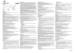
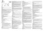
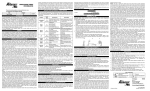
1
Do not use the test strip after the specified expiration date.
Tightly re‑cap the container immediately after removing a test strip.
Combur⁹ Test
04510038 191
50
04510046 040
100
Specimen collection and preparation
For specimen collection and preparation only use suitable tubes or collection containers.
Use fresh urine that has not been centrifuged. The urine specimen should not stand for more
than 2 hours before testing. In case of longer standing, mix before use.
Use only clean, well-rinsed vessels to collect urine.
Do not add preservatives to the urine.
Materials provided
▪
04510038191, package with 50 test strips
▪
04510046040, package with 100 test strips
Materials required (but not provided)
▪ General laboratory equipment
Assay
For optimum performance of the assay follow the directions given in this document.
English
Intended use
Nine‑patch test strip for the semi‑quantitative determination of leukocytes, nitrite, pH, protein,
glucose, ketone bodies, urobilinogen, bilirubin and blood in urine by visual reading.
For professional use only.
Summary
Urine test strips are used to measure certain constituents in urine which are significant of
renal, urinary, hepatic and metabolic disorders.
Test principle
Leukocytes (LEU): The test reveals the presence of granulocyte esterases. These
esterases cleave an indoxyl ester, and the indoxyl so liberated reacts with a diazonium salt
to produce a violet dye. Bacteria, trichomonads or erythrocytes present in the urine do not
affect the reaction.
Nitrite (NIT): The test is based on the principle of the Griess test and is specific for nitrite.
The reaction reveals the presence of nitrite and hence indirectly nitrite-forming bacteria in
the urine by a pink-to-red coloration of the test patch. Even a slight pink coloration is
indicative of significant bacteriuria.
pH: The test paper contains the indicators methyl red, phenolphthalein and bromothymol
blue and reacts specifically with H+-ions. The most frequent pH values of fresh urine from
healthy subjects lie between 5 and 6.
Protein (PRO): The test is based on the principle of the protein error of a pH indicator. It is
particularly sensitive to albumin. Quinine, quinidine, chloroquine, tolbutamide and an
elevated pH (up to 9) do not affect the test.
Glucose (GLU): The glucose determination is based on the specific glucoseoxidase/peroxidase reaction (GOD/POD method). The test is independent of the pH and
specific gravity of the urine and is not affected by the presence of ketone bodies.
Ketone bodies (KET): This test is based on the principle of Legal’s test and is more
sensitive to acetoacetic acid than to acetone.
Urobilinogen (UBG): A stable diazonium salt reacts almost immediately with urobilinogen to
give a red azo dye. The test is specific for urobilinogen and is not susceptible to the
interfering factors known to affect the Ehrlich’s test.
Bilirubin (BIL): The test is based on the coupling of bilirubin with a diazonium salt. Even the
slightest pink coloration constitutes a positive, i.e. pathologic, result. Other urinary
constituents produce a more or less intense yellow coloration.
Blood (ERY/Hb): The peroxidase-like action of hemoglobin and myoglobin specifically
catalyzes the oxidation of the indicator by means of the organic hydroperoxide contained in
the test paper to give a blue-green coloration.
Reagents
Each test contains per 1 cm2 test patch area the following:
Leukocytes: Indoxylcarbonic acid ester 15.5 μg; methoxymorpholinobenzene diazonium
salt 5.5 μg
Nitrite: 3-hydroxy-1,2,3,4-tetrahydro-7,8-benzoquinoline 33.5 μg; sulfanilamide 29.1 μg
pH: Bromothymol blue 13.9 μg; methyl red 1.2 μg; phenolphthalein 8.6 μg
Protein: 3’,3’’,5’,5’’-tetrachlorophenol-3,4,5,6-tetrabromosulfophthalein 13.9 μg
Glucose: 3,3’,5,5’-tetramethylbenzidine 103.5 μg; GOD 6 U, POD 35 U
Ketone bodies: Sodium nitroprusside 157.2 μg
Urobilinogen: 4-methoxybenzene-diazonium-tetrafluoroborate 67.7 μg
Bilirubin: 2,6-dichlorobenzene-diazonium-tetrafluoroborate 16.7 μg
Blood: 3,3’,5,5’-tetramethylbenzidine 52.8 μg; 2,5-dimethyl-2,5-dihydroperoxyhexane
297.2 μg
Precautions and warnings
For in vitro diagnostic use.
Exercise the normal precautions required for handling all laboratory reagents.
Disposal of all waste material should be in accordance with local guidelines.
Safety data sheet available for professional user on request.
The stopper of the test strip vial contains a non-toxic silicate-based desiccant which must not
be removed. If ingested by accident, drink large quantities of water.
Reagent handling
Test strips are ready for use.
Storage and stability
Store the package at 2-30 °C. The test strips are stable up to the expiration date specified on
the box, when stored in the original container.
1. Use fresh urine that has not been centrifuged. Thoroughly mix the urine sample. The
sample should be at room temperature when the test is performed and should not have
been standing for more than 2 hours.
2. Take a test strip out of the container. Close the container again with the original
desiccant stopper immediately after removal of the strip. This is important as otherwise
the test areas may become discolored due to environmental influences such as moisture
or nitrite gases in the air. Incorrect results may be obtained.
3. Briefly (about 1 second) dip the test strip into the urine making sure that all test areas are
moistened.
4. When withdrawing the test strip, wipe the edge against the rim of the vessel to remove
excess urine.
5. After 60 seconds (60-120 seconds for the leukocyte test area), compare the reaction
colors of the test areas with the colors on the label and assign always the value of the
nearest color block.
▪ Compare the 9th (blood) test area with both color scales as separate color scales are
given for erythrocytes and hemoglobin.
Any color changes appearing only along the edges of the test areas, or developing after
more than 2 minutes, do not have any diagnostic significance.
Quality control
The control intervals and limits should be adapted to each laboratory’s individual
requirements. Values obtained should fall within the defined limits. Each laboratory should
establish corrective measures to be taken if values fall outside the defined limits.
Follow the applicable government regulations and local guidelines for quality control.
For quality control, use commercially available urine controls, or other suitable control
material.
Important note for reporting results (for professional users).
According to the regulations from the German Medical Association for quality assurance of
medical laboratory analyses dated 11/23/2007, the decision to classify a laboratory test
result to either part B1 or B2 depends on the way the test results are expressed in the report
(scale level).
The specification in the report defines whether a determination is quantitative or qualitative
and therefore which legal requirements for quality assurance (B1 for quantitative or B2 for
qualitative) need to be followed. Examples of qualitative characteristics include titer levels,
concentrations/color ranges (+ to +++) or a defined range of values. A characteristic of a
quantitative value is when the value has a corresponding measured unit value.
Limitations - interference
Leukocytes: Formaldehyde (stabilizer) and medication with imipenem, meropenem and
clavulanic acid may cause false-positive reactions. If the urine specimen has a pronounced
intrinsic color (for example due to the presence of bilirubin or nitrofurantoin), the reaction
color may be intensified due to an additive effect. Urinary protein excretions in excess of
500 mg/dL and urinary glucose excretions in excess of 1 g/dL may diminish the intensity of
the reaction color, as can cephalexin and gentamicin if administered in high daily doses, or
boric acid if used as a preservative.
Nitrite: Prolonged urinary retention in the bladder (4‑8 hours) is essential in order to obtain
an accurate result. Administration of antibiotics or chemical drugs should be discontinued
3 days before the test. Large amounts of ascorbic acid decrease the sensitivity of the test.
Attention: Nitrogen oxides present in the atmosphere may have an influence on the stability
of the nitrite test pad.
Protein: False-positive readings may be found after infusion of polyvinylpyrrolidone (blood
substitute), or when the urine collection vessel contains chlorohexidine or traces of
disinfectants possessing quaternary ammonium groups.
Glucose: The effect of ascorbic acid has been largely eliminated so that at glucose
concentrations of 100 mg/dL and above even high ascorbic acid concentrations are not likely
to give false-negative results.
Ketone bodies: Phenylketones and phthalein compounds produce red colors on the test
patch; they are, however, quite distinct from the violet colors produced by ketone bodies and
can lead to false-positive results. Captopril, mesna (2‑Mercaptoethanesulfonic acid sodium
salt) and other substances containing sulfhydryl groups may produce false-positive results.
Urobilinogen: Nitrite concentrations above 5 mg/dL or formaldehyde (stabilizer) above
200 mg/dL may cause a decrease in the color reaction.
Bilirubin: Large amounts of ascorbic acid lower the sensitivity of the test.
Do not expose urine specimens to sunlight as this induces oxidation of bilirubin and
urobilinogen and hence leads to artificially low results for these two parameters.
Drugs that turn red in an acid environment (e.g. phenazopyridine) may produce
false‑positive readings or reddish colorations on the test patches for nitrite, protein,
urobilinogen and bilirubin.
Blood: The values appearing on the printout refer to intact erythrocytes. At concentrations of
about 5‑50 Ery/μL, significant hemolysis (such as may occur on prolonged standing of the
urine) leads to values which are higher than the corresponding concentrations given for
intact erythrocytes. Ascorbic acid has virtually no effect on the test. In women the test for
blood may be falsified from 3 days before to 3 days after a period. It is therefore advisable
not to perform the test during this time. After physical activity, e.g. strenuous jogging, raised
values for erythrocytes and protein may occur without being signs of disease.
False‑positive readings, particulary for erythrocytes, glucose and protein can result from
residues of strongly oxidizing disinfectants in the specimen collection vessel.
Knowledge of the effects of drugs or their metabolites upon the individual tests is not yet
complete. In doubtful cases, it is therefore advisable to repeat the test after discontinuing a
particular drug.
For diagnostic purposes, the results should always be assessed in conjunction with the
patient’s medical history, clinical examination and other findings.
Limits and ranges
By visual reading
▪
Measuring range
Leukocytes: NEG – ~ 500 LEU/μL (3+), Nitrite: NEG – POS (1+), pH: 5‑9, Protein:
NEG - 500 mg/dL (5 g/L; 3+), Glucose: NORM – 1000 mg/dL (55 mmol/L; 4+), Ketone
bodies: NEG – 150 mg/dL (15 mmol/L; 3+), Urobilinogen: NORM – 12 mg/dL
(200 μmol/L; 4+), Bilirubin: NEG – ~ 6 mg/dL (100 μmol/L; 3+), Blood: NEG
– ~ 250 ERY/μL (4+).
▪ Lower limits of measurement
Lower detection limit
Leukocytes: 10‑25 LEU/μL, Nitrite: 0.05 mg/dL (11 μmol/L), Protein: 6 mg albumin/dL,
Glucose: 40 mg/dL (2.2 mmol/L), Ketone bodies: For aceto-acetic acid 5 mg/dL
(0.5 mmol/L), Urobilinogen: 0.4 mg/dL (7 μmol/L), Bilirubin: 0.5 mg/dL (9 μmol/L),
Blood: intact erythrocytes: 5 ERY/μL, Hemoglobin or hemolysed erythrocytes:
corresponding to 10 ERY/μL.
▪ Accuracy
Leukocytes: ≥ 90 % compared with counting chamber, Nitrite: ≥ 90 % for 107 grampositive organisms compared with Griess test, pH: ≥ 95 % compared with pH-meter,
Protein: 90 % compared with radial immuno-diffusion, Glucose: ≥ 90 % compared with
hexokinase method, Ketone bodies: ≥ 85 % compared with photometric enzymatic
determination of acetate, Urobilinogen: ≥ 95 % compared with Watson & Henry
method, Bilirubin: ≥ 85 % compared with total bilirubin determination by Jendrassik's
method (direct bilirubin), Blood: ≥ 90 % compared with counting chamber.
Expected values
Each laboratory should investigate the transferability of the expected values to its own
patient population and if necessary determine its own reference ranges.
For further information, please refer to the appropriate operator’s manual for the instrument
concerned, and the Method Sheets of all necessary components.
A point (period/stop) is always used in this Method Sheet as the decimal separator to mark
the border between the integral and the fractional parts of a decimal numeral. Separators for
thousands are not used.
Deutsch
Anwendungszweck
Neunfach-Teststreifen zur semiquantitativen Bestimmung von Leukozyten, Nitrit, pH,
Protein, Glucose, Keton, Urobilinogen, Bilirubin und Blut im Urin mittels visueller Ablesung.
Nur für den Gebrauch durch Fachpersonal.
Zusammenfassung
Urinteststreifen dienen zur Messung bestimmter Urinbestandteile, die bei Nieren-,
Harnwegs-, Leber- und Stoffwechselerkrankungen eine wesentliche Rolle spielen.
Testprinzip
Leukozyten (LEU): Der Test weist Esterasenaktivität von Granulozyten nach. Diese
Esterasen spalten einen Indoxylester zu Indoxyl, das mit einem Diazoniumsalz zu einem
violetten Farbstoff reagiert. Im Urin vorkommende Bakterien, Trichomonaden oder
Erythrozyten beeinflussen die Reaktion nicht.
Nitrit (NIT): Der Test beruht auf dem Prinzip der Griess’schen Probe und ist spezifisch für
Nitrit. Er weist Nitrit und damit indirekt nitritbildende Keime im Urin durch eine rosa bis rote
Verfärbung des Testfeldes nach. Bereits eine schwache Rosafärbung zeigt eine signifikante
Bakteriurie an.
pH: Das Testpapier enthält die Indikatoren Methylrot, Phenolphtalein sowie Bromthymolblau
und reagiert spezifisch mit H+-Ionen. In frischem Urin von Gesunden liegen die pH-Werte am
häufigsten zwischen 5 und 6.
Protein (PRO): Der Test beruht auf dem Prinzip des Proteinfehlers eines pH-Indikators. Er
reagiert besonders empfindlich auf Albumin. Chinin, Chinidin, Chloroquin, Tolbutamid und
ein hoher pH-Wert (bis pH 9) beeinflussen den Test nicht.
Glucose (GLU): Der Glucose-Nachweis erfolgt nach der spezifischen
Glucoseoxidase/Peroxidase-Reaktion (GOD/POD-Methode). Der Test reagiert unabhängig
vom pH-Wert und dem spezifischen Gewicht des Urins und wird nicht durch Ketonkörper
gestört.
Keton (KET): Der Nachweis beruht auf dem Prinzip der Probe nach Legal und reagiert auf
Acetessigsäure stärker als auf Aceton.
Urobilinogen (UBG): Ein stabiles Diazoniumsalz reagiert nahezu sofort mit Urobilinogen zu
einem roten Azofarbstoff. Der Test ist spezifisch für Urobilinogen und unterliegt nicht den
bekannten Störungen der Probe nach Ehrlich.
Bilirubin (BIL): Der Nachweis beruht auf der Kupplung von Bilirubin mit einem
Diazoniumsalz. Schon geringste Rosatöne sind als positiv und damit pathologisch zu
werten. Andere Urinbestandteile rufen eine mehr oder weniger intensive Gelbfärbung hervor.
Blut (ERY/Hb): Ähnlich wie die Peroxidase katalysieren Hämoglobin bzw. Myoglobin
spezifisch die Oxidation des Indikators durch das im Testpapier enthaltene organische
Hydroperoxid, wobei eine bau-grüne Färbung entsteht.
Reagenzien
Jeder Test enthält pro cm2 Testfeld folgende Bestandteile:
Leukozyten: Indoxylcarbonsäureester 15.5 µg; Methoxymorpholinobenzoldiazoniumsalz
5.5 µg
Nitrit: 3-Hydroxy-1,2,3,4-tetrahydro-7,8-benzochinolin 33.5 µg; Sulfanilamid 29.1 µg
pH: Bromthymolblau 13.9 μg, Methylrot 1.2 μg, Phenolphthalein 8.6 μg
Protein: 3’,3’’,5’,5’’-Tetrachlorphenol-3,4,5,6-tetrabromsulfophthalein 13.9 µg
Glucose: 3,3’,5,5’-Tetramethylbenzidin 103.5 µg; GOD 6 U, POD 35 U
Ketonkörper: Natriumnitroprussid 157.2 µg
Urobilinogen: 4-Methoxybenzoldiazoniumtetrafluoroborat 67.7 µg
Bilirubin: 2,6-Dichlorbenzoldiazoniumtetrafluoroborat 16.7 µg
Blut: 3,3’,5,5’-Tetramethylbenzidin 52.8 µg; 2,5-Dimethyl-2,5-dihydroperoxyhexan 297.2 µg
Vorsichtsmaßnahmen und Warnhinweise
In-vitro-Diagnostikum.
Die beim Umgang mit Laborreagenzien üblichen Vorsichtsmaßnahmen beachten.
Die Entsorgung aller Abfälle ist gemäß den lokalen Richtlinien durchzuführen.
Sicherheitsdatenblatt auf Anfrage für berufsmäßige Benutzer erhältlich.
Der Stopfen der Teststreifenröhre enthält ein ungiftiges Trockenmittel auf Silikatbasis, das
nicht entfernt werden darf. Falls es versehentlich verschluckt wurde, reichlich Wasser
nachtrinken.
Reagenz-Handhabung
Die Teststreifen sind gebrauchsfertig.
Lagerung und Haltbarkeit
Die Packung bei 2-30 °C aufbewahren. Bei Aufbewahrung im Originalbehälter sind die
Teststreifen bis zu dem auf der Packung angegebenen Verfallsdatum haltbar.
Teststreifen nicht über das angegebene Verfallsdatum hinaus verwenden.
Röhre nach Entnahme eines Teststreifens sofort wieder fest verschließen.
Probenentnahme und Vorbereitung
Zur Probenentnahme und -vorbereitung nur geeignete Röhrchen oder Sammelgefäße
verwenden.
Frischen, unzentrifugierten Urin verwenden. Die Urinprobe bis zur Durchführung des Tests
nicht länger als 2 Stunden stehen lassen. Wird diese Zeit überschritten, Probe vor Gebrauch
mischen.
Nur saubere, gut gespülte Gefäße zur Urinsammlung verwenden.
Keine Urinkonservierungsmittel verwenden.
Gelieferte Materialien
▪
04510038191, Packung mit 50 Teststreifen
▪
04510046040, Packung mit 100 Teststreifen
Zusätzlich benötigte Materialien
▪ Allgemein übliche Laborausrüstung
Testdurchführung
Um eine einwandfreie Funktion des Tests sicherzustellen, sind die Anweisungen in diesem
Dokument zu befolgen.
1. Frischen, unzentrifugierten Urin verwenden. Die Urinprobe gründlich mischen. Die Probe
sollte bei der Testdurchführung Raumtemperatur haben und nicht länger als 2 Stunden
gestanden haben.
2. Einen Teststreifen aus der Röhre entnehmen. Teststreifenröhre nach Entnahme sofort
mit dem Originaltrockenmittelstopfen verschließen, da sonst Fehlmessungen durch
Verfärbung der Testfelder aufgrund von Umwelteinflüssen wie Feuchtigkeit oder
Nitritgase in der Luft nicht auszuschließen sind.
3. Teststreifen kurz (ca. 1 Sekunde) in den Urin eintauchen. Hierbei müssen alle Testfelder
benetzt werden.
4. Beim Herausnehmen des Streifens die seitliche Kante am Gefäßrand abstreifen, um
überschüssigen Urin zu entfernen.
5. Nach 60 Sekunden (Leukozytentestfeld nach 60–120 Sekunden) Reaktionsfarben der
Testfelder auf dem Streifen mit den Farben auf dem Etikett vergleichen und den Wert
des Farbblocks zuordnen, welcher der beobachteten Farbe am ähnlichsten ist.
▪ Vergleichen Sie das 9. (Blut-) Testfeld mit beiden Farbreihen, da für Erythrozyten und
Hämoglobin getrennte Farbskalen angegeben sind.
Farbveränderungen, die nur an den Rändern der Testbezirke oder nach mehr als 2 Minuten
auftreten, sind diagnostisch ohne Bedeutung.
Qualitätskontrolle
Die Kontrollintervalle und Kontrollgrenzen sind den individuellen Anforderungen jedes
Labors anzupassen. Die Ergebnisse müssen innerhalb der definierten Bereiche liegen.
Jedes Labor sollte Korrekturmaßnahmen für den Fall festlegen, dass Werte außerhalb der
festgelegten Grenzen liegen.
05866669001
2014-04
V 2.0
Bei der Qualitätskontrolle die entsprechenden Gesetzesvorgaben und Richtlinien beachten.
Zur Qualitätskontrolle handelsübliche Urinkontrollen oder anderes geeignetes
Kontrollmaterial einsetzen.
Wichtiger Hinweis zur Angabe des Ergebnisses (für Fachpersonal)
Nach der Richtlinie der Bundesärztekammer zur Qualitätssicherung
laboratoriumsmedizinischer Untersuchungen vom 23.11.2007 ist entscheidend für die
Zuordnung einer laboratoriumsmedizinischen Untersuchung zum Teil B1 oder B2, wie das
Ergebnis im Bericht angegeben wird (Skalenniveau).
Die Angabe im Bericht bestimmt darüber, ob es sich um eine quantitative oder qualitative
Bestimmung handelt und somit darüber, welche Regelungen für die Qualitätssicherung (B1
für quantitative Untersuchungen oder B2 für qualitative Untersuchungen) eingehalten
werden müssen. Qualitative Merkmale sind z.B. Titerstufe, + bis +++, Angabe eines
Wertebereichs. Ein Merkmal ist quantitativ, wenn dessen Wert einer Skala zugeordnet ist,
auf der Abstände definiert sind (metrische oder Kardinalskala).
Einschränkungen des Verfahrens - Interferenzen
Leukozyten: Formaldehyd (Stabilisator) und Medikation mit Imipenem, Meropenem und
Clavulansäure können falsch-positive Reaktionen verursachen. Weist die Urinprobe eine
starke Eigenfarbe auf (z.B. durch Bilirubin oder Nitrofurantoin), kann die Reaktionsfarbe
überdeckt werden. Proteinausscheidungen über 500 mg/dL und Glucoseausscheidungen
über 1 g/dL können ebenso wie Cephalexin und Gentamicin in hohen Tagesdosen oder
Borsäure als Konservierungsmittel zu einer Abschwächung der Reaktionsfarbe führen.
Nitrit: Eine längere Verweildauer des Urins in der Blase (4‑8 Stunden) ist Voraussetzung für
genaue Ergebnisse. Eine Antibiotika- oder Chemotherapie sollte 3 Tage vor Durchführung
des Tests unterbrochen werden. Große Mengen Ascorbinsäure verringern die Sensitivität
des Tests. Achtung: Nitrogenoxide in der Atmosphäre können die Haltbarkeit des
Nitrittestfeldes beeinflussen.
Protein: Falsch-positive Resultate können nach Infusionen mit Polyvinylpyrrolidon
(Blutersatzmittel) oder Chlorhexidin sowie Spuren von Desinfektionsmitteln mit quartären
Ammoniumgruppen im Uringefäß erhalten werden.
Glucose: Der Einfluss von Ascorbinsäure wurde weitgehend beseitigt, so dass bei
Glucosekonzentrationen ab 100 mg/dL auch mit hohen Ascorbinsäurekonzentrationen
praktisch keine falsch-negativen Testergebnisse zu erwarten sind.
Keton: Phenylketone und Phtaleinverbindungen erzeugen auf dem Testfeld rote Farbtöne,
die sich jedoch deutlich von den durch Keton hervorgerufenen violetten Farben
unterscheiden und zu falsch-positiven Befunden führen können. Captopril, Mesna
(Na-2‑Mercaptoethansulfonat) und andere Substanzen, die Sulfhydrylgruppen enthalten,
können falsch-positive Befunde ergeben.
Urobilinogen: Nitritkonzentrationen über 5 mg/dL oder Formaldehyd (Stabilisator) über
200 mg/dL können die Farbreaktion abschwächen.
Bilirubin: Große Mengen Ascorbinsäure verringern die Sensitivität des Tests.
Die Urinproben vor Sonnenlicht schützen, da dieses zur Oxidation von Bilirubin und
Urobilinogen und somit zu falsch niedrigen Ergebnissen bei diesen beiden Parametern führt.
Medikamente, die auf sauren Testfeldern rot werden (z.B. Phenazopyridin), können zu
falsch positiven Ergebnissen oder rötlichen Verfärbungen auf den Testfeldern für Nitrit,
Protein, Urobilinogen und Bilirubin führen.
Blut: Die ausgedruckten Konzentrationsangaben gelten für intakte Erythrozyten. Bei
Konzentrationen von ca. 5‑50 Ery/µL führt eine verstärkte Hämolyse (wie sie bei längerem
Stehen des Urins eintreten kann) zu Werten, die höher als die entsprechenden, für intakte
Erythrozyten angegebenen Konzentrationen sind. Ascorbinsäure hat keinen Einfluss auf den
Test. Bei Frauen kann der Test auf Blut 3 Tage vor bis 3 Tage nach der Periode verfälscht
werden. Deshalb empfiehlt es sich, den Test in dieser Zeit nicht durchzuführen. Nach
körperlichen Aktivitäten, wie z. B. intensivem Jogging, können erhöhte Werte bei
Erythrozyten und Protein auftreten, ohne ein Zeichen einer Erkrankung zu sein.
Reste von Detergenz oder stark oxidierenden Desinfektionsmitteln im Urinsammelgefäß
können zu falsch positiven Ergebnissen insbesondere bei Erythrozyten, Glucose und Protein
führen.
Es liegen noch keine vollständigen Erkenntnisse über die Auswirkung von Medikamenten
oder ihren Metaboliten auf die einzelnen Tests vor. In Zweifelsfällen ist es daher ratsam, den
Test nach Absetzen eines bestimmten Medikaments zu wiederholen.
Für diagnostische Zwecke sind die Ergebnisse stets im Zusammenhang mit der
Patientenvorgeschichte, der klinischen Untersuchung und anderen
Untersuchungsergebnissen zu werten.
Grenzen und Bereiche
Visuelle Ablesung
▪
▪
Messbereich
Leukozyten: NEG – ~ 500 LEU/μL (3+), Nitrit: NEG – POS (1+), pH: 5‑9, Protein: NEG
- 500 mg/dL (5 g/L; 3+), Glucose: NORM – 1000 mg/dL (55 mmol/L; 4+), Keton: NEG –
150 mg/dL (15 mmol/L; 3+), Urobilinogen: NORM – 12 mg/dL (200 µmol/L; 4+),
Bilirubin: NEG – ~ 6 mg/dL (100 µmol/L; 3+), Blut: NEG – ~ 250 ERY/μL (4+).
Untere Messgrenzen
Untere Nachweisgrenze
Leukozyten: 10‑25 LEU/μL, Nitrit: 0.05 mg/dL (11 µmol/L), Protein: 6 mg Albumin/dL,
Glucose: 40 mg/dL (2.2 mmol/L), Keton: Für Acetessigsäure 5 mg/dL (0.5 mmol/L);
Urobilinogen: 0.4 mg/dL (7 µmol/L), Bilirubin: 0.5 mg/dL (9 μmol/L), Blut: intakte
Erythrozyten: 5 ERY/μL, Hämoglobin oder hämolysierte Erythrozyten: entsprechend
10 ERY/μL.
▪
Richtigkeit
Leukozyten: ≥ 90 % im Vergleich zur Kammerzählung, Nitrit: ≥ 90 % bei 107
grampositiven Keimen bezogen auf die Griess'sche Probe, pH: ≥ 95 % bezogen auf pHMeter, Protein: 90 % im Vergleich zur Radialimmundiffusion; Glucose: ≥ 90 % zur
Hexokinasemethode, Keton: ≥ 85 % zur photometrischen, enzymatischen Bestimmung
von Acetat, Urobilinogen: ≥ 95 % zur Watson & Henry-Methode, Bilirubin: ≥ 85 % zur
Gesamtbilirubinbestimmung nach Jendrassik (direktes Bilirubin), Blut: ≥ 90 % im
Vergleich zur Kammerzählung
Referenzwerte
Jedes Labor sollte die Übertragbarkeit der Referenzbereiche für die eigenen
Patientengruppen überprüfen und gegebenenfalls selbst ermitteln.
Weitergehende Informationen siehe Bedienungshandbuch des jeweiligen Gerätes sowie die
Methodenblätter aller erforderlichen Komponenten.
Um die Grenze zwischen dem ganzzahligen Teil und dem gebrochenen Teil einer Zahl
anzugeben, wird in diesem Methodenblatt immer ein Punkt als Dezimaltrennzeichen
verwendet. Tausendertrennzeichen werden nicht verwendet.
Español
Uso previsto
Tira reactiva para la determinación simultánea semicuantitativa de nueve parámetros en
orina mediante lectura visual: leucocitos, nitrito, pH, proteína, glucosa, cuerpos cetónicos,
urobilinógeno, bilirrubina y sangre.
Destinado exclusivamente al uso profesional.
Características
Tiras reactivas de orina para la determinación de determinadas sustancias urinarias que
desempeñan un papel importante en trastornos renales, urinarios, hepáticos y metabólicos.
Principio del test
Leucocitos (LEU): El test revela la existencia de esterasas de granulocitos. Estas
esterasas segmentan un éster indoxilo cuyo indoxilo liberado reacciona con una sal de
diazonio para producir un colorante violeta. Las bacterias, las tricomonas o los eritrocitos
presentes en la orina no afectan la reacción.
Nitrito (NIT): El test se basa en el principio del ensayo de Griess y es específico para el
nitrito. La reacción revela la presencia de nitrito y por lo tanto indirectamente la existencia en
orina de bacterias formadoras de nitrito tiñendo la zona reactiva de color rosa rojizo. La más
leve coloración rosada indica una bacteriuria significativa.
pH: El papel de ensayo contiene los indicadores rojo de metilo, fenolftaleína y azul de
bromotimol y reacciona específicamente con los iones H+. El pH de la orina fresca de
personas sanas se sitúa normalmente entre 5 y 6.
Proteína (PRO): El test se basa en el principio de error proteico de un indicador del pH. De
particular sensibilidad frente a la albúmina. La quinina, quinidina, cloroquina, tolbutamida y
un pH elevado (hasta 9) no afectan el test.
Glucosa (GLU): La determinación de glucosa se basa en la reacción específica de la
glucosa-oxidasa/peroxidasa (método GOD/POD). El ensayo no depende del pH ni de la
densidad específica de la orina ni se ve afectado por la presencia de cuerpos cetónicos.
Cuerpos cetónicos (KET): El ensayo se basa en el principio del test de Legal y presenta
una mayor sensibilidad frente al ácido acetoacético que a la acetona.
Urobilinógeno (UBG): Una sal de diazonio estable reacciona casi inmediatamente con el
urobilinógeno dando lugar a la formación de un colorante azoico rojo. La presente prueba es
específica para el urobilinógeno y no se ve afectada por los factores interferentes que se
sabe afectan el ensayo de Ehrlich.
Bilirrubina (BIL): El ensayo se basa en la unión de la bilirrubina a una sal diazoica. La más
leve coloración rosada indica un resultado positivo, es decir patológico. Otros elementos de
la orina producen una coloración amarilla de diversa intensidad.
Sangre (ERY/Hb): La hemoglobina y la mioglobina actúan de forma similar a la peroxidasa
catalizando específicamente la oxidación del indicador por el hidroperóxido orgánico
contenido en la tira de papel que proporciona una coloración azul-verdosa.
Reactivos
Cada ensayo contiene por cm2 de zona de test:
Leucocitos: 15.5 μg de éster de ácido indoxilocarbónico; 5.5 μg de sal de
metoximorfolinobencenodiazonio
Nitrito: 33.5 µg de 3-hidroxi-1,2,3,4-tetrahidro-7,8-benzoquinolina; 29.1 µg de sulfanilamida
pH: 13.9 μg de azul de bromotimol, 1.2 μg de rojo de metilo, 8.6 μg de fenolftaleína
Proteína: 13.9 µg de 3’,3’’,5’,5’’‑tetraclorofenol-3,4,5,6-tetrabromosulfoftaleína
Glucosa: 103.5 µg de 3,3’,5,5’-tetrametilbencidina; 6 U de GOD, 35 U de POD
Cuerpos cetónicos: 157.2 μg de nitroprusiato de sodio
Urobilinógeno: 67.7 μg de tetrafluoroborato de 4-metoxibencenodiazonio
Bilirrubina: 16.7 μg de tetrafluoroborato de 2,6-diclorobencenodiazonio
Sangre: 52.8 µg de 3,3’,5,5’-tetrametilbencidina; 297.2 µg de
2,5-dimetil-2,5-dihidroperoxihexano
Medidas de precaución y advertencias
Sólo para el uso diagnóstico in vitro.
Observe las medidas de precaución usuales para la manipulación de reactivos.
Elimine los residuos según las normas locales vigentes.
Ficha de datos de seguridad a la disposición del usuario profesional que la solicite.
El tapón del tubo de tiras de ensayo contiene un desecante no tóxico a base de silicato que
no debe quitarse. En caso de ingestión accidental, beber agua en gran cantidad.
Preparación de los reactivos
Las tiras reactivas están listas para el uso.
Conservación y estabilidad
Conservar el tubo a una temperatura entre 2 °C y 30 °C. Las tiras reactivas permanecen
estables en su envase original sin abrir hasta la fecha de caducidad especificada en la caja.
No emplear la tira reactiva expirada la fecha de caducidad.
Cerrar bien el tubo inmediatamente después de sacar una tira reactiva.
Obtención y preparación de las muestras
Emplear únicamente tubos o recipientes adecuados para recoger y preparar las muestras.
Use orina fresca sin centrifugar. No deben pasar más de 2 horas antes de analizar la
muestra de orina. En caso de dejar reposar por más tiempo, mezclar bien antes de
analizarla.
Emplear sólo recipientes limpios y bien enjuagados para recoger la orina.
No añadir conservantes a la orina.
Material suministrado
▪
04510038191 tubo con 50 tiras reactivas
▪
04510046040 tubo con 100 tiras reactivas
Material requerido adicionalmente (no suministrado)
▪ Equipo usual de laboratorio
Realización del test
Para asegurar el funcionamiento óptimo del test, siga atentamente las instrucciones dadas
en la presente metódica.
1. Use orina fresca sin centrifugar. Mezcle bien la muestra de orina. Analice la muestra a
temperatura ambiente y dentro de las dos horas posteriores a la extracción.
2. Saque una tira reactiva del tubo. Tape el tubo con el tapón desecante original
inmediatamente después de sacar la tira reactiva para evitar que, debido a factores
ambientales como humedad o gases nítricos, se obtengan resultados erróneos por
zonas de test descoloridas.
3. Sumerja brevemente la tira en la orina (aproximadamente 1 segundo) asegurándose de
mojar todas las zonas del test.
4. Al retirar la tira, escurra el exceso de orina en el borde del recipiente.
5. Al cabo de 60 segundos (60-120 segundos para la zona de test de leucocitos), compare
los colores de reacción de las zonas de test reactiva con la escala cromática indicada en
la etiqueta. Asigne al color de reacción observado el color más parecido del bloque de
colores.
▪ Compare la 9a zona reactiva (sangre) con ambas referencias cromáticas, puesto que
para los eritrocitos y la hemoglobina se indican dos escalas cromáticas diferentes.
Cualquier cambio del color que se produce únicamente en un lado de la zona de test o
solamente al cabo de 2 minutos no tiene ningún significado diagnóstico.
Control de calidad
Adaptar los intervalos y límites de control a los requisitos individuales del laboratorio. Los
resultados deben hallarse dentro de los límites definidos. Cada laboratorio debería
establecer medidas correctivas a seguir en caso de que los valores se sitúen fuera de los
límites definidos.
Cumplir con las regulaciones gubernamentales y las normas locales de control de calidad
pertinentes.
Para el control de calidad se recomienda emplear controles comerciales de orina o material
de control adecuado.
Nota importante para la indicación de los resultados (para usuarios profesionales):
Según la directiva de la asociación médica alemana del 23 de nov. de 2007 sobre la
garantía de calidad de análisis de laboratorios médicos, la manera de indicar los resultados
de test en el informe (escala de medida) depende de la clasificación de las pruebas de
laboratorio en B1 o B2.
La especificación en el informe define si una determinación es cuantitativa o cualitativa y de
esta manera decide sobre las normas de garantía de calidad a respectar (B1 para
determinaciones cuantitativas y B2 para determinaciones cualitativas). Las características
cualitativas incluyen por ejemplo niveles del título, concentraciones/intervalos de color (+ a
+++) o la indicación de un intervalo de valores. Un valor es cuantitativo si el valor
corresponde a una escala métrica o cardinal.
Limitaciones del análisis - interferencias
Leucocitos: Se pueden obtener reacciones falso-positivas por formaldehído (estabilizador)
y por medicación con imipenem, meropenem y ácido clavulánico. Si la muestra de orina
tiene un fuerte color propio (por ejemplo debido a la presencia de bilirrubina o
nitrofurantoína), la reacción cromática puede ser más intensa debido a un efecto aditivo. Las
excreciones de proteína urinaria que superen los 500 mg/dL y las excreciones de glucosa
en orina superiores a 1 g/dL pueden reducir la intensidad de la reacción cromática, de la
misma forma que lo hacen la cefalexina y la gentamicina si se las administra en altas dosis
diarias, o bien si se emplea el ácido bórico como conservante.
Nitrito: Se requiere una retención urinaria prolongada en la vejiga (4‑8 horas) como medida
esencial para obtener resultados correctos. La administración de antibióticos o
quimioterapéuticos debe suspenderse 3 días antes de efectuar el análisis. El ácido
ascórbico en grandes cantidades reduce la sensibilidad del ensayo. ¡Atención! Los óxidos
de nitrógeno presentes en la atmósfera pueden influir en la estabilidad del tampón del
ensayo de nitrito.
Proteína: Se pueden obtener lecturas falso-positivas tras infusiones de sucedáneos de la
sangre (polivinilpirrolidona), o bien si el recipiente de recolección de orina contiene
clorohexidina o restos de desinfectantes que contienen grupos de amoníaco cuaternario.
Glucosa: El efecto producido por el ácido ascórbico ha sido eliminado mayormente, de
modo que con concentraciones de glucosa de 100 mg/dL y superiores, no se obtienen
resultados falso-negativos, aún frente a altas concentraciones de ácido ascórbico.
Cuerpos cetónicos: Las fenilcetonas y los derivados de la ftaleína producen colores rojizos
en el tampón del test; sin embargo, se diferencian notablemente de las tonalidades violeta
producidas por los cuerpos cetónicos y pueden interpretarse como resultados positivos
falsos. El captopril, el mesna (la sal sódica del ácido 2‑mercaptoetanosulfónico) y otras
sustancias que contienen grupos sulfhidrilo pueden producir resultados positivos falsos.
Urobilinógeno: Las concentraciones de nitrito superiores a los 5 mg/dL o el formaldehído
(estabilizador) superior a los 200 mg/dL pueden provocar una reducción en la reacción
cromática.
Bilirrubina: El ácido ascórbico en grandes cantidades reduce la sensibilidad del ensayo.
No exponer las muestras de orina a la luz del sol ya que ésta induce la oxidación de la
bilirrubina y del urobilinógeno produciendo resultados artificialmente disminuidos para estos
parámetros.
Las drogas que se enrojecen en un medio ácido (p. ej. la fenazopiridina) pueden producir
lecturas falso-positivas o coloraciones rojizas en los tampones de test de nitrito, proteína,
urobilinógeno y bilirrubina.
Sangre: Los valores impresos indican los eritrocitos intactos. Una hemólisis significativa de
aproximadamente 5‑50 eritrocitos/µL (que puede producirse en el caso de conservación
prolongada de la orina) produce valores superiores a las concentraciones indicadas para
eritrocitos intactos. El ácido ascórbico no afecta el test de forma relevante. En mujeres
pueden obtenerse valores erróneos para el test de detección de sangre entre 3 días antes
hasta 3 días después de la menstruación. Por esto se recomienda no efectuar el test
durante este período de tiempo. Las actividades físicas extenuantes pueden incrementar los
valores de eritrocitos y proteína sin que indiquen la presencia de una enfermedad.
Los restos de detergentes o desinfectantes de gran poder oxidante en el recipiente de
recolección de muestras pueden arrojar resultados falso positivos, particularmente para los
eritrocitos, la glucosa y la proteína.
No se han concluido los estudios acerca de los efectos de fármacos y sus metabolitos sobre
cada ensayo en particular. En caso de que surjan dudas, se recomienda repetir el análisis
tras suspender la administración del fármaco en particular.
Para el diagnóstico, los resultados del test siempre deben interpretarse teniendo en cuenta
la anamnesis del paciente, el análisis clínico así como los resultados de otros exámenes.
Límites e intervalos
Lectura visual
▪
Intervalo de medición
Leucocitos: NEG – ~ 500 LEU/μL (3+), nitrito: NEG – POS (1+), pH: 5‑9, proteína:
NEG - 500 mg/dL (5 g/L; 3+), glucosa: NORM – 1000 mg/dL (55 mmol/L; 4+), cuerpos
cetónicos: NEG – 150 mg/dL (15 mmol/L; 3+), urobilinógeno: NORM – 12 mg/dL
(200 μmol/L; 4+), bilirrubina: NORM – 6 mg/dL (100 μmol/L; 3+), sangre: NEG
– ~ 250 ERY/μL (4+).
▪ Límites inferiores de medición
Límite inferior de detección
Leucocitos: 10‑25 LEU/μL, nitrito: 0.05 mg/dL (11 μmol/L), proteína: 6 mg/dL de
albúmina, glucosa: 40 mg/dL (2.2 mmol/L), cuerpos cetónicos: para el ácido
acetoacético 5 mg/dL (0.5 mmol/L), urobilinógeno: 0.4 mg/dL (7 μmol/L), bilirrubina:
0.5 mg/dL (9 μmol/L), sangre: eritrocitos intactos: 5 ERY/μL, hemoglobina o eritrocitos
hemolizados: correspondiente a 10 ERY/μL.
▪ Exactitud
Leucocitos: ≥ 90 % referido a la cámara de recuento, nitrito: ≥ 90 % para 107
organismos Gram positivos referido a la prueba de Griess, pH: ≥ 95 % referido al pHmetro, proteína: 90 % referido a la inmunodifusión radial, glucosa: ≥ 90 % referido al
método de la hexoquinasa, cuerpos cetónicos: ≥ 85 % referido a la determinación
enzimática fotométrica de acetato, urobilinógeno: ≥ 95 % referido al método de Watson
& Henry, bilirrubina: ≥ 85 % referido a la determinación de la bilirrubina total según el
método de Jendrassik (bilirrubina directa) sangre: ≥ 90 % referido a la cámara de
recuento.
Valores teóricos
Cada laboratorio debería comprobar si los intervalos de referencia pueden aplicarse a su
grupo de pacientes y, en caso necesario, establecer sus propios valores.
Para más información, consulte el manual del operador del instrumento correspondiente y
las metódicas de todo el material empleado.
En la presente metódica se emplea como separador decimal un punto para distinguir la
parte entera de la parte fraccionaria de un número decimal. No se utilizan separadores de
millares.
References / Literatur / Referencias bibliográficas
12254620, Compendium Visual Urinalysis with Test Strips
COBAS and COMBUR-TEST are trademarks of Roche.
Significant additions or changes are indicated by a change bar in the margin.
© 2013, Roche Diagnostics
Roche Diagnostics GmbH, Sandhofer Strasse 116, D-68305 Mannheim
www.roche.com
05866669001
2014-04
V 2.0